Esophageal lichen planus: Current knowledge, challenges and future perspectives
- PMID: 36405107
- PMCID: PMC9669830
- DOI: 10.3748/wjg.v28.i41.5893
Esophageal lichen planus: Current knowledge, challenges and future perspectives
Abstract
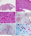
Lichen planus (LP) is a frequent, chronic inflammatory disease involving the skin, mucous membranes and/or skin appendages. Esophageal involvement in lichen planus (ELP) is a clinically important albeit underdiagnosed inflammatory condition. This narrative review aims to give an overview of the current knowledge on ELP, its prevalence, pathogenesis, clinical manifestation, diagnostic criteria, and therapeutic options in order to provide support in clinical management. Studies on ELP were collected using PubMed/Medline. Relevant clinical and therapeutical characteristics from published patient cohorts including our own cohort were extracted and summarized. ELP mainly affects middle-aged women. The principal symptom is dysphagia. However, asymptomatic cases despite progressed macroscopic esophageal lesions may occur. The pathogenesis is unknown, however an immune-mediated mechanism is probable. Endoscopically, ELP is characterized by mucosal denudation and tearing, trachealization, and hyperkeratosis. Scarring esophageal stenosis may occur in chronic courses. Histologic findings include mucosal detachment, T-lymphocytic infiltrations, epithelial apoptosis (Civatte bodies), dyskeratosis, and hyperkeratosis. Direct immuno-fluorescence shows fibrinogen deposits along the basement membrane zone. To date, there is no established therapy. However, treatment with topical steroids induces symptomatic and histologic improvement in two thirds of ELP patients in general. More severe cases may require therapy with immunosuppressors. In symptomatic esophageal stenosis, endoscopic dilation may be necessary. ELP may be regarded as a precancerous condition as transition to squamous cell carcinoma has been documented in literature. ELP is an underdiagnosed yet clinically important differential diagnosis for patients with unclear dysphagia or esophagitis. Timely diagnosis and therapy might prevent potential sequelae such as esophageal stenosis or development of invasive squamous cell carcinoma. Further studies are needed to gain more knowledge about the pathogenesis and treatment options.
Keywords: Budesonide; Dysphagia; Esophagitis; Lichen planus; Precancerosis; T-lymphocytes.
©The Author(s) 2022. Published by Baishideng Publishing Group Inc. All rights reserved.
Conflict of interest statement
Conflict-of-interest statement: All the authors report no relevant conflicts of interest for this article.
Figures


References
-
- Jansson-Knodell CL, Codipilly DC, Leggett CL. Making Dysphagia Easier to Swallow: A Review for the Practicing Clinician. Mayo Clin Proc. 2017;92:965–972. - PubMed
-
- Almashat SJ, Duan L, Goldsmith JD. Non-reflux esophagitis: a review of inflammatory diseases of the esophagus exclusive of reflux esophagitis. Semin Diagn Pathol. 2014;31:89–99. - PubMed
-
- Malone V, Sheahan K. Novel and rare forms of oesophagitis. Histopathology. 2021;78:4–17. - PubMed
-
- Ahuja NK, Clarke JO. Evaluation and Management of Infectious Esophagitis in Immunocompromised and Immunocompetent Individuals. Curr Treat Options Gastroenterol. 2016;14:28–38. - PubMed
Publication types
MeSH terms
LinkOut - more resources
Full Text Sources
Medical

