Review
. 2022 Nov;15(11 Suppl 1):S3-S15.

Using Gene Expression Profiling to Personalize Skin Cancer Management

Affiliations
Review

Using Gene Expression Profiling to Personalize Skin Cancer Management

Jo Ann LeQuang. J Clin Aesthet Dermatol. 2022 Nov.

Abstract

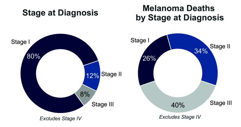
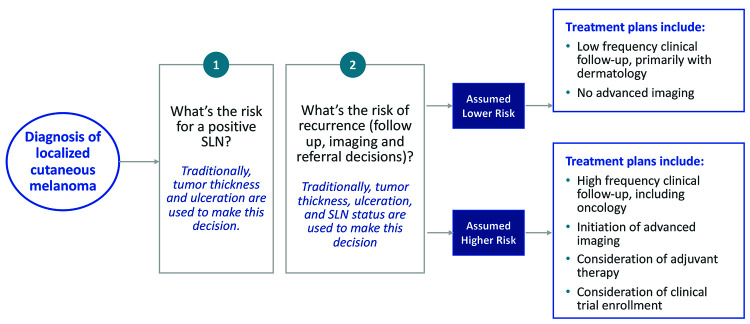
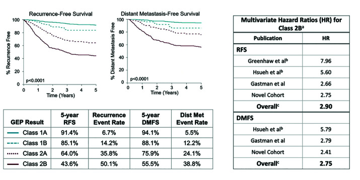
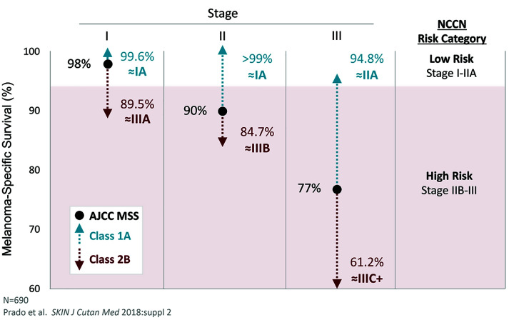
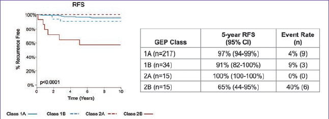
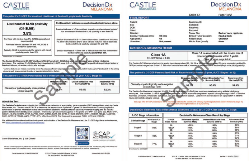
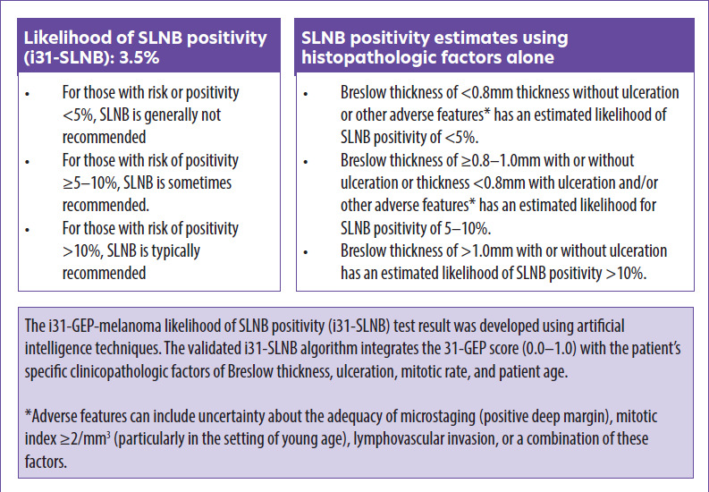
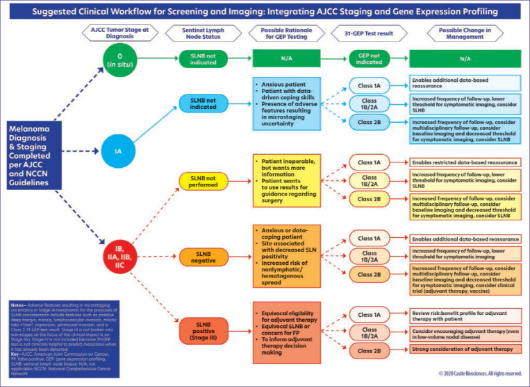
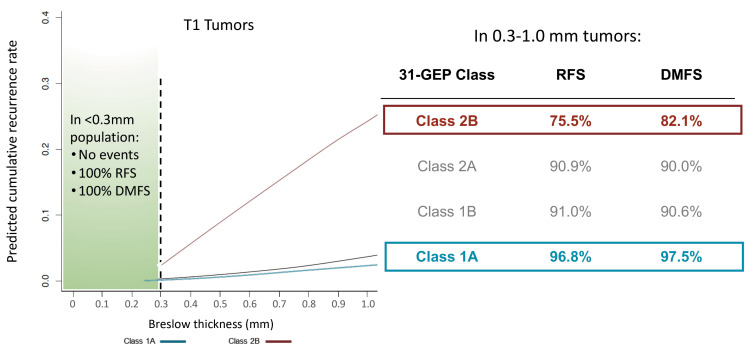
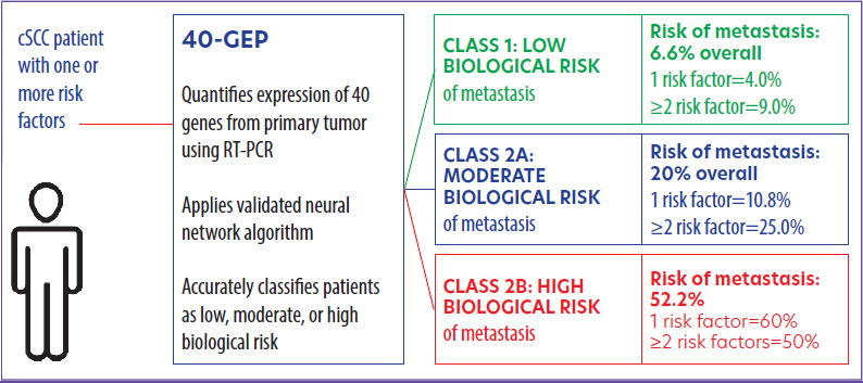
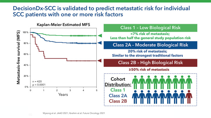
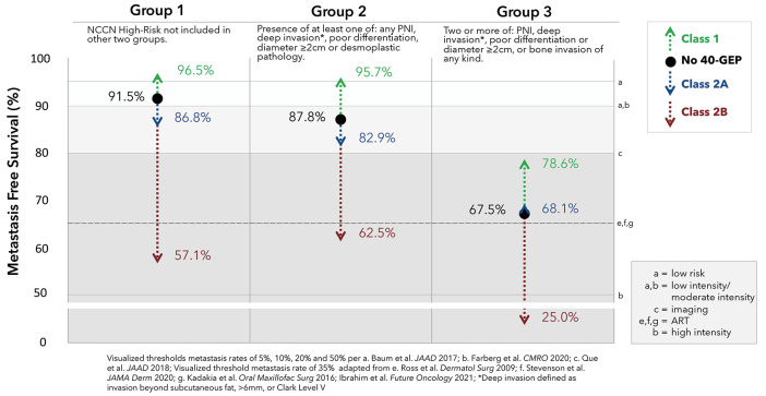
Risk-stratification of cancer, traditionally performed through staging, directs optimal disease management decisions with the result of improved patient outcomes. Many forms of cutaneous cancer have overall excellent survival rates, but conventional staging methods are imperfect in identifying high-risk patients. Gene expression profiling (GEP) is a clinically available, objective metric that can be used in conjunction with traditional clinicopathological staging to help clinicians stratify risk in patients with skin cancer, even in those who lack traditional risk markers. For patients with melanoma, the 31-GEP test provides personalized prognostic information that can guide risk-appropriate clinical management and surveillance decisions. The i31-GEP integrates 31-GEP results with clinicopathological features to provide a risk of recurrence (i31-GEP for ROR) and likelihood of having a positive sentinel lymph node biopsy (SLNB) (i31-GEP for SLNB) for patients with melanoma. For patients with cutaneous squamous cell carcinoma who have at least one risk factor, the 40-GEP test allows for better risk stratification by identifying the high-risk patients who are most likely to develop metastasis. These tests can be easily integrated into clinical practice to help guide treatment choices.

Keywords: 31-GEP; 40-GEP; cutaneous squamous cell carcinoma; gene expression profiling; i31-GEP; melanoma; prognosis; risk of recurrence; risk stratification; sentinel lymph node biopsy.


Conflict of interest statement

DISCLOSURES: Ms. LeQuang reports no conflicts of interest relevant to the content of this article.

Figures

FIGURE 1.
FIGURE 1.
Melanoma staged at diagnosis (left) and melanoma deaths by stage at diagnosis (right)—These diagrams exclude Stage IV. While American Joint Commission on Cancer (AJCC) clinicopathologic factors are helpful clinically, the majority of deaths occur in early stage disease in patients with cutaneous melanoma. Reprinted with permission. Kwatra SG, Hines H, Semenov YR, et al. A dermatologist’s guide to implementation of gene expression profiling in the management of melanoma. J Clin Aesthet Dermatol. 2020;13(11 Suppl 1):s3–s14.
FIGURE 2.
FIGURE 2.
A decision flowchart used in assessing risks and treatments for patients with cutaneous melanoma—According to National Comprehensive Cancer Network guidelines, physicians should discuss and consider a sentinel lymph node biopsy (SLNB) with patients who have 5–10% risk of positivity; for patients with >10% risk of positivity, physicians should recommend an SLNB.
FIGURE 3.
FIGURE 3.
A) Kaplan-Meier survival curves for recurrence-free survival (RFS) and distant-metastasis-free survival (DMFS) based on prospective studies and a review (Greenhaw et al, Hsueh et al, Gastman, and a novel cohort)—Note that the overall hazard ratios (HR) were the same using fixed-effect and random-effect models (B). aMultivariate model included all 31-GEP subclasses, age, Breslow thickness, ulceration, and node status; bProspective study; cSame hazard ratio with fixed effect and random effects models Adapted with permission under Creative Commons Attribution-NonCommercial-NoDerivatives 4.0 International (CC BY-NC-ND 4.0) (https://creativecommons.org/licenses/by-nc-nd/4.0/). Greenhaw BN, Covington KR, Kurley SJ, et al. Molecular risk prediction in cutaneous melanoma: a meta-analysis of the 31-gene expression profile (GEP) prognostic test in 1,479 patients. J Am Acad Dermatol. 2020 Sep;83(3):745–753.
FIGURE 4.
FIGURE 4.
31-GEP further informs risk obtained by AJCC-8 —AJCC melanoma 5-year survival rates (in black) alone and with the addition of 31-GEP test data (in blue and red), providing more robust data: AJCC: American Joint Committee on Cancer, 8th edition; GEP: gene expression profile; MSS: melanoma-specific survival; NCCN: National Comprehensive Cancer Network Adapted with permission: under Creative Commons Attribution-NonCommercial-NoDerivatives 4.0 International (CC BY-NC-ND 4.0) (https://creativecommons.org/licenses/by-nc-nd/4.0/). Wisco OJ, Marson JW, Litchman GH, et al. Improved cutaneous melanoma survival stratification through integration of 31-gene expression profile testing with the American Joint Committee on Cancer 8th Edition Staging. Melanoma Res. 2022;32(2):98–102.
FIGURE 5.
FIGURE 5.
The 31-GEP-melanoma test can identify the subset of patients with thin melanomas (≤1mm) at risk for recurrence or metastasis. In this study, GEP Class 2B had a hazard ratio of 9.34 (95% CI, 2.03–42.97, p=0.004) and was most predictive of a poor outcome; CI: confidence interval; GEP: gene expression profile; RFS: recurrence-free survival Adapted with permission under Creative Commons Attribution-NonCommercial-NoDerivatives 4.0 International (CC BY-NC-ND 4.0) (https://creativecommons.org/licenses/by-nc-nd/4.0/). Gastman BR, Gerami P, Kurley SJ, et al. Identification of patients at risk of metastasis using a prognostic 31-gene expression profile in subpopulations of melanoma patients with favorable outcomes by standard criteria. J Am Acad Dermatol. 2019;80(1):149–157.e4.
FIGURE 6.
FIGURE 6.
31-GEP-melanoma report provides a detailed and highly personalized risk assessment; GEP: gene expression profile; ROR: risk of recurrence
FIGURE 7.
FIGURE 7.
A sample i31-GEP report stating the likelihood that a particular patient will have a positive SLNB—The right-hand column shows clinicopathological features, while the left-hand column incorporates the 31-GEP report in its findings. This report also provides data on the patient’s risk of recurrence for both positive and negative SLNB findings. GEP: gene expression profiling; SLNB: sentinel lymph node biopsy
FIGURE 8.
FIGURE 8.
Kwatra et al developed a workflow paradigm for the use of 31-gene expression profile (GEP) and i31-GEP-risk of recurrence (ROR)-melanoma testing. Reprinted with permission.
FIGURE 9.
FIGURE 9.
Cumulative rate of recurrence and 5-year outcome results demonstrate the differences between 31-GEP Class 1A and Class 2B results. GEP: gene expression profile; RFS: recurrence-free survival; DMFS: distant metastasis-free survival
FIGURE 10.
FIGURE 10.
Overview of the analysis of the event rates for patients in the cohort who progressed to metastasis; GEP: gene expression profiling; RT-PCR: reverse transcriptase polymerase chain reaction; cSCC: cutaneous squamous cell carcinoma Adapted with permission under Creative Commons Attribution-NonCommercial-NoDerivatives 4.0 International (CC BY-NC-ND 4.0) (https://creativecommons.org/licenses/by-nc-nd/4.0/).Ibrahim SF, Kasprzak JM, Hall MA, et al. Enhanced metastatic risk assessment in cutaneous squamous cell carcinoma with the 40-gene expression profile test. Future Oncol. 2022;18(7):833–847.
FIGURE 11.
FIGURE 11.
The 40-GEP is validated to predict metastatic risk for individual patients with cSCC who have one or more risk factors. GEP: gene expression profile; cSCC: cutaneous squamous cell carcinoma Adapted with permission under Creative Commons Attribution-NonCommercial-NoDerivatives 4.0 International (CC BY-NC-ND 4.0) (https://creativecommons.org/licenses/by-nc-nd/4.0/). Ibrahim SF, Kasprzak JM, Hall MA, et al. Enhanced metastatic risk assessment in cutaneous squamous cell carcinoma with the 40-gene expression profile test. Future Oncol. 2022;18(7):833–847.
FIGURE 12.
FIGURE 12.
Using NCCN designated risk factors, tumors were stratified as high, higher, and highest risk (Groups 1, 2, and 3, respectively), which appear as black dots. Numbers in green show how a 40-GEP Class 1 test result can affect risk prediction, while percentages in blue and red represent Class 2A and Class 2B, respectively. GEP: gene expression profile; NCCN: National Comprehensive Cancer Network; cSCC: cutaneous squamous cell carcinoma

Similar articles

Cited by

References

    1. Weiss SA, Wolchok JD, Sznol M. Immunotherapy of melanoma: facts and hopes. Clin Cancer Res. 2019;25(17):5191–5201. - PMC - PubMed
    1. https://www.cancer.gov/news-events/cancer-currents-blog/2020/metastatic-... NCI staff. Deaths from metastatic melanoma drop substantially in the United States. 21 Apr 2020. National Institute of Health. National Cancer Institute. Accessed 13 Jun 2022.
    1. Islami F, Ward EM, Sung H et al. Annual report to the nation on the status of cancer, part 1: national cancer statistics. JNCI J Natl Cancer Inst. 2021;113(12):1658–1669. - PMC - PubMed
    1. Wolchok JD, Chiarion-Sileni V, Gonzalez R et al. Overall survival with combined nivolumab and ipilimumab in advanced melanoma. N Engl J Med. 2017;377:1345–1356. - PMC - PubMed
    1. Hodi FS, Chiarion-Sileni V, Gonzalez R et al. Nivolumab plus ipilimumab or nivolumab alone versus ipilimumab alone in advanced melanoma (CheckMate 067): 4-year outcomes of a multicentre, randomised, phase 3 trial. Lancet Oncol. 2018;19:1480–1492. - PubMed

LinkOut - more resources