. 2021 Aug 3;10(12):3279-3294.
doi: 10.1515/nanoph-2021-0241. eCollection 2021 Sep.

Repeated porphyrin lipoprotein-based photodynamic therapy controls distant disease in mouse mesothelioma via the abscopal effect

Affiliations

Repeated porphyrin lipoprotein-based photodynamic therapy controls distant disease in mouse mesothelioma via the abscopal effect

Jenny Lou et al. Nanophotonics. .

Abstract

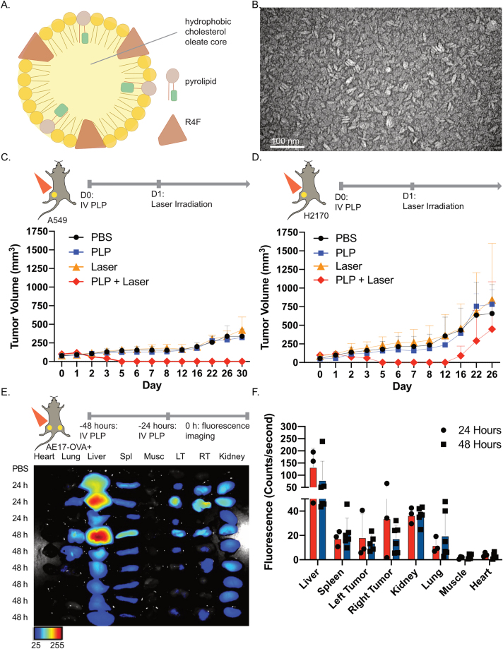
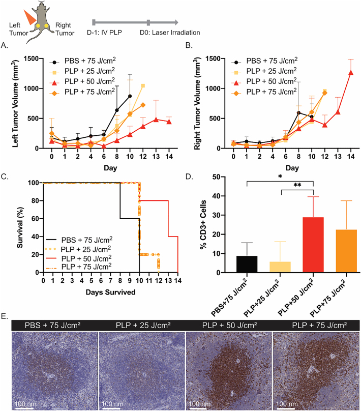
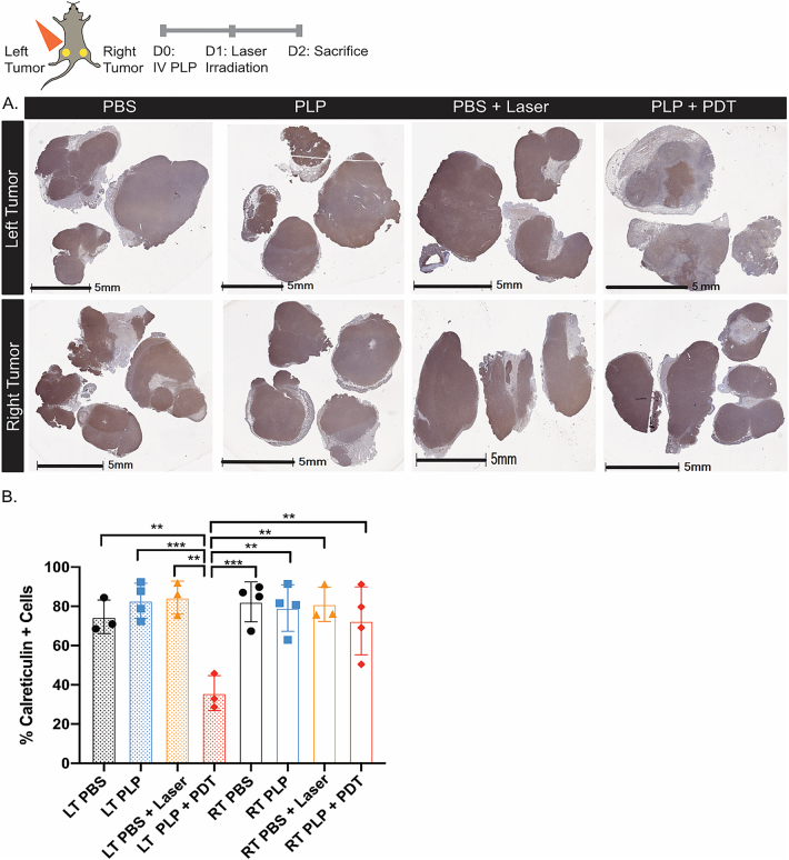
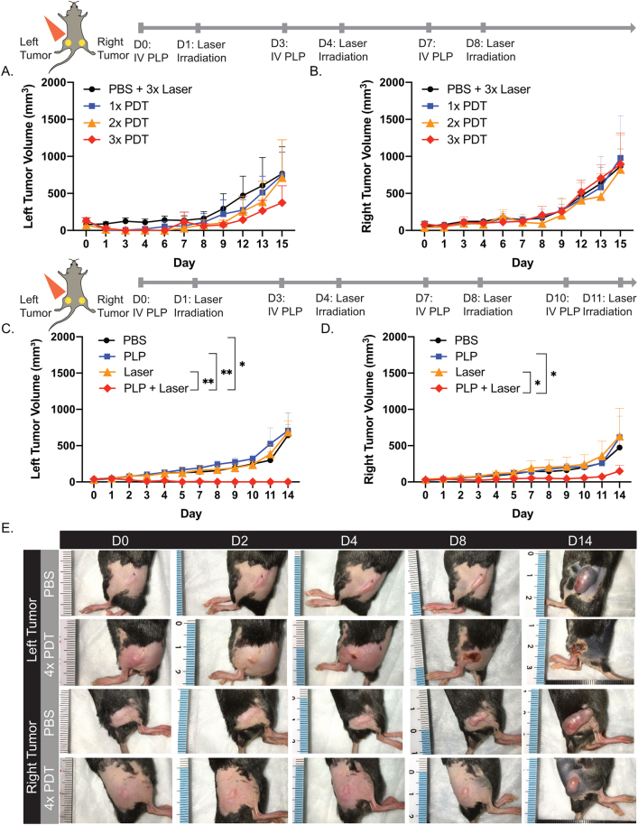
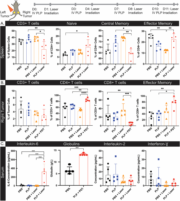
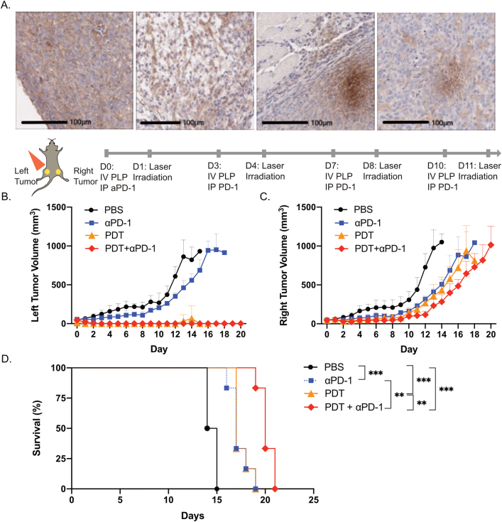
While photodynamic therapy (PDT) can induce acute inflammation in the irradiated tumor site, a sustained systemic, adaptive immune response is desirable, as it may control the growth of nonirradiated distant disease. Previously, we developed porphyrin lipoprotein (PLP), a ∼20 nm nanoparticle photosensitizer, and observed that it not only efficiently eradicated irradiated primary VX2 buccal carcinomas in rabbits, but also induced regression of nonirradiated metastases in a draining lymph node. We hypothesized that PLP-mediated PDT can induce an abscopal effect and we sought to investigate the immune mechanism underlying such a response in a highly aggressive, dual subcutaneous AE17-OVA+ mesothelioma model in C57BL/6 mice. Four cycles of PLP-mediated PDT was sufficient to delay the growth of a distal, nonirradiated tumor four-fold relative to controls. Serum cytokine analysis revealed high interleukin-6 levels, showing a 30-fold increase relative to phosphate-buffered solution (PBS) treated mice. Flow cytometry revealed an increase in CD4+ T cells and effector memory CD8+ T cells in non-irradiated tumors. Notably, PDT in combination with PD-1 antibody therapy prolonged survival compared to monotherapy and PBS. PLP-mediated PDT shows promise in generating a systemic immune response that can complement other treatments, improving prognoses for patients with metastatic cancers.

Keywords: PD-1; immune response; immunotherapy; photodynamic therapy; porphyrin; thoracic malignant tumor.


Figures

Figure 1:
Figure 1:
Porphyrin-lipoprotein (PLP) can be an effective photosensitizer for lung tumor models. (A) Schematic of PLP, the photosensitizer used for photodynamic therapy. (B) Transmission electron microscopy (TEM) image of PLP at 200 000× magnification. (C) Assessment of tumor burden in subcutaneous A549 tumors in nude mice, after treatment with phosphate-buffered solution (PBS), PLP (4 mg/kg), laser irradiation, or photodynmaic therapy (PDT) (PLP + laser). n = 3 per group. (D) Assessment of tumor burden in subcutaneous H2170 tumors in nude mice, after treatment with PBS, PLP (4 mg/kg), laser irradiation, or PDT (PLP + laser). n = 2 per group. Mice received light irradiation with a 671 nm laser at a light dose of 100 J/cm2 at 24 h postinjection of PLP. (E) Ex vivo fluorescence imaging of heart, lung, spleen (Spl), muscle (Musc), left (LT) and right (RT) tumors, and kidneys dissected from mice bearing dual subcutaneous AE17-OVA+ tumors. Mice received intravenous injections of PBS (n = 1), or PLP (4 mg/kg) at 24 h (n = 3) or 48 h (n = 6) prior to imaging. (F) Quantification of the fluorescence signal in various organs. Data are mean ± standard deviation.
Figure 2:
Figure 2:
Optimization of light dosage for immune priming. (A) Dual subcutaneous AE17-OVA+ tumor bearing mice were treated with either PBS or PLP (4 mg/kg). Twenty-four hours post-injection, the left hindlimb tumors on mice were irradiated with a 671 nm laser at a light dose of either 25 J/cm2, 50 J/cm2, or 75 J/cm2. Evaluation of tumor volumes of irradiated left hindlimb tumor volumes (n = 5). (B) Assessment of non-irradiated right hindlimb tumor volumes (n = 5). (C) Survival of mice treated with either PBS + 75 J/cm2, PLP + 25 J/cm2, PLP + 50 J/cm2, or PLP + 75 J/cm2 (n = 5). (D) Histological analysis of the percentage of CD3+ T cells in spleens of mice harvested at time of endpoint (n = 3). (E) Microscopic image of a CD3-stained spleen from one mouse in each treatment group. Data are mean ± standard deviation. Statistical significance was determined using a one-way ANOVA followed by a post-hoc Tukey test. *p < 0.05, **p < 0.01.
Figure 3:
Figure 3:
PDT reduced calreticulin expression in irradiated tumors. Dual AE17-OVA+ tumor bearing mice were treated once with either PBS, PLP, PBS + laser irradiation, or PLP + laser irradiation. Twenty-four hours after irradiation, irradiated and nonirradiated tumors were collected and processed for immunohistochemistry. (A) Whole slide scan of calreticulin stained tumors in mice for irradiated tumors on the left flank and nonirradiated tumors on the right flank. Scale bars represent 5 mm. (B) Quantification of the whole tissue slide by Halo (n = 3–4 per group) for the left (LT) and right (RT) tumors. Data are mean ± standard deviation. Statistical significance was determined using a one-way ANOVA followed by a post-hoc Tukey test. *p < 0.05, **p < 0.01, ***p < 0.001.
Figure 4:
Figure 4:
Four repeated cycles of PDT induced an abscopal effect. (A) Dual subcutaneous AE17-OVA+ tumor bearing mice were treated with either PBS and laser irradiation at 50 J/cm2 three times (n = 6), or PLP and laser irradiation at 50 J/cm2 one (n = 5), two (n = 6), or three times (n = 5). Evaluation of irradiated, left hindlimb tumor volumes. (B) Assessment of nonirradiated, right hindlimb tumor volumes. (C) Dual subcutaneous AE17-OVA+ tumor bearing mice were treated with four cycles of PBS, PLP (4 mg/kg), laser irradiation at 50 J/cm2, or PDT (PLP + laser). Tumors for irradiated, left tumor volumes were measured. (D) Nonirradiated, right hindlimb tumor volumes were measured (n = 4). (E) Photos of left, irradiated and right, nonirradiated hindlimb tumors of mice treated with either PBS or four cycles of PDT on days 0, 2, 4, 8, and 14. Experiments were repeated twice more with similar findings for left and right hindlimb tumor volumes. Data are mean ± standard deviation. Statistical significance was determined using a one-way ANOVA followed by a post hoc Tukey test. *p < 0.05, **p < 0.01.
Figure 5:
Figure 5:
The abscopal effect involved CD4+ and CD8+ T cells. (A) Dual subcutaneous AE17-OVA+ tumor bearing mice were treated with four cycles of PBS, PLP (4 mg/kg), laser irradiation at 50 J/cm2, or PDT (PLP + laser). Mice were sacrificed on day 14 and spleens and right tumors were harvested for flow cytometry, while serum was collected for cytokine analysis. Summary of the percentage of CD3+ T cells, naive, central memory, and effector memory CD4+ T cells in the spleen of treated mice. (B) Summary of the percentage of CD3+ T cells, CD4+ T cells, CD8+ T cells, and effector memory CD8+ T cells in the non-irradiated, right tumors of treated mice. (C) Assessment of the concentration of interleukin-6, globulins, interleukin-2, and interferon-gamma in serum of mice (n = 5). Data are mean ± standard deviation. Statistical significance was determined using a one-way ANOVA, followed by a post hoc Tukey test. n = 5, *p < 0.05, **p < 0.01, ***p < 0.001, ****p < 0.0001.
Figure 6:
Figure 6:
Combination therapy with PDT and αPD-1 antibody treatment improved survival relative to monotherapy. (A) Microscopic images of PD-1 stained AE17-OVA+ tumors. Dual subcutaneous AE17-OVA+ tumor bearing mice were treated with four cycles of PBS, αPD-1 antibody (12 mg/kg), PDT (PLP at 4 mg/kg), or combination αPD-1 antibody (12 mg/kg) and PDT (PLP at 4 mg/kg). Scale bars represent 100 μm. (B) Assessment of irradiated, left hindlimb tumors of mice receiving the aforementioned treatments. (C) Assessment of nonirradiated, right hindlimb tumors of mice receiving the aforementioned treatments. (D) Survival of mice receiving the aforementioned treatments. Data are mean ± standard deviation. Statistical significance of the survival of mice was determined using a one-way ANOVA, followed by a post hoc Tukey test (n = 5, **p < 0.01, ***p < 0.001).
Figure 7:
Figure 7:
Schematic of immune mechanism underlying the abscopal effect induced by PLP-mediated PDT.

References

    1. Obaid G., Broekgaarden M., Bulin A. L., et al. Photonanomedicine: a convergence of photodynamic therapy and nanotechnology. Nanoscale . 2016;8:12471–12503. doi: 10.1039/c5nr08691d. - DOI - PMC - PubMed
    1. Rajora M. A., Lou J. W. H., Zheng G. Advancing porphyrin’s biomedical utility: via supramolecular chemistry. Chem. Soc. Rev. . 2017;46:6433–6469. doi: 10.1039/c7cs00525c. - DOI - PubMed
    1. Nath S., Obaid G., Hasan T. The course of immune stimulation by photodynamic therapy: bridging fundamentals of photochemically induced immunogenic cell death to the enrichment of T-cell repertoire. Photochem. Photobiol. . 2019;95:1288–1305. doi: 10.1111/php.13173. - DOI - PMC - PubMed
    1. Falk-Mahapatra R., Gollnick S. O. Photodynamic therapy and immunity: an update. Photochem. Photobiol. . 2020;96:550–559. doi: 10.1111/php.13253. - DOI - PMC - PubMed
    1. Cecic I., Stott B., Korbelik M. Acute phase response-associated systemic neutrophil mobilization in mice bearing tumors treated by photodynamic therapy. Int. Immunopharm. . 2006;6:1259–1266. doi: 10.1016/j.intimp.2006.03.008. - DOI - PubMed

LinkOut - more resources