. 2022 Oct 28;55(5):169-184.
doi: 10.1267/ahc.22-00076. Epub 2022 Oct 25.

Role of Cancer Stem-like Cells in the Process of Invasion and Mesenchymal Transformation by a Reconstituted Triple-negative Breast Cancer Cell Population Resistant to p53-induced Apoptosis

Affiliations

Role of Cancer Stem-like Cells in the Process of Invasion and Mesenchymal Transformation by a Reconstituted Triple-negative Breast Cancer Cell Population Resistant to p53-induced Apoptosis

Sana Inoue et al. Acta Histochem Cytochem. .

Abstract

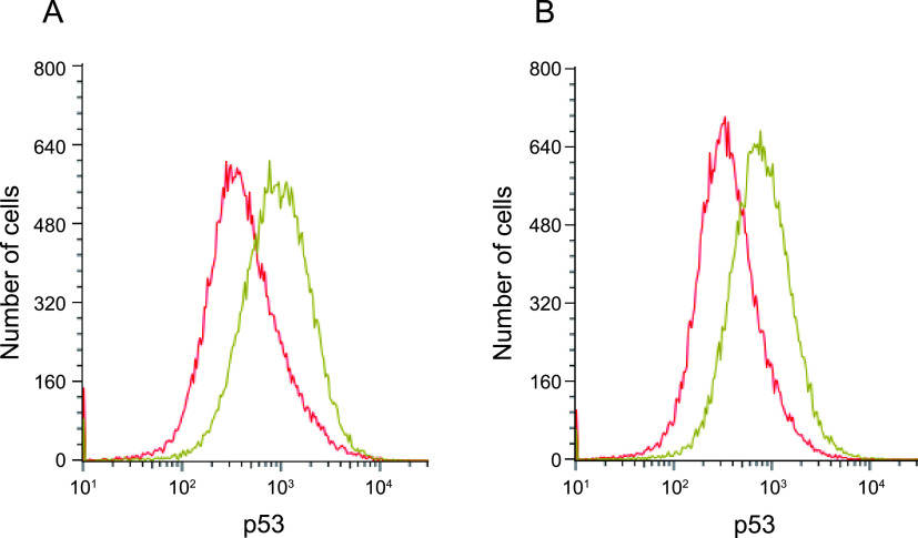
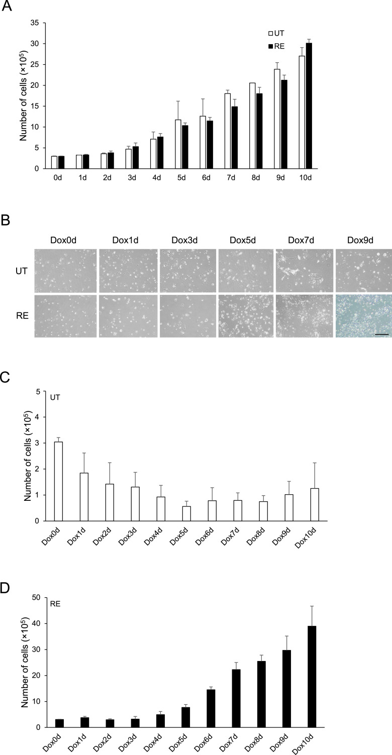
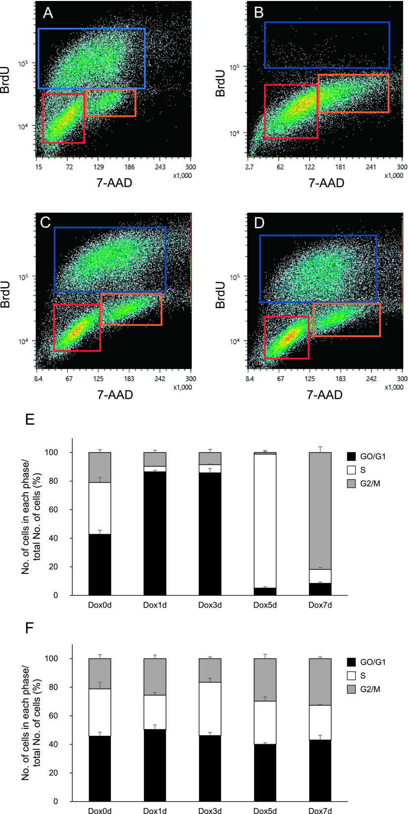
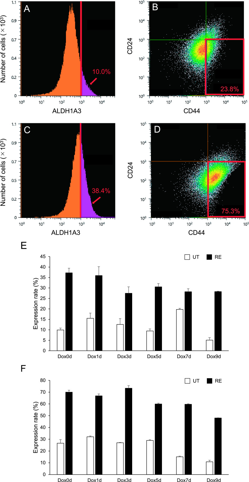
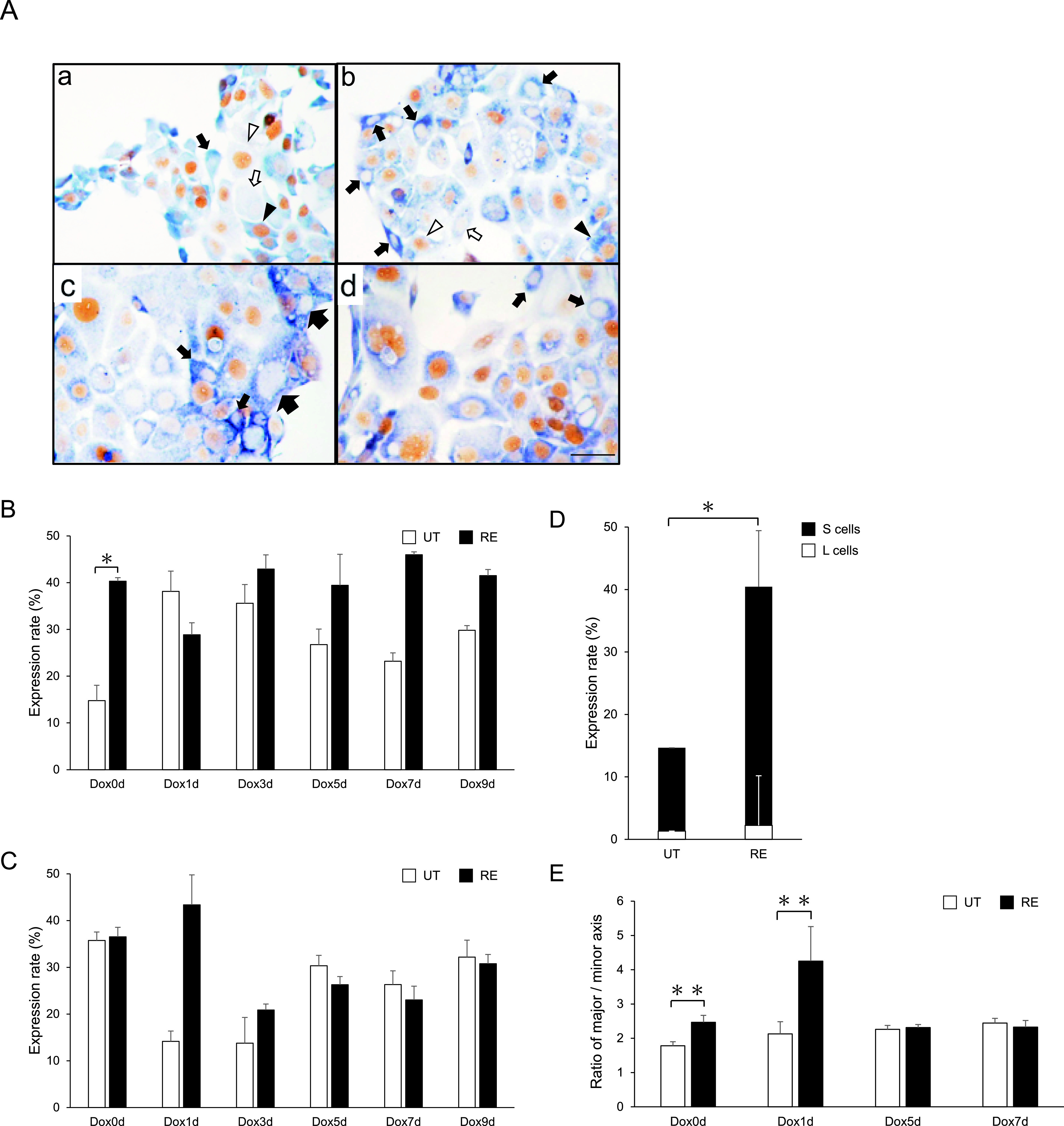
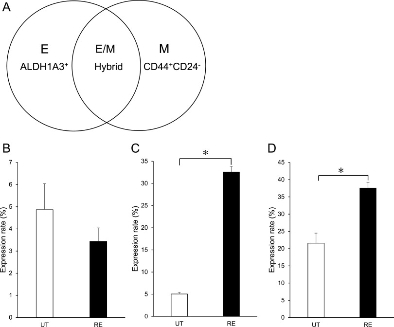
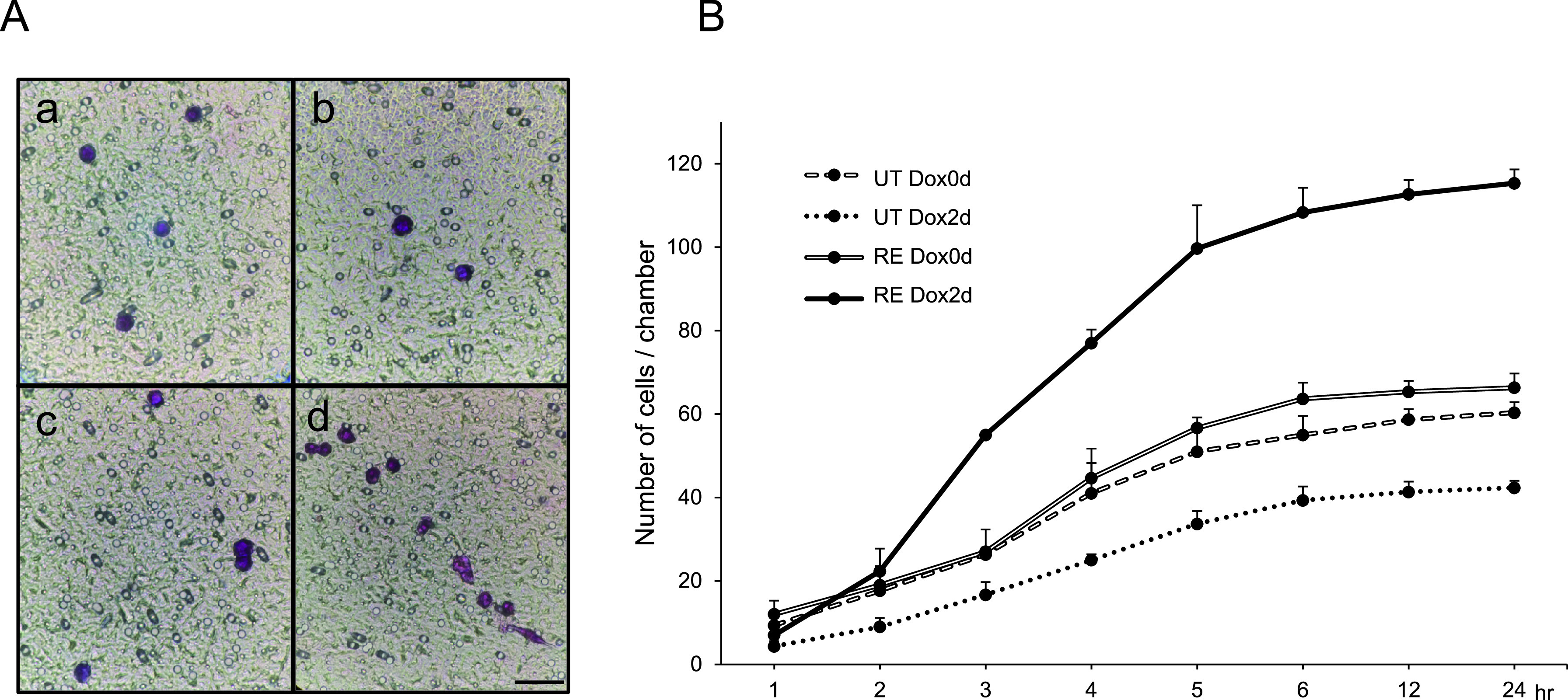
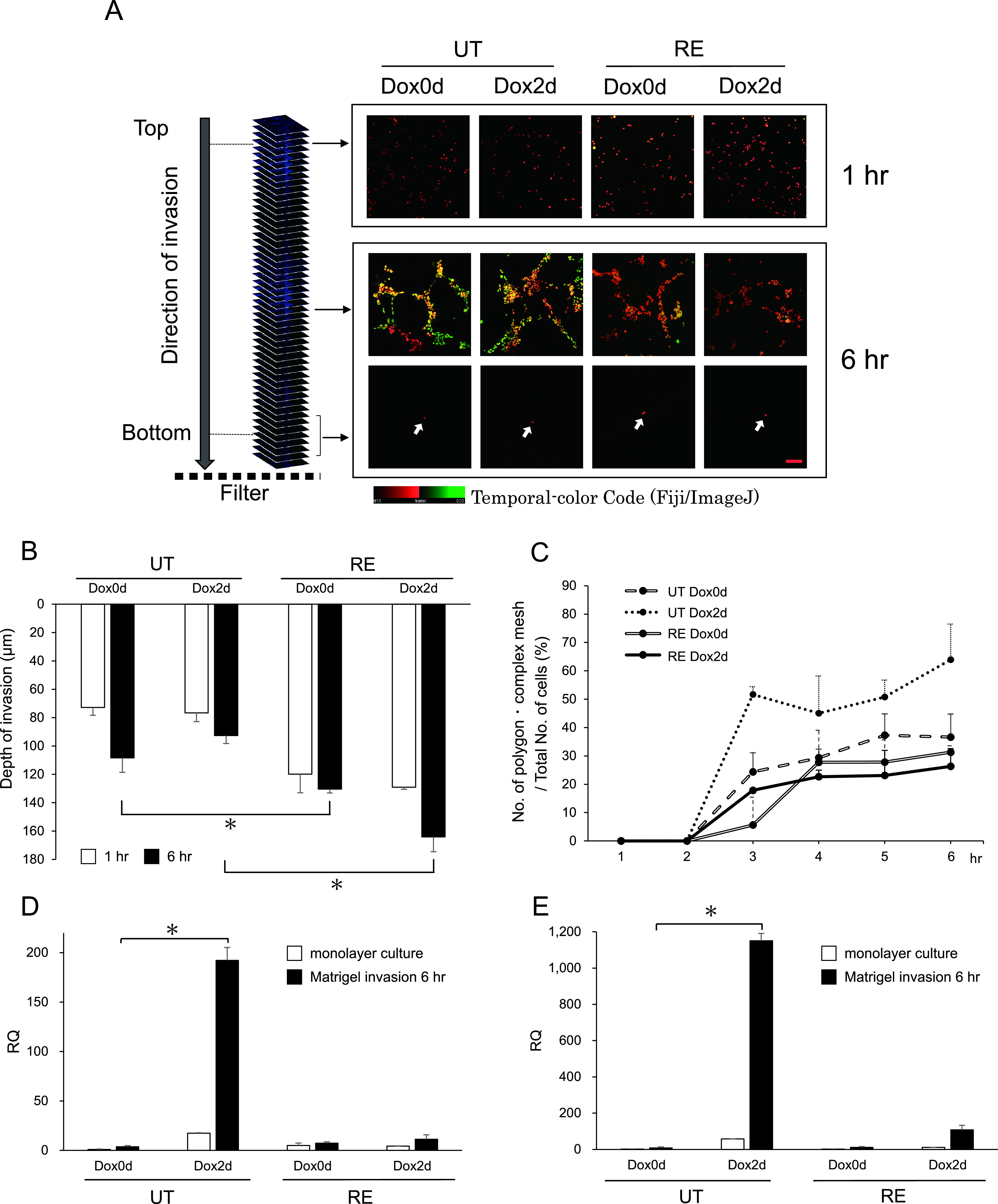
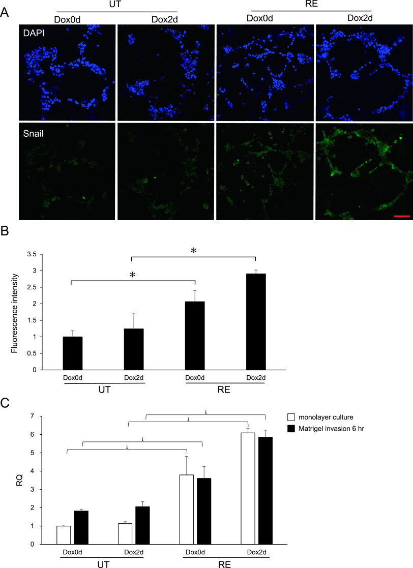
We investigated the role of cancer stem cells (CSCs) in a population of triple-negative breast cancer (TNBC) cells that are resistant to apoptosis. A human breast cancer cell population capable of inducing p53 expression with doxycycline (Dox) was created and used as an untreated control (UT). After the addition of Dox to UT for 5 days, the cell population reconstituted with cells showing resistance to apoptosis was named RE. Fluorescence-activated cell sorting (FACS) and immunostaining revealed that after the addition of Dox, the ratio of cells in the S and G2/M phases decreased in UT as apoptosis proceeded, but did not markedly change in apoptosis-resistant RE. CSC-like cells in RE exhibited a cell morphology with a larger ratio of the major/minor axis than UT. FACS showed that RE had a higher proportion of CSC-like cells and contained more CD44+CD24- mesenchymal CSCs than ALDH1A3+ epithelial-like CSCs. In a Matrigel invasion assay, UT was more likely to form a three-dimensional cell population, whereas RE exhibited a planar population, higher migration ability, and the up-regulated expression of epithelial-mesenchymal transition-related genes. These results provide insights into the mechanisms by which TNBC cells acquire treatment resistance at the time of recurrence.

Keywords: apoptosis resistance; breast cancer; cancer stem-like cell; invasion.


Conflict of interest statement

VThe authors declare that there are no conflicts of interest.

Figures

Fig. 1.
Fig. 1.
Dox-induced p53 expression by a FACS analysis. UT Dox0d (A) and RE Dox0d (B) are indicated by a red line, and UT Dox2d (A) and RE Dox2d (B) by a green line.
Fig. 2.
Fig. 2.
Time-course changes in the number of cells and effects of the addition of Dox on growth curves. A: Changes in the number of cells without Dox. B: Examples of cell growth patterns with Dox analyzed by an inverted phase-contrast microscope. Bar = 200 μm. C and D: Changes in the number of viable cells with Dox in UT (C) and RE (D). The number of UT is expressed on a 1/10 scale of RE.
Fig. 3.
Fig. 3.
Cell cycle analysis by FACS. A, B, C, and D: Each cell was pulse labeled with BrdU and nuclear DNA was stained with 7-AAD. The red open square indicates the G0/G1 phase, blue the S phase, and orange the G2/M phase. A: UT Dox0d, B: UT Dox3d, C: RE Dox0d, and D: RE Dox3d. E and F: Each phase of the cell cycle, the G0/G1, S, and G2/M phases, was expressed as a ratio to the total number of cells.
Fig. 4.
Fig. 4.
Detection of CSC-like cells in UT and the reconstituted RE cell population by a FACS analysis. A and C: Positive cells are defined as a fluorescent signal intensity of 103 or more, shown in purple areas in the histogram. A: UT Dox0d, C: RE Dox0d. B and D: CD24 is defined as 103 or less and CD44+ as 103 or more, which are indicated by the open red square. E and F: Time-course analysis of the expression patterns of ALDH1A3 (E) and CD44+CD24 (F) in UT and RE. The bar indicates means ± SE.
Fig. 5.
Fig. 5.
Analysis of the cell population constitution by immunocytochemical staining and morphological measurements. A: Double immunocytochemical staining. ALDH1A3 stains blue in the cytoplasm and Ki-67 stains brown in the nucleus. a and b: Examples of the expression patterns of ALDH1A3 and Ki-67 in Dox0d. a: UT Dox0d, b: RE Dox0d. solid arrow: ALDH1A3+Ki-67, solid arrowhead: ALDH1A3+Ki-67+, open arrowhead: ALDH1A3Ki-67+, and open arrow: ALDH1A3Ki67. c and d: Examples of L (≥ 50 μm) and S cells (< 50 μm), which are distinguished by the length of their major axis. Large arrows: L cells, small arrows: S cells. c: UT Dox1d, d: RE Dox1d. Bar = 50 μm. B and C: Time-course analysis of expression patterns of the CSC marker, ALDH1A3+Ki-67 (B) and the cancer precursor cell marker, ALDH1A3+Ki-67+ (C) indicated by the expression rate with the addition of Dox. The bar indicates means ± SE. *p < 0.01. D: The rate of ALDH1A3+Ki-67 cells without Dox sorted by cell size. The ratios of S cells and L cells divided by the total number of cells are shown. The bar indicates means ± SE. *p < 0.01 UT versus RE. E: The ratio of the major/minor axis of mesenchymal-like elongated cells in ALDH1A3+Ki-67 cells. **p < 0.05 (the Student’s t-test).
Fig. 6.
Fig. 6.
Epithelial-like and mesenchymal-like CSCs by a FACS analysis. A: The representation of cell subpopulations in the diagram indicates CSCs with the epithelial-like ALDH1A3+ (E), mesenchymal-like CD44+CD24 (M), and hybrid epithelial/mesenchymal ALDH1A3+/CD44+CD24 (E/M) phenotypes. B: Ratio of cells from ALDH1A3+ cells (E) minus hybrid ALDH1A3+/CD44+CD24 cells (E/M). C: Ratio of hybrid ALDH1A3+/CD44+CD24 cells (E/M). The bar indicates means ± SE. *p < 0.01 UT versus RE. D: Ratio of cells from CD44+CD24 cells (M) minus hybrid ALDH1A3+/CD44+CD24 cells (E/M). The bar indicates means ± SE. *p < 0.01 UT versus RE.
Fig. 7.
Fig. 7.
Quantification of cell invasion and migration. A: Cells reaching the filter pores after a 6-hr culture in Matrigel were stained by Giemsa and photographed. Bar = 50 μm. B: Time-course analysis of the total number of invading cells per chamber. Each point indicates the average value and standard deviation of 3 chambers.
Fig. 8.
Fig. 8.
Depth and morphological characteristics of invading cells in Matrigel. A: Fluorescence images of cells infiltrating Matrigel by a confocal laser scanning microscope. Cells cultured in the Matrigel chamber were stained with DAPI and color-coded as follows. Regarding the three-dimensional construction of cells, Z-stack images are represented on a color code scale using Fiji/ImageJ software (version 1.52 g, Java 1.80_172, NIH). Bar = 100 μm. B: Depth of invasion in Matrigel. The distance from the surface of Matrigel to the invasion front of cell populations in Matrigel cultures for 1 hr and 6 hr was measured by a confocal laser scanning microscope. The values at each point are indicated by the mean and standard deviation (n = 5). *p < 0.01 (the Student’s t-test). C: Percentages of cell populations that form polygon and complex meshes during infiltration in Matrigel. The definition of the polygon and complex mesh was previously described (Aranda and Owen 2009 [2]). D and E: Expression levels of VM marker mRNA in infiltrating cells in Matrigel. D: MMP-9, E: VE-cadherin). p < 0.01 (the Student’s t-test).
Fig. 9.
Fig. 9.
Snail expression patterns in the process of Matrigel infiltration. Cells in the Matrigel culture for 6 hr were immunofluorescently stained with Snail. A: Fluorescence images of Snail expression patterns in Matrigel using a confocal laser scanning microscope. The nucleus is shown by DAPI (blue) and Snail expression is observed in the nucleus and cytoplasm (green). Bar = 100 μm. B: A quantitative analysis was performed on the Snail fluorescence channel using Fiji/ImageJ software. The average value of fluorescence intensity in 9 randomly selected fields of view where cells exist was measured, and the values of three wells were combined. The bar indicates means ± SE. *p < 0.01 UT versus RE. C: Semiquantitative analysis of Snail mRNA expression. RT-PCR was performed using RNA extracted from cells in the conventional monolayer culture and in Matrigel.

Similar articles

Cited by

References

    1. Al-Hajj, M., Wicha, M. S., Benito-Hernandez, A., Morrison, S. J. and Clarke, M. F. (2003) Prospective identification of tumorigenic breast cancer cells. Proc. Natl. Acad. Sci. U S A 100; 3983–3988. - PMC - PubMed
    1. Aranda, E. and Owen, G. I. (2009) A semi-quantitative assay to screen for angiogenic compounds and compounds with angiogenic potential using the EA.hy926 endothelial cell line. Biol. Res. 42; 377–389. - PubMed
    1. Biddle, A., Liang, X., Gammon, L., Fazil, B., Harper, L. J., Emich, H., et al. (2011) Cancer stem cells in squamous cell carcinoma switch between two distinct phenotypes that are preferentially migratory or proliferative. Cancer Res. 71; 5317–5326. - PubMed
    1. Bocci, F., Gearhart-Serna, L., Boareto, M., Ribeiro, M., Ben-Jacob, E., Devi, G. R., et al. (2019) Toward understanding cancer stem cell heterogeneity in the tumor microenvironment. Proc. Natl. Acad. Sci. U S A 116; 148–157. - PMC - PubMed
    1. Bomken, S., Fiser, K., Heidenreich, O. and Vormoor, J. (2010) Understanding the cancer stem cell. Br. J. Cancer 103; 439–445. - PMC - PubMed