. 2022 Oct 28;55(5):159-168.
doi: 10.1267/ahc.22-00043. Epub 2022 Oct 25.

Optimized Mouse-on-mouse Immunohistochemical Detection of Mouse ESR2 Proteins with PPZ0506 Monoclonal Antibody

Affiliations

Optimized Mouse-on-mouse Immunohistochemical Detection of Mouse ESR2 Proteins with PPZ0506 Monoclonal Antibody

Mina Ozawa et al. Acta Histochem Cytochem. .

Abstract

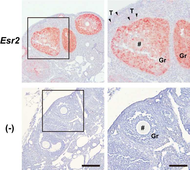
Despite the physiological significance of ESR2, a lack of well-validated detection systems for ESR2 proteins has hindered progress in ESR2 research. Thus, recent identification of a specific anti-human ESR2 monoclonal antibody (PPZ0506) and its specific cross-reactivity against mouse and rat ESR2 proteins heightened momenta toward development of appropriate immunohistochemical detection systems for rodent ESR2 proteins. Building upon our previous optimization of ESR2 immunohistochemical detection in rats using PPZ0506, in this study, we further aimed to optimize mouse-on-mouse immunohistochemical detection using PPZ0506. Our assessment of several staining conditions using paraffin-embedded ovary sections revealed that intense heat-induced antigen retrieval, appropriate blocking, and appropriate antibody dilutions were necessary for optimization of mouse-on-mouse immunohistochemistry. Subsequently, we applied the optimized immunostaining method to determine expression profiles of mouse ESR2 proteins in peripheral tissues and brain subregions. Our analyses revealed more localized distribution of mouse ESR2 proteins than previously assumed. Moreover, comparison of these results with those obtained in humans and rats using PPZ0506 revealed interspecies differences in ESR2 expression. We expect that our optimized methodology for immunohistochemical staining of mouse ESR2 proteins will help researchers to solve multiple lines of controversial evidence concerning ESR2 expression.

Keywords: ERβ; ESR2; PPZ0506; estrogen receptor β; immunohistochemistry.


Conflict of interest statement

VThe authors declare that they have no conflicts of interest to declare.

Figures

Fig. 1.
Fig. 1.
Quantification of mouse Esr2 expression levels. Expression levels of mouse Esr2 transcripts were quantified using RT-qPCR. Gapdh was used as a reference gene for normalization. Data are presented as mean ± SD (n = 4).
Fig. 2.
Fig. 2.
Distribution of mouse Esr2 transcripts in ovary. The distribution of mouse Esr2 transcripts in ovary was analyzed using RNAScope in situ hybridization assays. Paraffin-embedded ovary sections were reacted with RNAScope probes against mouse Esr2 transcripts. Non-specific signals were evaluated using RNAScope Negative Control Probes (−). #, Oocyte cells; Gr, granulosa cells; T, theca cells. Bars = 200 μm in left panels and 100 μm in right panels. Similar staining patterns were obtained in three separate experiments (n = 3).
Fig. 3.
Fig. 3.
Effects of blocking reagents on immunohistochemical detection of mouse ESR2 proteins with the PPZ0506 antibody. Effects of different blocking reagents on immunohistochemical detection with the PPZ0506 antibody were evaluated. Paraffin-embedded sections were prepared from ovary and spleen, and immunostained after treatment with different blocking reagents. (A) Sections were stained without primary antibody reaction. (B–D) Sections were immunoreacted after blocking in a mouse-on-mouse blocking reagent (B), 1% BSA/PBST (C), and 0.1 mg/mL goat anti-mouse IgG antibody/PBST (D). Bars = 200 μm in left panels and 100 μm in right panels. Similar immunostaining patterns were obtained in three separate experiments (n = 3).
Fig. 4.
Fig. 4.
Effects of heat-induced antigen retrieval on immunohistochemical detection of mouse ESR2 proteins with the PPZ0506 antibody. Ovary sections with (A) and without (B) heat-induced antigen retrieval were immunostained with the PPZ0506 antibody. Heat-induced antigen retrieval was performed by autoclaving at 121°C for 10 min in 10 mM citrate buffer (pH 6.0). Sections were treated with PPZ0506 antibody at 1:4,000. Bar = 100 μm. Similar immunostaining patterns were obtained in three separate experiments (n = 3).
Fig. 5.
Fig. 5.
Titration of PPZ0506 antibody for immunostaining of paraffin-embedded ovary sections. Optimal concentrations of PPZ0506 primary antibody were assessed for immunodetection of mouse ESR2 proteins in paraffin-embedded ovary sections. (A) The stock PPZ0506 solution (1 mg/mL) was diluted in the range of 1:100 to 1:64,000 for immunohistochemical staining. The dilution ratio is indicated in the upper left of each panel. (B) A wider image of the section stained at 1:4,000 antibody dilution is shown. (−), Omission of primary antibody reaction; #, oocyte cell; CL, corpus luteum; Gr, granulosa cells; T, theca cells. Bars = 100 μm in panel A and 200 μm in panel B. Similar immunostaining patterns were obtained in three separate experiments (n = 3).
Fig. 6.
Fig. 6.
Immunohistochemical detection of mouse ESR2 proteins in peripheral tissues. Optimized immunohistochemical procedures were applied to analyze the distribution of mouse ESR2 proteins in peripheral tissues. Paraffin-embedded sections prepared from mouse peripheral tissues were immunostained with PPZ0506. (−), Omission of primary antibody reaction. Bars = 200 μm in left panels and 100 μm in middle and right panels. Similar immunostaining patterns were obtained in three separate experiments (n = 3).
Fig. 7.
Fig. 7.
Distribution of mouse ESR2 proteins and Esr2 transcripts in brain subregions. Distributions of mouse ESR2 proteins and Esr2 transcripts in brain subregions were analyzed using optimized immunohistochemical staining (A) and RNAScope in situ hybridization (B) assays, respectively. (A) Frozen brain sections including the amygdala, BNST, and PVN were immunostained with PPZ0506. (−), Omission of primary antibody reaction. (B) Frozen brain sections including the amygdala, BNST, and PVN were reacted with RNAScope probes against mouse Esr2 transcripts. Non-specific signals were evaluated using RNAScope Negative Control Probes-DapB (−). 3V, third ventricle; AC, anterior commissure; BNST, bed nucleus of the stria terminalis; f, fornix; LV, lateral ventricle; opt, optic tract; PVN, paraventricular hypothalamic nucleus. Bars = 200 μm in left panels and 50 μm in middle and right panels. Similar staining patterns were obtained in three separate experiments (n = 3).

Similar articles

Cited by

References

    1. Andersson, S., Sundberg, M., Pristovsek, N., Ibrahim, A., Jonsson, P., Katona, B., et al. (2017) Insufficient antibody validation challenges oestrogen receptor beta research. Nat. Commun. 8; 15840. - PMC - PubMed
    1. Antonson, P., Apolinário, L. M., Shamekh, M. M., Humire, P., Poutanen, M., Ohlsson, C., et al. (2020) Generation of an all-exon Esr2 deleted mouse line: Effects on fertility. Biochem. Biophys. Res. Commun. 529; 231–237. - PubMed
    1. Cacioppo, J. A., Koo, Y., Lin, P. C., Osmulski, S. A., Ko, C. D. and Ko, C. (2016) Generation of an estrogen receptor beta-iCre knock-in mouse. Genesis 54; 38–52. - PMC - PubMed
    1. Cheng, G., Weihua, Z., Warner, M. and Gustafsson, J. A. (2004) Estrogen receptors ER alpha and ER beta in proliferation in the rodent mammary gland. Proc. Natl. Acad. Sci. U S A 101; 3739–3746. - PMC - PubMed
    1. Couse, J. F., Lindzey, J., Grandien, K., Gustafsson, J. A. and Korach, K. S. (1997) Tissue distribution and quantitative analysis of estrogen receptor-alpha (ERalpha) and estrogen receptor-beta (ERbeta) messenger ribonucleic acid in the wild-type and ERalpha-knockout mouse. Endocrinology 138; 4613–4621. - PubMed