

Efficient-ECGNet framework for COVID-19 classification and correlation prediction with the cardio disease through electrocardiogram medical imaging
- PMID: 36405585
- PMCID: PMC9672089
- DOI: 10.3389/fmed.2022.1005920
Efficient-ECGNet framework for COVID-19 classification and correlation prediction with the cardio disease through electrocardiogram medical imaging
Abstract
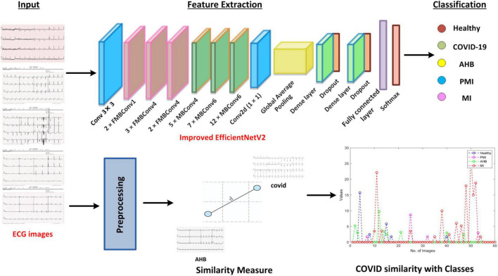
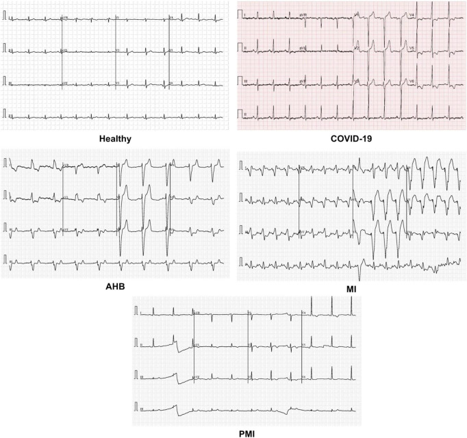
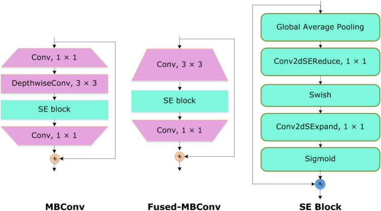
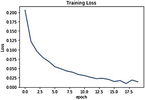
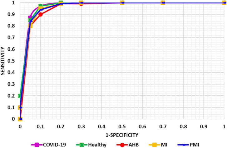
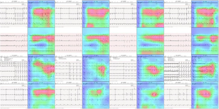
In the last 2 years, we have witnessed multiple waves of coronavirus that affected millions of people around the globe. The proper cure for COVID-19 has not been diagnosed as vaccinated people also got infected with this disease. Precise and timely detection of COVID-19 can save human lives and protect them from complicated treatment procedures. Researchers have employed several medical imaging modalities like CT-Scan and X-ray for COVID-19 detection, however, little concentration is invested in the ECG imaging analysis. ECGs are quickly available image modality in comparison to CT-Scan and X-ray, therefore, we use them for diagnosing COVID-19. Efficient and effective detection of COVID-19 from the ECG signal is a complex and time-taking task, as researchers usually convert them into numeric values before applying any method which ultimately increases the computational burden. In this work, we tried to overcome these challenges by directly employing the ECG images in a deep-learning (DL)-based approach. More specifically, we introduce an Efficient-ECGNet method that presents an improved version of the EfficientNetV2-B4 model with additional dense layers and is capable of accurately classifying the ECG images into healthy, COVID-19, myocardial infarction (MI), abnormal heartbeats (AHB), and patients with Previous History of Myocardial Infarction (PMI) classes. Moreover, we introduce a module to measure the similarity of COVID-19-affected ECG images with the rest of the diseases. To the best of our knowledge, this is the first effort to approximate the correlation of COVID-19 patients with those having any previous or current history of cardio or respiratory disease. Further, we generate the heatmaps to demonstrate the accurate key-points computation ability of our method. We have performed extensive experimentation on a publicly available dataset to show the robustness of the proposed approach and confirmed that the Efficient-ECGNet framework is reliable to classify the ECG-based COVID-19 samples.
Keywords: COVID-19; ECG; Efficient-ECGNet; computer vision; deep learning; medical imaging.
Copyright © 2022 Nawaz, Nazir, Javed, Malik, Saudagar, Khan, Abul Hasanat, AlTameem and AlKhathami.
Conflict of interest statement
The authors declare that the research was conducted in the absence of any commercial or financial relationships that could be construed as a potential conflict of interest.
Figures

References
LinkOut - more resources
Full Text Sources