

Retroperitoneal alveolar rhabdomyosarcoma intruding into spinal canal: A case report and literature review
- PMID: 36405590
- PMCID: PMC9670135
- DOI: 10.3389/fmed.2022.1019964
Retroperitoneal alveolar rhabdomyosarcoma intruding into spinal canal: A case report and literature review
Abstract
Background: Rhabdomyosarcoma (RMS) is the most frequent soft sarcoma in children and adolescents. Alveolar rhabdomyosarcoma (ARMS) is a relatively rare subtype that is characterized by aggressive behavior and an unsatisfactory prognosis. An ARMS can arise anywhere but most commonly occurs at extremity sites with a very small fraction in the retroperitoneum. The utility of 2-Deoxy-2-[fluorine-18]-fluoro-D-glucose (18F-FDG) positron emission tomography combined with computed tomography (PET/CT) remains to be established in ARMS.
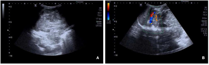
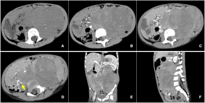
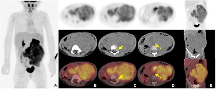
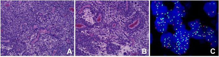
Case report: A 3-year-old female child was accidentally found with a large left upper abdominal mass for a day. CT examination indicated a huge soft tissue mass in the left retroperitoneum extending superiorly to the level of the left hilus renalis and inferiorly to the left acetabulum in the pelvic cavity, with intrusion into the lumbar foramens. 18F-FDG PET/CT found a mass in the left retroperitoneum from the level of T12 to the left acetabulum, with the maximum standardized uptake value (SUVmax) of about 7.0, and a CT value of about 39 HU, invading the left L3-5 intervertebral foramina and protruding into the spinal canal, with unclear boundary with the spinal cord. Retroperitoneal tumor resection and the repair operation of vascular exploration were performed. An ARMS was confirmed by postoperative biopsy, immunohistochemical staining, and genetic detection with the rupture of the fork head in rhabdomyosarcoma (FKHR). The patient received chemotherapy and was in a good condition with no recurrence and obvious complications.
Conclusion: Retroperitoneal ARMS is rare and indicates a poor outcome with the potential to involve vital organs and intrude into the spinal canal. Accurate diagnosis and staging using PET/CT would contribute to better risk stratifications and appropriate treatment individually.
Keywords: 18F-FDG; PET/CT; alveolar rhabdomyosarcoma; case report; retroperitoneum; spinal canal intrusion.
Copyright © 2022 Zhang, Huang, Li, Qiu, Jiao, Chen, Yang, Song and Kang.
Conflict of interest statement
The authors declare that the research was conducted in the absence of any commercial or financial relationships that could be construed as a potential conflict of interest. Written informed consent was obtained from the minor's legal guardian for the publication of any potentially identifiable images or data included in this article.
Figures
Similar articles
-
Primary perianal alveolar rhabdomyosarcoma with uncommon metastatic sites: a case report and follow-up using 18F-FDG PET/CT.Front Med (Lausanne). 2024 Dec 17;11:1474698. doi: 10.3389/fmed.2024.1474698. eCollection 2024. Front Med (Lausanne). 2024. PMID: 39744536 Free PMC article.
-
Multimodality imaging evaluation of nasal sinus alveolar rhabdomyosarcoma: Two case reports.Front Med (Lausanne). 2022 Nov 10;9:1047464. doi: 10.3389/fmed.2022.1047464. eCollection 2022. Front Med (Lausanne). 2022. PMID: 36438027 Free PMC article.
-
More advantages in detecting bone and soft tissue metastases from prostate cancer using 18F-PSMA PET/CT.Hell J Nucl Med. 2019 Jan-Apr;22(1):6-9. doi: 10.1967/s002449910952. Epub 2019 Mar 7. Hell J Nucl Med. 2019. PMID: 30843003
-
The Role of 18F-FDG-PET/CT in Pediatric Sarcoma.Semin Nucl Med. 2017 May;47(3):229-241. doi: 10.1053/j.semnuclmed.2016.12.004. Epub 2017 Jan 18. Semin Nucl Med. 2017. PMID: 28417853 Review.
-
Pediatric cervical kyphosis in the MRI era (1984-2008) with long-term follow up: literature review.Childs Nerv Syst. 2022 Feb;38(2):361-377. doi: 10.1007/s00381-021-05409-z. Epub 2021 Nov 22. Childs Nerv Syst. 2022. PMID: 34806157 Review.
Cited by
-
Primary perianal alveolar rhabdomyosarcoma with uncommon metastatic sites: a case report and follow-up using 18F-FDG PET/CT.Front Med (Lausanne). 2024 Dec 17;11:1474698. doi: 10.3389/fmed.2024.1474698. eCollection 2024. Front Med (Lausanne). 2024. PMID: 39744536 Free PMC article.
References
-
- Ries LAG . Cancer Incidence and Survival among Children and Adolescents: United States Seer Program, 1975–1995. Bethesda: National Cancer Institute; (1999).
Publication types
LinkOut - more resources
Full Text Sources
Research Materials
Miscellaneous