. 2022 Nov 3:13:1005937.
doi: 10.3389/fimmu.2022.1005937. eCollection 2022.

Interleukin-6 and indoleamine-2,3-dioxygenase as potential adjuvant targets for Papillomavirus-related tumors immunotherapy

Affiliations

Interleukin-6 and indoleamine-2,3-dioxygenase as potential adjuvant targets for Papillomavirus-related tumors immunotherapy

Roberta Liberato Pagni et al. Front Immunol. .

Abstract

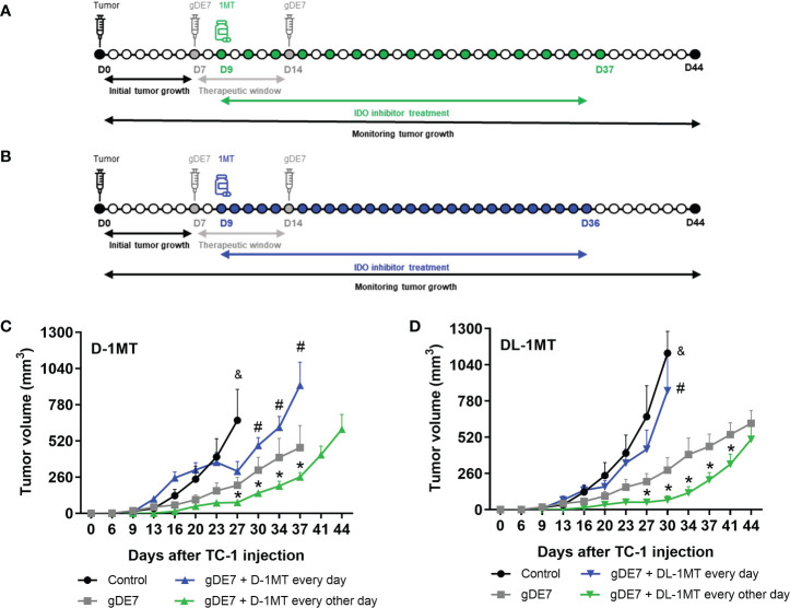
High-risk Human papillomavirus (HPV) infections represent an important public health issue. Nearly all cervical malignancies are associated with HPV, and a range of other female and male cancers, such as anogenital and oropharyngeal. Aiming to treat HPV-related tumors, our group developed vaccines based on the genetic fusion of the HSV-1 glycoprotein D (gD) with the HPV-16 E7 oncoprotein (gDE7 vaccines). Despite the promising antitumor results reached by gDE7 vaccines in mice, combined therapies may increase the therapeutic effects by improving antitumor responses and halting immune suppressive mechanisms elicited by tumor cells. Considering cancer immunosuppressive mechanisms, indoleamine-2,3-dioxygenase (IDO) enzyme and interleukin-6 (IL-6) stand out in HPV-related tumors. Since IL-6 sustained the constitutive IDO expression, here we evaluated the therapeutic outcomes achieved by the combination of active immunotherapy based on a gDE7 protein-based vaccine with adjuvant treatments involving blocking IDO, either by use of IDO inhibitors or IL-6 knockout mice. C57BL/6 wild-type (WT) and transgenic IL-6-/- mice were engrafted with HPV16-E6/E7-expressing TC-1 cells and treated with 1-methyl-tryptophan isoforms (D-1MT and DL-1MT), capable to inhibit IDO. In vitro, the 1MT isoforms reduced IL-6 gene expression and IL-6 secretion in TC-1 cells. In vivo, the multi-targeted treatment improved the antitumor efficacy of the gDE7-based protein vaccine. Although the gDE7 immunization achieves partial tumor mass control in combination with D-1MT or DL-1MT in WT mice or when administered in IL-6-/- mice, the combination of gDE7 and 1MT in IL-6-/- mice further enhanced the antitumor effects, reaching total tumor rejection. The outcome of the combined therapy was associated with an increased frequency of activated dendritic cells and decreased frequencies of intratumoral polymorphonuclear myeloid-derived suppressor cells and T regulatory cells. In conclusion, the present study demonstrated that IL-6 and IDO negatively contribute to the activation of immune cells, particularly dendritic cells, reducing gDE7 vaccine-induced protective immune responses and, therefore, opening perspectives for the use of combined strategies based on inhibition of IL-6 and IDO as immunometabolic adjuvants for immunotherapies against HPV-related tumors.

Keywords: HPV; IDO; IL-6; cancer; immunotherapy; vaccine.


Conflict of interest statement

BP and LA hold ownership interest, including patents, in ImunoTera Soluções Terapêuticas Ltda. The remaining authors declare that the research was conducted in the absence of any commercial or financial relationships that could be construed as a potential conflict of interest.

Figures

Figure 1
Figure 1
The administration regimen of 1MT isoforms affects the therapeutic antitumor effects of gDE7. Female WT mice were inoculated with TC-1 (D0) cells. Seven (D7) and fourteen (D14) days after the tumor engraftment, the animals were immunized with gDE7 (30μg, subcutaneously). Two days after the first dose (D9), mice were treated with 1MT isoforms (A, B), either daily with 8mg/animal (A) (n = 10) or 10 mg/animal every other day (B) (n = 10) for four weeks, until day 36 (D36) or day 37 (D37), respectively. The experimental groups were followed for 60 days. Tumor volumes were followed up to 44 days in mice treated with the different treatments using (C) D-1MT or (D) DL-1MT. The tumor growth “endpoint data” for each group was plotted up to the date when at least 80% of the mice were alive. The data represent the average of two independent experiments and were analyzed by ANOVA. (&) p <0.05, statistical significance of control group concerning all the others; (#) p <0.05, statistical significance of gDE7 + 1MT every day group concerning all the others; (*) p <0.05, statistical significance of gDE7 + 1MT every other day group concerning all the others. The results were confirmed through multiple comparisons by Turkey’s test (three or four groups) or Sidak’s test (two groups), comparing each group mean with the other group mean at the same time point. D0 - day zero; D7 - day 7; D9 - day 9; D14 - day 14; D36 - day 36; D37 - day 37; D44 - day 44.
Figure 2
Figure 2
The combination of gDE7 and 1MT improves mice survival depending on their administration regimen. (A–D) The data represent the average of two independent experiments (n=5, total n=10). (A, B) Survival curves. Data were analyzed by Kaplan-Meyer test. (A) Two mice from gDE7 group and from gDE7 + DL-1MT survived until day 60. (B) Two, one, and four mice from gDE7, gDE7 + D-1MT, and gDE7 + DL-1MT groups survived until day 60, respectively. (C, D) Tumor-free mouse curves. Data were analyzed by ANOVA test* p <0.05. (E) Gating strategy for immune cell analyses in the tumor microenvironment, evaluated at day 21 after the tumor engraftment. Doublets were initially excluded by FSC-H versus FSC-W parameters, followed by SSC-H versus SSC-W parameters. Cells were gated by the expression of CD45+ and successively analyzed for: CD8+, followed by CD8+ IFN-g+; CD4+, followed by CD25+ and FoxP3+ CD25+ (T regulatory cells); CD11chigh MCH-IIhigh (dendritic cells); and Gr1high CD11b+. (F) Frequency of CD45+ cells. (G) Frequency of Gr1high CD11b+ cells. (H) Frequency of CD11chigh MCH-IIhigh cells. (I) Frequency of CD8+ T cells. (J) Frequency of E7-specific CD8+ IFN-g+/CD8+ cells. (#) p<0.05 represent the statistical significance of stimulated (red dots) versus non-stimulated (white dots) cells inside each experimental group. (K) Frequency of CD4+ T cells. (L) Frequency of FoxP3+ CD25+ CD4+ cells. Data representative of two independently performed experiments (n=6). Statistical significance: (*) p<0.05, (**) p<0.01 by ANOVA. (#) p<0.05 (###) p<0.001 represents the ANOVA statistical significance of stimulated (red dots) versus non-stimulated (white dots) cells inside each experimental group.
Figure 3
Figure 3
Expression of IL-6 affects immune cells and negatively impacts gDE7 antitumor effects. (A) Wild-type (WT) and IL-6-/-mice were subcutaneously inoculated with 1 x 10⁵ TC-1 cells and tumor growth was monitored until day 30 (D30) when at least 80% of the mice from each group were alive. Immune cell analyses of the tumor microenvironment were evaluated at day 21 after the tumor engraftment. (B) Frequency of CD45+ cells. (C) Frequency of CD11chigh MCH-IIhigh cells. (D) Frequency of CD8+ T cells. (E) Frequency of CD4+ T cells. (F) Frequency of FoxP3+ CD25+ T cells. Data from one experiment (n=9) were pooled and analyzed by unpaired t-test. (G, H) Effects of 1MT isoforms on the expression IL-6 by TC-1 cells. (G) Real-time PCR assay - Relative gene expression of IL-6 in TC-1 cells with or without 1mM D-1MT or DL-1MT treatment for 24h (n=3). (H) CBA assay - IL-6 release by TC-1 cells with or without exposure to 1mM D-1MT or DL-1MT for 24h (n=3). I Wild-type (WT) and IL-6-/- mice were subcutaneously inoculated with 1 x 10⁵ TC-1 cells and vaccinated with two doses (D7 and D14) of gDE7 (30µg per animal). The experimental groups were followed for 60 days, but the “endpoint data” for each group was plotted up to the date when at least 80% of the mice were alive. (J-L) The antitumor effects of gDE7 in WT IL-6-/-mice were measured by (J) tumor volume (mm3), (K) percentage of mice survival and (L) percentage of tumor-free mice. (M) Frequency of circulating E7-specific CD8+ IFN-γ+/CD8+ T cells on day 21 (D21) after overnight ex-vivo stimulation of cells with the HPV-16 E7 Kb MHC class I-restricted immunodominant epitope peptide. (B-F) Data from one experiment (n=9) were pooled and analyzed by unpaired t-test. (A, J-M) data represent means ± SD from two independently performed experiments (n = 12) with comparable results and analyzed by ANOVA or by Kaplan-Meyer test (exclusively for survival assay). (K) One and four mice from WT + gDE7 and IL-6-/-+ gDE7 groups survived until day 60, respectively. (&) p <0.05, statistical significance of wild type (WT) group concerning all the others; (#) p <0.05, statistical significance of IL-6-/-+ gDE7 group concerning all the others; (*) p<0.05, (***) p<0.001, statistical significance of one experimental group concerning the other group. Regarding tumor volume graphs, the results were confirmed through multiple comparisons by Turkey’s test (three or four groups) or Sidak’s test (two groups), comparing each group mean with the other group mean at the same time point.
Figure 4
Figure 4
Lack of IL-6 combined with IDO inhibition augment immunotherapy control mediated by gDE7 on TC-1 cells engrafted in mice. (A) IL-6-/-mice were subcutaneously inoculated with 1 x 10⁵ TC-1 cells and vaccinated with two doses (D7 and D14) of gDE7 (30µg per animal). Two days after the first dose (D9), mice were treated with 1MT at a concentration of 10 mg/animal every other day for four weeks, until D37. The experimental groups were followed for 60 days, but the “endpoint data” for each group was plotted up to the date when at least 80% of the mice were alive. (B-D) Data represent means ± SD from two (groups IL-6-/- and IL-6-/- + gDE7) (n=6, total n=12) or three (groups IL-6-/- + gDE7 + 1MT) (n=6 or 7, total n=19) independently performed experiments with comparable results and analyzed by ANOVA or by Kaplan-Meyer test (exclusively for survival assay). The antitumor effects of gDE7 combined with 1MT isoforms were followed by (B) tumor volume (mm3), (C) mice survival, and (D) presence of tumor-free mice. (C) Four, fourteen, and nine mice from IL-6-/- + gDE7, IL-6-/- + gDE7 + D-1MT, and IL-6-/- + gDE7 + DL-1MT groups survived until day 60, respectively. (E) Frequency of circulating E7-specific CD8+ IFN-γ+/total CD8+ T cells on D28 after overnight ex-vivo stimulation of cells with the HPV-16 E7 Kb MHC class I-restricted immunodominant epitope peptide (n=12). (F) Gate strategy of circulating IFN-γ producing CD8+ T cells by flow cytometry. (&) p <0.05, statistical significance of IL-6-/- control group concerning all the others; (Φ) p <0.05, statistical significance IL-6-/-+ gDE7 group concerning all the others; (δ) p <0.05, statistical significance of IL-6-/-+ gDE7 + DL-1MT group concerning all the others. (*) p<0.05, (**) p<0.01, statistical significance of one experimental group in comparison with the other groups. (#) p<0.05 represents the statistical significance of stimulated (red dots) versus non-stimulated (white dots) cells inside each experimental group. Regarding tumor volume graphs, the results were confirmed through multiple comparisons by Turkey’s test (three or four groups), comparing each group mean with the other group mean at the same time point.
Figure 5
Figure 5
IDO inhibition increases activation of intratumoral DCs and decreases immune suppressive PMN-MDSC in tumor microenvironment of IL-6-/- mice immunized with gDE7. (A) Gating strategy for immune cell analyses of the tumor microenvironment, evaluated at day 21 after the tumor engraftment. Cells were gated by the expression of CD45+ and successively analyzed according to the expression of CD8+ (CD8+ T lymphocytes), CD4+ (CD4+ T lymphocytes) followed by CD25+ and FoxP3+ CD25+ (T regulatory cells); CD11chigh MCH-IIhigh (dendritic cells), CD11bint Ly6Cint Gr1- (resident monocytes), CD11bint Ly6Chigh Ly6G- (inflammatory monocytes) or CD11bhigh Ly6Cint Ly6G+ (PMN-MDSC). Antigen-presenting cells were considered activated by the expression of the co-stimulatory molecule CD86, analyzed by the median of fluorescence intensity (MFI) in the gated subsets cells. (B) Frequencies of CD45+ cells. (C) Frequencies of CD8+ cells. (D) Frequencies of CD4+ cells. (E) Frequency of FoxP3+ CD25+ CD4+ cells. (F) Frequencies of CD11chigh MCH-IIhigh cells. (G) CD86 MFI in CD11chigh MCH-IIhigh cells. (H) Frequencies of Gr1+ (Ly6C+/Ly6G- or Ly6C-/Ly6G+) CD11b+ cells. (I) CD86 MFI in Gr1+ CD11b+ subsets cells. Data representative of two independently performed experiments (n=6). Statistical significance: (*) p<0.05, (**) p<0.01, (***) p<0.001 by ANOVA. (#) p<0.05 and (##) p<0.01 represent the statistical significance of control group versus other groups.

Similar articles

Cited by

References

    1. de Martel C, Plummer M, Vignat J, Franceschi S. Worldwide burden of cancer attributable to HPV by site, country and HPV type. Int J Cancer (2017) 141:664–70. doi: 10.1002/ijc.30716 - DOI - PMC - PubMed
    1. Sung H, Ferlay J, Siegel RL, Laversanne M, Soerjomataram I, Jemal A, et al. . Global cancer statistics 2020: GLOBOCAN estimates of incidence and mortality worldwide for 36 cancers in 185 countries. CA Cancer J Clin (2021) 71:209–49. doi: 10.3322/caac.21660 - DOI - PubMed
    1. Colturato LF, Signorini Filho RC, Fernandes RCM, Gebrim LH, Oliani AH. Lymph node micrometastases in initial stage cervical cancer and tumoral recurrence. Int J Gynecol Obstet (2016) 133:69–75. doi: 10.1016/j.ijgo.2015.08.019 - DOI - PubMed
    1. Signorini Filho RC, Colturato LF, Barbosa GB, Rosa TSF, Gebrim LH. Clinical and histopathological comparison of two historical series of 142 wertheim-meigs operations performed in a reference center in Brazil. Int J Women’s Heal Wellness (2017) 3:55. doi: 10.23937/2474-1353/1510055 - DOI
    1. Abu-Rustum NR, Yashar CM, Bean S, Bradley K, Campos SM, Chon HS, et al. . NCCN guidelines insights: Cervical cancer, version 1.2020. J Natl Comp Cancer Netw (2020) 18(6):660–6. doi: 10.6004/jnccn.2020.0027 - DOI - PubMed

Publication types

Substances