

Integrating bulk and single-cell sequencing reveals the phenotype-associated cell subpopulations in sepsis-induced acute lung injury
- PMID: 36405762
- PMCID: PMC9666384
- DOI: 10.3389/fimmu.2022.981784
Integrating bulk and single-cell sequencing reveals the phenotype-associated cell subpopulations in sepsis-induced acute lung injury
Abstract
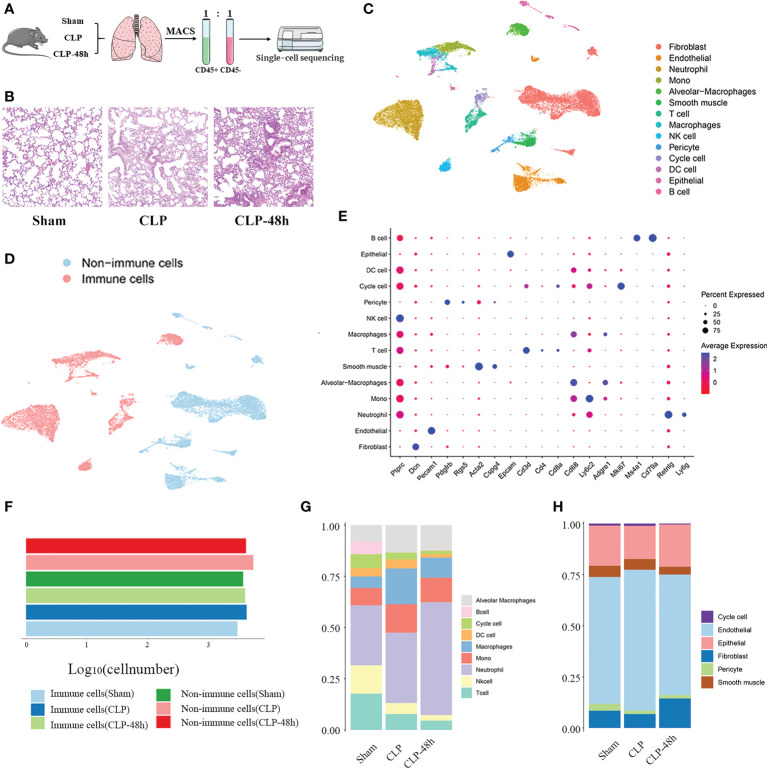
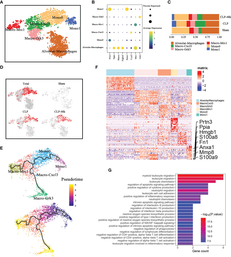
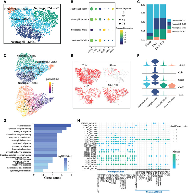
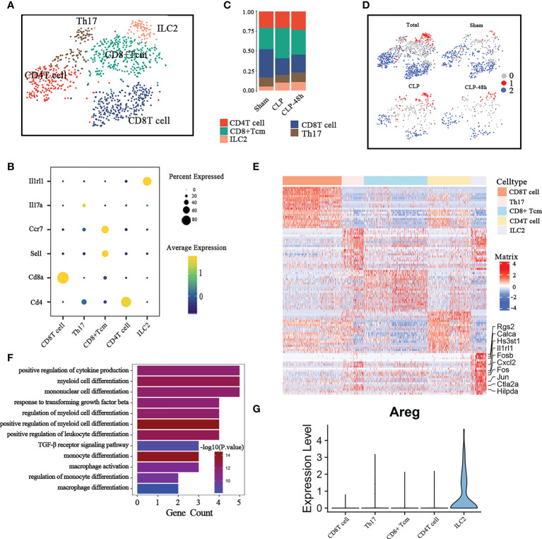
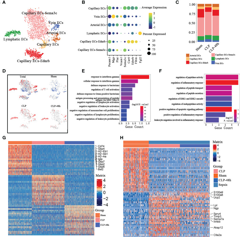
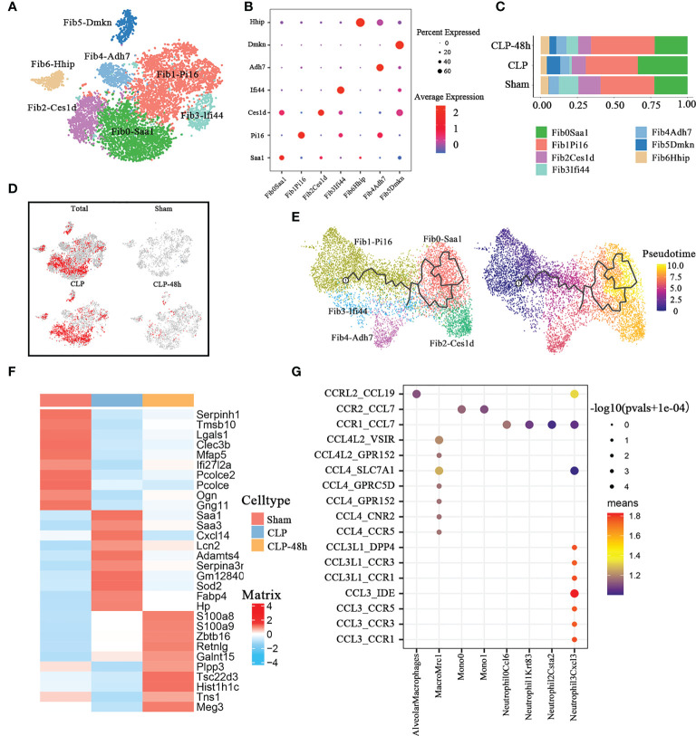
The dysfunctional immune response and multiple organ injury in sepsis is a recurrent theme impacting prognosis and mortality, while the lung is the first organ invaded by sepsis. To systematically elucidate the transcriptomic changes in the main constituent cells of sepsis-injured lung tissue, we applied single-cell RNA sequencing to the lung tissue samples from septic and control mice and created a comprehensive cellular landscape with 25044 cells, including 11317 immune and 13727 non-immune cells. Sepsis alters the composition of all cellular compartments, particularly neutrophils, monocytes, T cells, endothelial, and fibroblasts populations. Our study firstly provides a single-cell view of cellular changes in septic lung injury. Furthermore, by integrating bulk sequencing data and single-cell data with the Scissors-method, we identified the cell subpopulations that are most associated with septic lung injury phenotype. The phenotypic-related cell subpopulations identified by Scissors-method were consistent with the cell subpopulations with significant composition changes. The function analysis of the differentially expressed genes (DEGs) and the cell-cell interaction analysis further reveal the important role of these phenotype-related subpopulations in septic lung injury. Our research provides a rich resource for understanding cellular changes and provides insights into the contributions of specific cell types to the biological processes that take place during sepsis-induced lung injury.
Keywords: Scissors-method; cellular landscape; lung injury; sepsis; single-cell sequencing.
Copyright © 2022 Wang, Chen, Ma, Wang, Wang, Xia, Zhang and Yao.
Conflict of interest statement
The authors declare that the research was conducted in the absence of any commercial or financial relationships that could be construed as a potential conflict of interest.
Figures
Similar articles
-
Single-Cell Sequencing Reveals the Regulatory Role of Maresin1 on Neutrophils during Septic Lung Injury.Cells. 2022 Nov 23;11(23):3733. doi: 10.3390/cells11233733. Cells. 2022. PMID: 36496993 Free PMC article.
-
Role of activated neutrophils in chest trauma-induced septic acute lung injury.Shock. 2012 Jul;38(1):98-106. doi: 10.1097/SHK.0b013e318254be6a. Shock. 2012. PMID: 22552016
-
Role of pulmonary microvascular endothelial cell apoptosis in murine sepsis-induced lung injury in vivo.Respir Res. 2015 Sep 16;16(1):109. doi: 10.1186/s12931-015-0266-7. Respir Res. 2015. PMID: 26376777 Free PMC article.
-
Immunohistochemical detection of sepsis-induced lung injury in human autopsy material.Leg Med (Tokyo). 2003 Jun;5(2):73-86. doi: 10.1016/s1344-6223(03)00010-5. Leg Med (Tokyo). 2003. PMID: 12935535 Review.
-
[Advances in the study of the relationship between autophagy and sepsis-induced lung injury].Zhonghua Shao Shang Za Zhi. 2014 Aug;30(4):325-8. Zhonghua Shao Shang Za Zhi. 2014. PMID: 25429812 Review. Chinese.
Cited by
-
Macrophage‑driven pathogenesis in acute lung injury/acute respiratory disease syndrome: Harnessing natural products for therapeutic interventions (Review).Mol Med Rep. 2025 Jan;31(1):16. doi: 10.3892/mmr.2024.13381. Epub 2024 Nov 8. Mol Med Rep. 2025. PMID: 39513609 Free PMC article. Review.
-
Histone lactylation-regulated METTL3 promotes ferroptosis via m6A-modification on ACSL4 in sepsis-associated lung injury.Redox Biol. 2024 Aug;74:103194. doi: 10.1016/j.redox.2024.103194. Epub 2024 May 16. Redox Biol. 2024. PMID: 38852200 Free PMC article.
-
S100A8/A9hi neutrophils induce mitochondrial dysfunction and PANoptosis in endothelial cells via mitochondrial complex I deficiency during sepsis.Cell Death Dis. 2024 Jun 28;15(6):462. doi: 10.1038/s41419-024-06849-6. Cell Death Dis. 2024. PMID: 38942784 Free PMC article.
-
Deficiency of S100A8/A9 attenuates pulmonary microvascular leakage in septic mice.Respir Res. 2023 Nov 17;24(1):288. doi: 10.1186/s12931-023-02594-0. Respir Res. 2023. PMID: 37978525 Free PMC article.
-
Heterogeneity of immune cells and their communications unveiled by transcriptome profiling in acute inflammatory lung injury.Front Immunol. 2024 Apr 30;15:1382449. doi: 10.3389/fimmu.2024.1382449. eCollection 2024. Front Immunol. 2024. PMID: 38745657 Free PMC article.
References
-
- Rudd KE, Johnson SC, Agesa KM, Shackelford KA, Tsoi D, Kievlan DR, et al. . Global, regional, and national sepsis incidence and mortality, 1990-2017: analysis for the global burden of disease study. Lancet (London England) (2020) 395(10219):200–11. doi: 10.1016/S0140-6736(19)32989-7 - DOI - PMC - PubMed
Publication types
MeSH terms
LinkOut - more resources
Full Text Sources
Medical