Review
. 2022 Aug 22;25(4):200-206.
doi: 10.1002/ajum.12314. eCollection 2022 Nov.

Paediatric orbital ultrasound: Tips and tricks

Affiliations
Review

Paediatric orbital ultrasound: Tips and tricks

Amit Gupta et al. Australas J Ultrasound Med. .

Abstract

Background: The orbital structures are ideally suited for ultrasound examination due to their superficial location and cystic composition of the eye. However, orbital ultrasound remains an underutilised modality due to preference for other cross-sectional modalities in general practice.

Aim: In this article, we review the basic principles, clinical uses and technique of orbital ultrasound in peadiatric patients.

Materials and methods: The clinical utility of orbital ultrasound in peadiatric patients is demonstrated using selected cases.

Results: Ultrasound is useful in the diagnosis of various posterior segment pathologies, especially in conditions causing opacification of light-conducting media of the eye. It is also beneficial in diagnosing various orbital pathologies, particularly in differentiating solid from cystic lesions.

Discussion: The added advantages of its use in children include lack of ionising radiation and reduced requirement of sedation or general anesthesia. Ultrasound is the most practical initial investigation in cases where ophthalmoscopy is limited by opacification of ocular media. The addition of color Doppler on ultrasound can give additional information about the vascularity of the lesion.

Conclusion: Use of ultrasound can be streamlined into the workup of various orbital and ocular pathologies either as an initial investigation or as a problem-solving tool in cases with a diagnostic dilemma on other modalities.

Keywords: orbital imaging; paediatric radiology; ultrasound.


Conflict of interest statement

There is no conflict of interest.

Figures

Figure 1
Figure 1
Child presented with cataract in right eye, obscuring the ophthalmologist's view of the posterior chamber. (a) Right orbital ultrasound shows echogenic appearance of lens (arrows) consistent with congenital cataract. (b) Contralateral eye lens (arrows) shows normal anechoic appearance with thin echogenic capsules. No abnormality is noted in posterior chamber of both eyes.
Figure 2
Figure 2
Child with right leukocoria. (a) Right eye lens shows echogenic appearance (dotted arrows) suggestive of cataractous changes. Echogenic calcified retinal leaves are seen with hyperechoic soft‐tissue (arrows) in posterior chamber—consistent with neglected retinal detachment. (b) Colour Doppler examination showed no internal vascularity in the posterior chamber soft‐tissue.
Figure 3
Figure 3
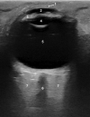
Normal ultrasonographic appearance of the orbital structures as detailed in Table 1. 1—Cornea, 2—Anterior chamber, 3—Ciliary bodies, 4—Lens, 5—Vitreous chamber, 6—Optic nerve, 7—Retrobulbar fat.
Figure 4
Figure 4
(a) Orbital ultrasound shows the presence of an echogenic retrolental mass (arrows) suspicious for a neoplasm. (b) A linear anechoic structure (arrows) is seen within the mass extending from the posterior wall of lens. (c) Presence of colour flow (arrow) on Doppler examination confirms it to be a vascular channel suggestive of persistent hyaloid artery. This imaging appearance can confidently diagnose a persistent fetal vasculature. Note is also made of shallow anterior chamber (dotted arrow) and rounded morphology of the lens (asterisk) (a). [Colour figure can be viewed at wileyonlinelibrary.com]
Figure 5
Figure 5
(a) Axial contrast‐enhanced MRI image shows bulky hyperenhancing medial rectus muscle (white arrows) in the left orbit with a hypoenhancing lesion in the centre (black arrow). This appearance may suggest a diagnosis of orbital cellulitis. (b) Orbital ultrasound of the same patient shows increased bulk of left medial rectus muscle with a well‐defined intramuscular cystic lesion (arrow) with the presence of an eccentric echogenic scolex (dotted arrow) within it. This finding is pathognomonic for myocysticercus. (c) Colour Doppler shows increased vascularity in the surrounding muscle belly (arrow). [Colour figure can be viewed at wileyonlinelibrary.com]
Figure 6
Figure 6
(a) In a known case of bilateral retinoblastoma post‐chemotherapy, axial contrast‐enhanced MRI image shows subtle retinal thickening and enhancement (arrow) in the left globe. Note is made of right ocular implant. (b) Orbital ultrasound shows easily appreciable diffuse nodular retinal thickening (arrows) in the left globe suggestive of retinoblastoma recurrence. (c) Colour Doppler examination shows increased vascularity (arrows) in the thickened retinal soft‐tissue. [Colour figure can be viewed at wileyonlinelibrary.com]

References

    1. Agarwal K, Vinekar A, Chandra P, Padhi TR, Nayak S, Jayanna S, et al. Imaging the pediatric retina: an overview. Indian J Ophthalmol 2021; 69(4): 812–23. - PMC - PubMed
    1. McNicholas MM, Brophy DP, Power WJ, Griffin JF. Ocular sonography. AJR Am J Roentgenol 1994; 163(4): 921–6. - PubMed
    1. Nagaraj UD, Koch BL. Imaging of orbital infectious and inflammatory disease in children. Pediatr Radiol 2021; 51(7): 1149–61. - PubMed
    1. Lorente‐Ramos RM, Armán JA, Muñoz‐Hernández A, Gómez JMG, de la Torre SB. US of the eye made easy: a comprehensive how‐to review with ophthalmoscopic correlation. Radiographics 2012; 32(5): E175–200. - PubMed
    1. Silva CT, Brockley CR, Crum A, Mandelstam SA. Pediatric ocular sonography. Semin Ultrasound CT MR 2011; 32(1): 14–27. - PubMed