

Periprosthetic Joint Infection With Actinomyces radingae May Lead to the Identification of a Neglected Source of Intraoperative Contamination
- PMID: 36405864
- PMCID: PMC9672404
- DOI: 10.1016/j.artd.2022.08.023
Periprosthetic Joint Infection With Actinomyces radingae May Lead to the Identification of a Neglected Source of Intraoperative Contamination
Abstract
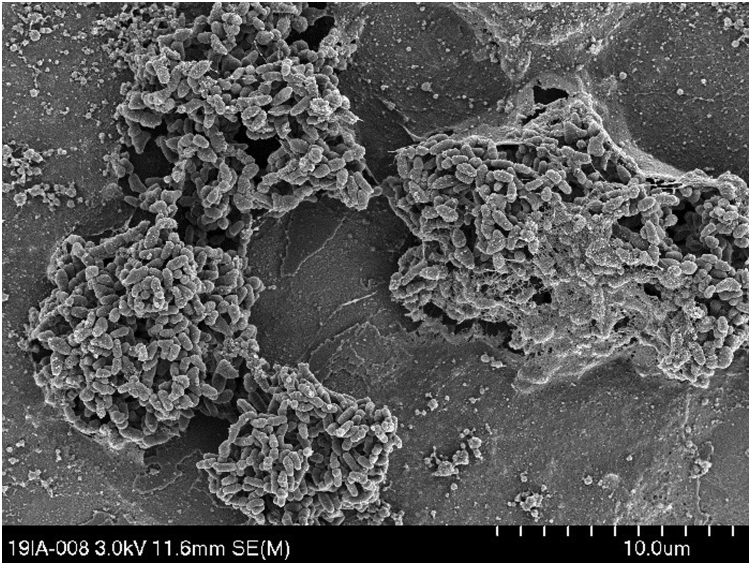
Periprosthetic joint infection remains a major complication in arthroplasty. We present the first description of a case of periprosthetic joint infection with Actinomyces radingae, microorganism that is mostly found on the skin of the upper body and might cause particular challenges as it is difficult to culture and specify. Furthermore, a thorough microbiologic workup may indicate the source of infection. In this case, it is possible that perspiration from the surgeon was the source of intraoperative contamination. Intraoperative contamination through perspiration may be important and should be avoided by all means.
Keywords: Actinomyces radingae; PJI; Periprosthetic joint infection; THA; Total hip arthroplasty.
© 2022 The Authors.
Figures

Similar articles
-
A first case of prosthetic joint infection with Actinomyces radingae.Anaerobe. 2023 Apr;80:102662. doi: 10.1016/j.anaerobe.2022.102662. Epub 2023 Jan 18. Anaerobe. 2023. PMID: 36681233
-
Lower Urinary Tract Infection and Periprosthetic Joint Infection after Elective Primary Total Hip Arthroplasty.Hip Pelvis. 2017 Mar;29(1):30-34. doi: 10.5371/hp.2017.29.1.30. Epub 2017 Mar 6. Hip Pelvis. 2017. PMID: 28316960 Free PMC article.
-
High incidence of early periprosthetic joint infection following total hip arthroplasty with concomitant or previous hardware removal.Arch Orthop Trauma Surg. 2019 Aug;139(8):1051-1056. doi: 10.1007/s00402-019-03149-z. Epub 2019 Feb 18. Arch Orthop Trauma Surg. 2019. PMID: 30778724
-
Management of Periprosthetic Hip Joint Infection.Hip Pelvis. 2015 Jun;27(2):63-71. doi: 10.5371/hp.2015.27.2.63. Epub 2015 Jun 30. Hip Pelvis. 2015. PMID: 27536605 Free PMC article. Review.
-
Identification of periprosthetic joint infection after total hip arthroplasty.J Orthop Translat. 2014 Nov 12;3(1):21-25. doi: 10.1016/j.jot.2014.10.001. eCollection 2015 Jan. J Orthop Translat. 2014. PMID: 30035036 Free PMC article. Review.
Cited by
-
Actinomyces radingae as an emerging pathogen in breast implant surgery: First reported case.JPRAS Open. 2025 Jun 4;45:199-202. doi: 10.1016/j.jpra.2025.05.014. eCollection 2025 Sep. JPRAS Open. 2025. PMID: 40678268 Free PMC article.
References
-
- Gundtoft P.H., Overgaard S., Schønheyder H.C., Møller J.K., Kjærsgaard-Andersen P., Pedersen A.B. The “true” incidence of surgically treated deep prosthetic joint infection after 32,896 primary total hip arthroplasties. Acta Orthop. 2015;86:326–334. doi: 10.3109/17453674.2015.1011983. - DOI - PMC - PubMed
Publication types
LinkOut - more resources
Full Text Sources
