

Updates on the Diagnosis and Management of Glaucoma
- PMID: 36405987
- PMCID: PMC9673042
- DOI: 10.1016/j.mayocpiqo.2022.09.007
Updates on the Diagnosis and Management of Glaucoma
Abstract
Glaucoma is the leading cause of blindness throughout the world (after cataracts); therefore, general physicians should be familiar with the diagnosis and management of affected patients. Glaucomas are usually categorized by the anatomy of the anterior chamber angle (open vs narrow/closed), rapidity of onset (acute vs chronic), and major etiology (primary vs secondary). Most glaucomas are primary (ie, without a contributing comorbidity); however, several coexisting ophthalmic conditions may serve as the underlying etiologies of secondary glaucomas. Chronic glaucoma occurs most commonly; thus, regular eye examinations should be performed in at-risk patients to prevent the insidious loss of vision that can develop before diagnosis. Glaucoma damages the optic nerve and retinal nerve fiber layer, leading to peripheral and central visual field defects. Elevated intraocular pressure (IOP), a crucial determinant of disease progression, remains the only modifiable risk factor; thus, all current treatments (medications, lasers, and operations) aim to reduce the IOP. Pharmacotherapy is the usual first-line therapy, but noncompliance, undesirable adverse effects, and cost limit effectiveness. Laser and surgical treatments may lower IOP significantly over long periods and may be more cost effective than pharmacotherapy, but they are plagued by greater procedural risks and frequent treatment failures. Traditional incisional procedures have recently been replaced by several novel, minimally invasive glaucoma surgeries with improved safety profiles and only minimal decreases in efficacy. Minimally invasive glaucoma surgeries have dramatically transformed the surgical management of glaucoma; nevertheless, large, randomized trials are required to assess their long-term efficacy.
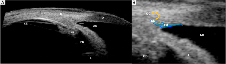
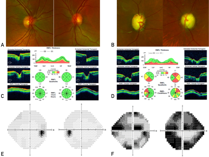
Keywords: ACA, anterior chamber angle; ACG, angle-closure glaucoma; AIT, ab-interno trabeculotomy; CAI, carbonic anhydrase inhibitor; CE, cataract extraction; GDD, glaucoma drainage device; IOP, intraocular pressure; KDB, Kahook Dual Blade; MIGS, minimally invasive glaucoma surgery; MMC, mitomycin C; OAG, open-angle glaucoma; OCT, optical coherence tomography; ONH, optic nerve head; PGA, prostaglandin analog; PGI, PAUL glaucoma implant; POAG, primary open-angle glaucoma; RNFL, retinal nerve fiber layer; SLT, selective laser trabeculoplasty; TM, trabecular meshwork.
© 2022 The Authors.
Figures
References
-
- Kang J.M., Tanna A.P. Glaucoma. Med Clin North Am. 2021;105(3):493–510. - PubMed
-
- Tham Y.C., Li X., Wong T.Y., Quigley H.A., Aung T., Cheng C.Y. Global prevalence of glaucoma and projections of glaucoma burden through 2040: a systematic review and meta-analysis. Ophthalmology. 2014;121(11):2081–2090. - PubMed
-
- Hollands H., Johnson D., Hollands S., Simel D.L., Jinapriya D., Sharma S. Do findings on routine examination identify patients at risk for primary open-angle glaucoma? The rational clinical examination systematic review. JAMA. 2013;309(19):2035–2042. - PubMed
-
- Stein J.D., Khawaja A.P., Weizer J.S. Glaucoma in adults-screening, diagnosis, and management: a review. JAMA. 2021;325(2):164–174. - PubMed
Publication types
LinkOut - more resources
Full Text Sources
Medical
