

Expression and Prognostic Significance of PDIA3 in Cervical Cancer
- PMID: 36406049
- PMCID: PMC9674421
- DOI: 10.1155/2022/4382645
Expression and Prognostic Significance of PDIA3 in Cervical Cancer
Abstract
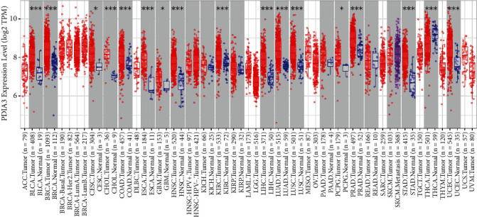
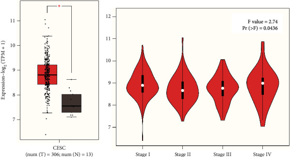
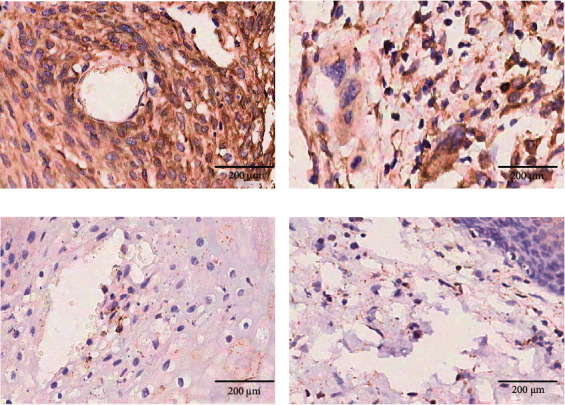
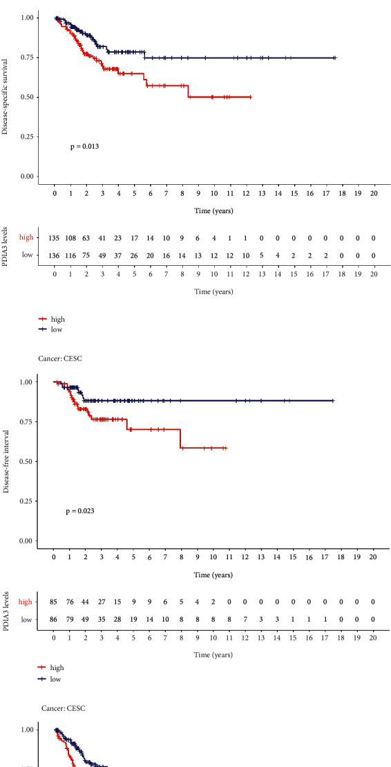
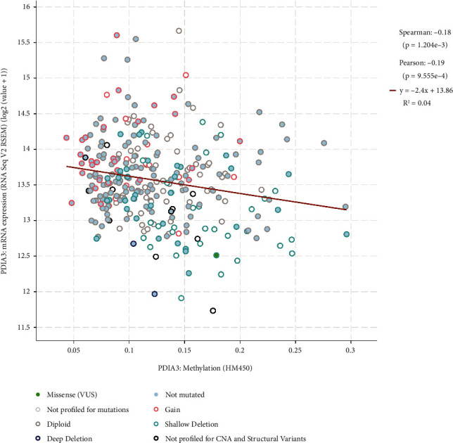
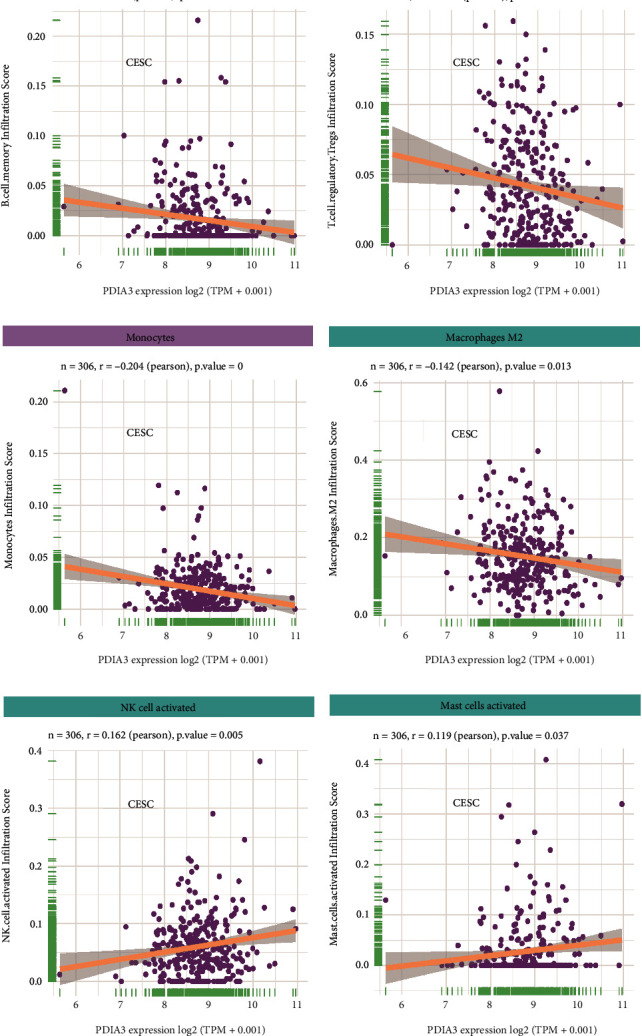
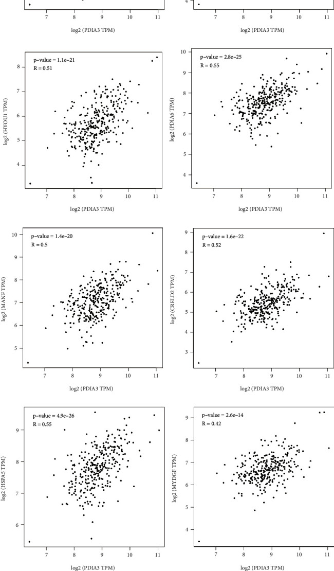
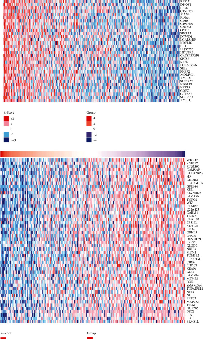
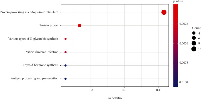
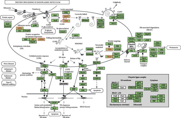
To investigate the expression of protein disulfide isomerase A3 (PDIA3/ERP57) in cervical cancer and its clinical prognostic significance as well as its function and possible action mechanism in the progression of cervical cancer. Based on TIMER2.0 database, the human protein map (Human Protein Atlas) was used to determine the expression level of PDIA3 protein for the analysis of PDIA3 expression in 39 The Cancer Genome Atlas (TCGA) tumors. The PDIA3 expression in cervical cancer tissues in the TCGA and Genotype-Tissue Expression databases was further verified based on the GEPIA2 database to analyze the relationship between the PDIA3 expression and the pathological stage of cervical cancer patients. Immunohistochemistry was used to detect the PDIA3 expression in cervical cancer tissue microarray, including 111 cancer tissue samples and 24 adjacent cancer tissue samples, and the relationship between PDIA3 protein expression and clinical characteristics of patients with cervical cancer was analyzed. The Kaplan-Meier method and log-rank test were used for survival analysis. Based on the cBioPortal database, the Spearman's and Pearson's methods were used to analyze the correlation between PDIA3 expression and DNA methylation. The correlation between PDIA3 expression and the infiltration levels of each immune cell in cervical cancer was evaluated. The STRING was used to construct protein interaction network. Based on LinkedOmics database, the Spearman's method was used to analyze the co-expressed genes of PDIA3 in TCGA cervical cancer. The gene ontology functional enrichment analysis was performed on Top 50 differentially co-expressed genes based on DAVID database. The PDIA3 expression in cervical cancer tissues was significantly higher than that in normal tissues, which (F = 2.74, PR (>F) = 0.0436) was significantly increased with the progression of tumor stage, and PDIA3 showed strong immunoreactivity in cervical cancer tissues. In cervical cancer patients, overall survival (P = 0.014), disease-specific survival (P = 0.013), disease-free interval (P = 0.023), and progression-free interval (P = 0.001) in those with high expression of PDIA3 were significantly lower than those with low expression, suggesting that high expression of PDIA3 was associated with poor prognosis. In cervical cancer, high expression of PDIA3 was associated with DNA methylation and negatively correlated with B cell memory (r = -0.132, P = 0.021), T cell regulatory (r = -0.127, P = 0.026), monocytes (r = -0.204, P = 0), and macrophages M2 (r = -0.142, P = 0.013), whereas positively correlated with levels of NK cell activated (r = 0.162, P = 0.005) and mast cells activated (r = 0.119, P = 0.037). The genes positively correlated with PDIA3 expression included HSPA5 and PPIB, which were mainly enriched in biological processes, such as endoplasmic reticulum (ER) protein folding and ER stress response. PDIA3 can be used as a marker of poor prognosis of cervical cancer. The expression level of PDIA3 is closely related to the survival and prognosis of cervical cancer patients, DNA methylation, and immune cell infiltration.
Copyright © 2022 Jing Zhang et al.
Conflict of interest statement
The authors declare that they have no conflicts of interest.
Figures
Similar articles
-
Protein Disulfide-Isomerase A3 Is a Robust Prognostic Biomarker for Cancers and Predicts the Immunotherapy Response Effectively.Front Immunol. 2022 Mar 25;13:837512. doi: 10.3389/fimmu.2022.837512. eCollection 2022. Front Immunol. 2022. PMID: 35401558 Free PMC article.
-
Pan-Cancer Analysis of PDIA3: Identifying It as a Potential Biomarker for Tumor Prognosis and Immunotherapy.Oxid Med Cell Longev. 2022 Aug 22;2022:9614819. doi: 10.1155/2022/9614819. eCollection 2022. Oxid Med Cell Longev. 2022. PMID: 36046686 Free PMC article.
-
A pan-cancer analysis to determine the prognostic analysis and immune infiltration of HSPA5.Curr Cancer Drug Targets. 2023 May 8. doi: 10.2174/1568009623666230508111721. Online ahead of print. Curr Cancer Drug Targets. 2023. PMID: 37157205
-
Functional analysis of tumor-derived immunoglobulin lambda and its interacting proteins in cervical cancer.BMC Cancer. 2023 Oct 2;23(1):929. doi: 10.1186/s12885-023-11426-9. BMC Cancer. 2023. PMID: 37784026 Free PMC article.
-
Protein disulfide isomerase A3 as novel biomarker for endometrial cancer.Front Oncol. 2023 Oct 16;13:1247446. doi: 10.3389/fonc.2023.1247446. eCollection 2023. Front Oncol. 2023. PMID: 37909009 Free PMC article.
Cited by
-
PDIA3 driven STAT3/PD-1 signaling promotes M2 TAM polarization and aggravates colorectal cancer progression.Aging (Albany NY). 2024 May 17;16(10):8880-8897. doi: 10.18632/aging.205847. Epub 2024 May 17. Aging (Albany NY). 2024. PMID: 38761176 Free PMC article.
-
The role of PDIA3 in oral squamous cell carcinoma and its value as A diagnostic and prognostic biomarker.Heliyon. 2023 Nov 22;9(12):e22596. doi: 10.1016/j.heliyon.2023.e22596. eCollection 2023 Dec. Heliyon. 2023. PMID: 38213579 Free PMC article.
-
Clinicopathological significance of protein disulphide isomerase A3 and phosphorylated signal transducer and activator of transcription 3 in cervical carcinoma.Contemp Oncol (Pozn). 2024;28(1):51-62. doi: 10.5114/wo.2024.139368. Epub 2024 May 3. Contemp Oncol (Pozn). 2024. PMID: 38800530 Free PMC article.
References
LinkOut - more resources
Full Text Sources
Other Literature Sources
Research Materials
Miscellaneous
