Case Reports
. 2022 Nov 4:9:1020166.
doi: 10.3389/fvets.2022.1020166. eCollection 2022.

Case report: Mixed infection of bovine papillomaviruses associated with squamous papilloma of the upper alimentary tract in a dairy cow

Affiliations
Case Reports

Case report: Mixed infection of bovine papillomaviruses associated with squamous papilloma of the upper alimentary tract in a dairy cow

Kamila Peruchi Fernandes et al. Front Vet Sci. .

Abstract

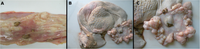
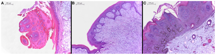
Bovine papillomavirus (BPV) infection can induce neoplastic lesions in both cutaneous and mucosal epithelia in cattle. This study describes the BPV types associated with proliferative lesions with diverse histopathological features present in the upper alimentary tract of a dairy cow suffering from chronic diarrhea from Midwestern Brazil. At autopsy, warts and plaques composed of multiple spherical nodules were observed in the esophageal mucosa, the areas surrounding and constricting the opening of the cardia, and the rumen pillars. One esophageal papillomatous proliferative lesion and a smooth-surfaced proliferative lesion located at the rumen entrance were evaluated by histopathological and molecular analyses. PCR amplification of partial fragments of the BPV L1 and E1 genes was performed followed by sequencing of the obtained amplicons. Upon histopathological evaluation, the esophageal lesion was classified as a squamous papilloma, whereas the other ruminal proliferative lesion consisted of a fibropapilloma. Direct sequencing of PCR products obtained from ruminal fibropapilloma DNA revealed the presence of BPV2. Sequencing of inserts from selected clones containing partial fragments of the BPV L1 and E1 genes revealed a mixed infection of BPV types 2 and 4 in the esophageal squamous papilloma. The findings reported in our investigation reinforce the association of BPV with benign lesions of the bovine alimentary tract in both single and mixed infections, as previously demonstrated to occur in a buffalo. In addition, this report represents the documentation of the occurrence of massive alimentary papillomatosis associated with BPV types 2 and 4 in cattle raised on lands without infestation by bracken fern in Midwestern Brazil.

Keywords: BPV; PCR; cattle; esophagus; rumen; sequencing.


Conflict of interest statement

The authors declare that the research was conducted in the absence of any commercial or financial relationships that could be construed as a potential conflict of interest.

Figures

Figure 1
Figure 1
Macroscopic features of a fibropapilloma and papillomas in the esophagus, rumen, and cardia: (A) esophageal mucosa, multiple pedunculated nodules with a verrucous cauliflower-like appearance, consistent with papillomas, and others with spherical to plaque-like conformation and smooth surface, characterizing fibropapillomas; (B) rumen and reticulum, note in the mucosa of the rumen multifocal spherical and plaque-like nodules with a smooth, shiny white surface, distributed overwhelmingly along the rumen pillars and around the cardia; a single papillomatous nodule can be seen near the cardia; (C) rumen, a closer view of the cardia with numerous nodules with a smooth shiny surface and pearly-white aspect are disposed all around the periphery of the cardia. There is also a single papilloma near to the cardia.
Figure 2
Figure 2
(A) Proximal esophagus, exophytic proliferation of papillary projections of stratified squamous epithelium forming thick fronds supported by a thin core of fibrovascular stroma converging to a common peduncle. Hematoxylin-eosin (HE) 4×. (B) Ruminal fibropapilloma with neoplastic proliferation of squamous epithelial cells and mesenchymal cells forming streams, expanding the submucosa and elevating the overlying proliferated mucosa. The epithelial component forms numerous acanthomatous projections into the mesenchymal component. Hematoxylin-eosin (HE) 10×. (C) Skin, face, a thick proliferation of squamous epithelial cells is seen forming a broad-based well-delimited nodule with moderate hyperkeratosis.

Similar articles

Cited by

References

    1. Daudt C, da Silva FRC, Lunardi M, Alves CBDT, Weber MN, Cibulski SP, et al. . Papillomaviruses in ruminants: an update. Transbound Emerg Dis. (2018) 65:1381–95. 10.1111/tbed.12868 - DOI - PubMed
    1. Wilson VG, West M, Woytek K, Rangasamy D. Papillomavirus E1 proteins: form, function, and features. Virus Genes. (2002) 24:275–90. 10.1023/A:1015336817836 - DOI - PubMed
    1. Favre M. Structural polypeptides of rabbit, bovine, and human papillomaviruses. J Virol. (1975) 15:1239–47. 10.1128/jvi.15.5.1239-1247.1975 - DOI - PMC - PubMed
    1. Roden RB, Weissinger EM, Henderson DW, Booy F, Kirnbauer R, Mushinksi JF, et al. . Neutralization of bovine papillomavirus by antibodies to L1 and L2 capsid proteins. J Virol. (1994) 68:7570–4. 10.1128/jvi.68.11.7570-7574.1994 - DOI - PMC - PubMed
    1. López-Bueno A, Mavian C, Labella AM, Castro D, Borrego JJ, Alcami A, et al. . Concurrence of iridovirus, polyomavirus, and a unique member of a new group of fish papillomaviruses in lymphocystis disease-affected gilthead sea bream. J Virol. (2016) 90:8768–79. 10.1128/JVI.01369-16 - DOI - PMC - PubMed

Publication types

LinkOut - more resources