. 2022 Nov 12:23:187-205.
doi: 10.1016/j.bioactmat.2022.11.001. eCollection 2023 May.

From liver fibrosis to hepatocarcinogenesis: Role of excessive liver H2O2 and targeting nanotherapeutics

Affiliations

From liver fibrosis to hepatocarcinogenesis: Role of excessive liver H2O2 and targeting nanotherapeutics

Meiyu Shao et al. Bioact Mater. .

Abstract

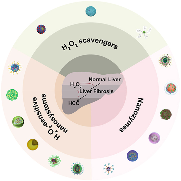
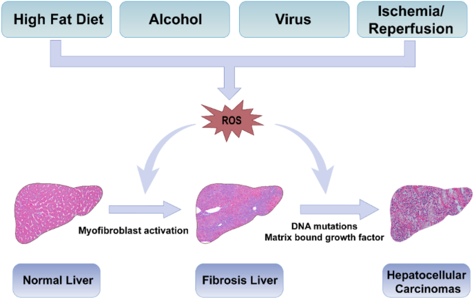
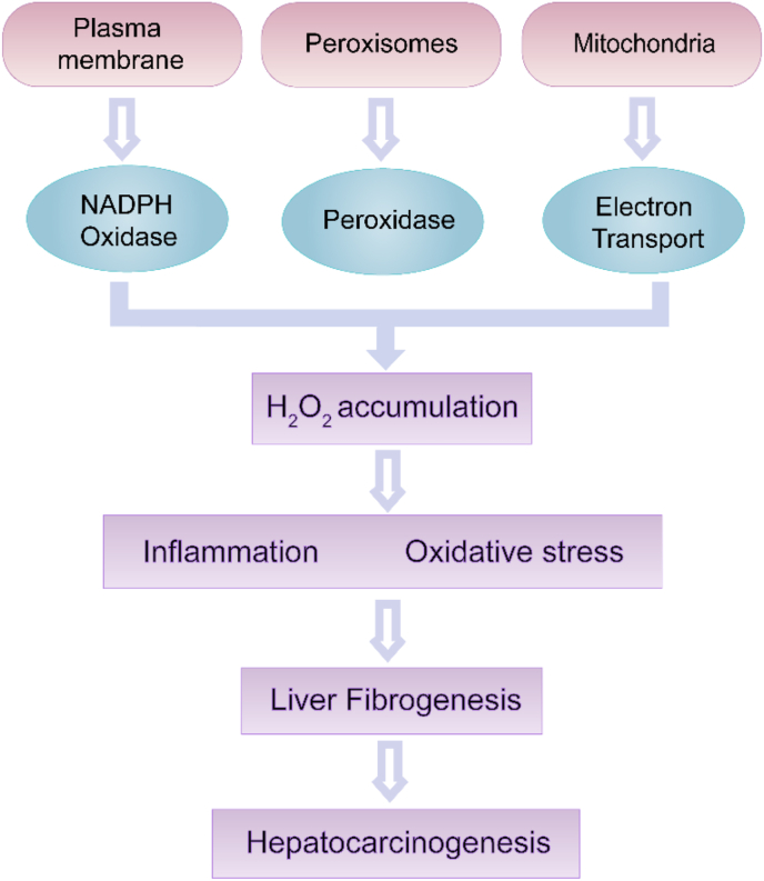
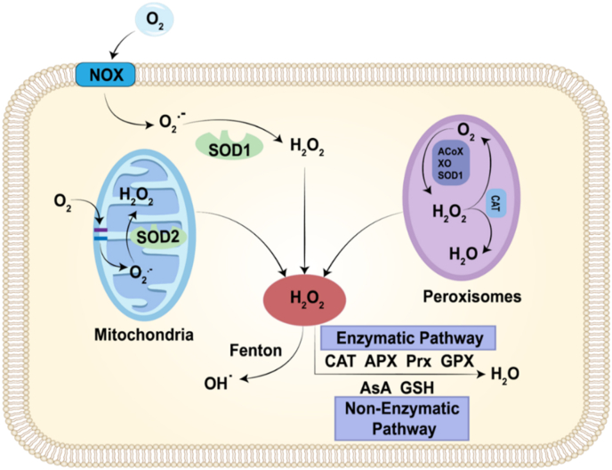
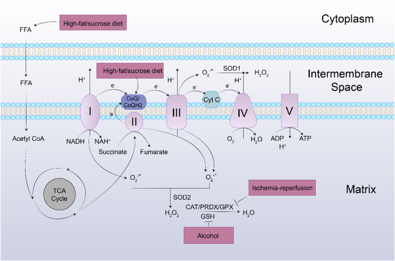
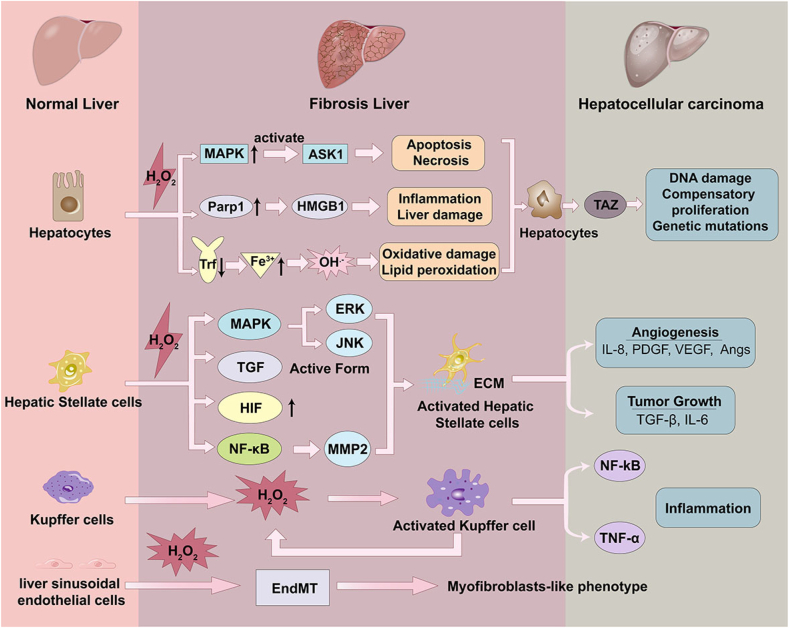
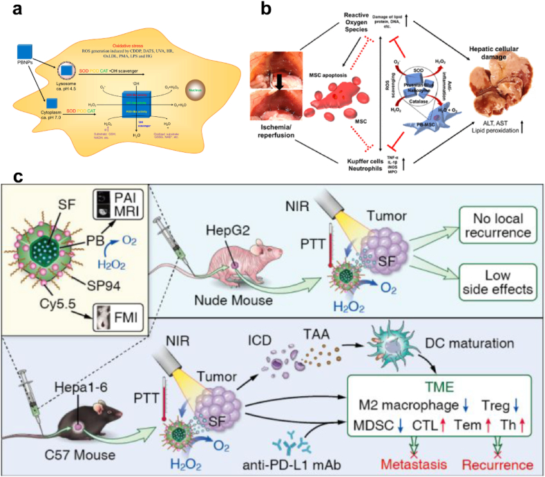
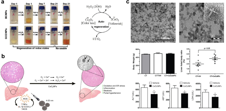
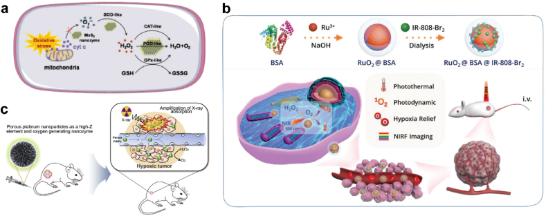
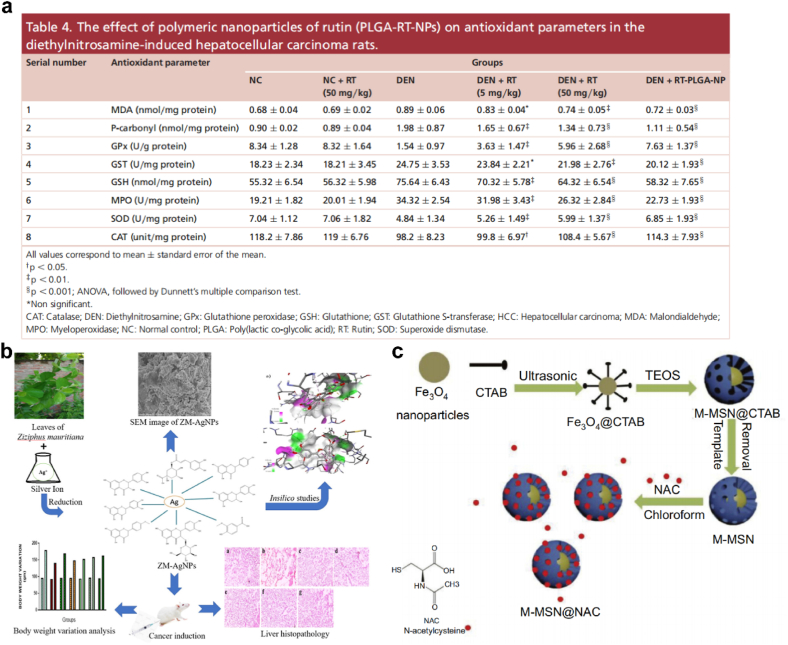
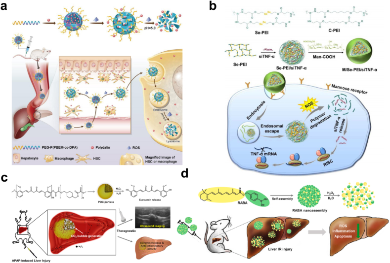
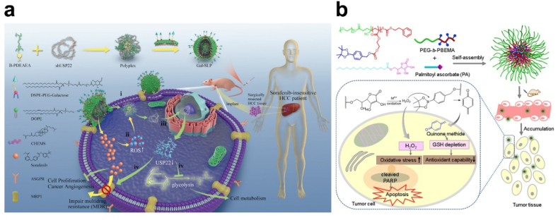
Liver fibrosis and hepatocellular carcinoma (HCC) have been worldwide threats nowadays. Liver fibrosis is reversible in early stages but will develop precancerosis of HCC in cirrhotic stage. In pathological liver, excessive H2O2 is generated and accumulated, which impacts the functionality of hepatocytes, Kupffer cells (KCs) and hepatic stellate cells (HSCs), leading to genesis of fibrosis and HCC. H2O2 accumulation is associated with overproduction of superoxide anion (O2 •-) and abolished antioxidant enzyme systems. Plenty of therapeutics focused on H2O2 have shown satisfactory effects against liver fibrosis or HCC in different ways. This review summarized the reasons of liver H2O2 accumulation, and the role of H2O2 in genesis of liver fibrosis and HCC. Additionally, nanotherapeutics targeting H2O2 were summarized for further consideration of antifibrotic or antitumor therapy.

Keywords: H2O2 accumulation; Hepatocarcinogenesis; Liver fibrosis; Nanotherapeutics; Oxidative stress.


Conflict of interest statement

The authors declare no competing interests.

Figures

Image 1
Graphical abstract
Fig. 1
Fig. 1
Scheme of pathological changes of normal liver to the fibrosis owing to ROS stimuli. As illustrated above, high fat diet, alcohol, virus, drugs, ischemia-reperfusion injury and other harmful substances make the hepatic tissues vulnerable to ROS related oxidative stress, which will trigger liver injury and lead to liver fibrosis, and further induce precancerosis of HCC.
Fig. 2
Fig. 2
Role of H2O2 in liver fibrosis. Endogenous H2O2 sources include NADPH oxidases, peroxidase and the mitochondria electron transport. The excessive H2O2 accumulation leads to oxidative stress and oxidative damage of biomolecules, eventually induces the fibrosis and HCC.
Fig. 3
Fig. 3
Different routes of H2O2 production and scavenging. The major sources of H2O2 are the NOXs and the mitochondrial respiratory chain, as well as a considerable number of oxidases in peroxisome. H2O2 is cleared by pathways involving antioxidant enzyme systems, such as CAT, APX, PRX and GPX. In non-enzymatic pathway, H2O2 is scavenged by ascorbate (AsA), glutathione (GSH). The generated H2O2 can further convert to OH• via Fenton reaction.
Fig. 4
Fig. 4
Mitochondria-derived ROS generation and their elimination by the antioxidant system. O2•− is generated predominantly at complex I, III of the respiratory chain. O2•− is converted to H2O2 by two dismutases including SOD2 in mitochondrial matrix and SOD1 in mitochondrial intermembrane space. Under normal physiological conditions, the generated H2O2 is then eliminated by CAT, and PRDXs, GPXs. During the process of liver fibrosis, the harmful stimuli alter the balance between generation and scavenging of H2O2 due to high H2O2 production and low antioxidant capacity of mitochondria. Additionally, excessive free fatty acid can interfere with ETC through promoting the TCA cycle.
Fig. 5
Fig. 5
Illustration of the effect of accumulated H2O2 on hepatocyte, HSCs, KCs, and LSECs during fibrogenesis. During the continuous H2O2 stimuli, the functionality of several liver-specific cells, such as hepatocytes, KCs and HSCs was impacted leading to the generation and development of liver fibrosis. The excess H2O2 induces apoptosis and necrosis of hepatocytes, promotes HSCs activation, exacerbates the inflammatory responses involved the activation of resident KCs, and induces the endothelial-to-mesenchymal transition (EndMT) process of LSECs, which contributing to liver fibrosis and eventually inducing HCC.
Fig. 6
Fig. 6
(a) Illustration of the POD-like activity of PB NPs. Adapted with permission [136]. Copyright 2016, American Chemical Society. (b) PB impregnated mesenchymal stem cells for hepatic I/R injury alleviation. Adapted with permission [141]. Copyright 2021, American Chemical Society. (c) Scheme illustrating the SP94-PB-SF-Cy5.5 NPs treatment regimen for HCC [138]. Copyright 2020, American Chemical Society.
Fig. 7
Fig. 7
(a) Free radical scavenging activity and autoregenerative properties of GCCNPs. Adapted with permission [145]. Copyright 2017, American Chemical Society. (b) Scheme illustrating the CeO2NPs administration protecting against chronic liver injury by reducing liver steatosis and portal hypertension and markedly attenuating the intensity of the inflammatory response. Adapted with permission [143]. Copyright 2016, Elsevier. (c) CeO2NPs treatment increased liver regeneration and cell proliferation. Adapted with permission [148]. Copyright 2019, Springer Nature.
Fig. 8
Fig. 8
(a) Schematic illustration of tumor targeting of Bac@MnO2 and its tumor inhibition mechanism. Adapted with permission [154]. Copyright 2020, Wiley-VCH. (b) Step-by-step synthesis of drug loaded hollow MnO2 nanoparticles and (c) pH-responsive drug delivery and oxygen generation in H2O2 solution. Adapted with permission [155]. Copyright 2017, Springer Nature. (d) Mn3O4 scavenging of O2•− compared with CeO2 NPs, SOD and (e) reaction with H2O2 compared with CeO2 NPs, CAT. Adapted with permission [156]. Copyright 2018, The Royal Society of Chemistry.
Fig. 9
Fig. 9
(a-c) Illustration of MnO2/BPD synthesis, reduction and self-assembly in vivo. O2 was produced and supported PDT efficiency. Adapted with permission [160]. Copyright 2020, American Chemical Society. (d) Schematic representation of the mechanism by which NanoMnSor can serve as a theranostic anticancer agent. Adapted with permission [161]. Copyright 2020, American Chemical Society.
Fig. 10
Fig. 10
(a) Synthesis and biomedical application of DOX@Fe-HMON-Tf NPs. Adapted with permission [164]. Copyright 2021, Springer Nature. (b) The carboxy‐functional iron oxide nanoparticles (Fe2O3@DMSA) significantly impact on the iron transport system and promotes the retention of intracellular iron, resulting in excessive ROS‐induced tumoricidal autophagy. Adapted with permission [165]. Copyright 2021, Springer Nature.
Fig. 11
Fig. 11
(a) Schematic illustration of the cancer-cell membrane-coated self-assembled nanocomposites for multimodal synergistic breast cancer therapy. Adapted with permission [173]. Copyright 2022, Elsevier. (b) Schematic illustrating the chemical structures, synthetic route and application of the hybrid nanomedicine PEG-Au/FeMOF@CPT. Adapted with permission [174]. Copyright 2020, Wiley-VCH.
Fig. 12
Fig. 12
(a) Schematic illustration of the cellular cascade process of MoS2 for ROS scavenging. Adapted with permission [175]. Copyright 2021, The Royal Society of Chemistry. (b) Schematic illustrating the synthetic route and application of multifunctional RuO2@BSA@IR-808-Br2. Adapted with permission [176]. Copyright 2020, The Royal Society of Chemistry. (c) Schematic illustration of enhanced radiotherapy using porous Pt NPs. Adapted with permission [178]. Copyright 2019, Elsevier.
Fig. 13
Fig. 13
(a) The effect of polymeric nanoparticles of rutin (PLGA-RT-NPs) on antioxidant parameters in the diethylnitrosamine-induced hepatocellular carcinoma rats. Adapted with permission [183]. Copyright 2018, Future Medicine. (b) Scheme illustrating the biosynthesis of silver nanoparticles loaded with biomolecules presented in Ziziphus mauritiana extract with anticancer activity evaluation against hepatic cancer. Adapted with permission [184]. Copyright 2022, Elsevier. (c) Schematic diagram of synthesis of M-MSN@NAC. Adapted with permission [192]. Copyright 2019, Dovepress.
Fig. 14
Fig. 14
(a) Schematic illustration of PD-MC as a ROS and pH dual-responsive nanodrug to regulate multiple cell types for the treatment of liver fibrosis. Adapted with permission [15]. Copyright 2020, Wiley-VCH. (b) Formation and intracellular kinetics of M/Se-PEI/siTNF-α polyplexes and ROS-triggered siRNA release. Adapted with permission [193]. Copyright 2018, The Royal Society of Chemistry. (c) Schematic illustration of POC as an ultrasound imaging and therapeutic agent for acute liver failure. Adapted with permission [194]. Copyright 2019, Elsevier. (d) Schematic illustration of H2O2-activatable hybrid prodrug nanoassemblies as a pure nanodrug for hepatic ischemia/reperfusion injury. Adapted with permission [195]. Copyright 2022, Elsevier.
Fig. 15
Fig. 15
(a) Schematic illustration of a self‐activated cascade‐responsive co‐delivery system (Gal‐SLP) for synergetic cancer therapy. Adapted with permission [196]. Copyright 2021, Wiley-VCH. (b) Schematic illustration of integrated micellar nanoparticles with tumor-specific H2O2 generation to elevate the oxidative stress in tumor tissue and simultaneous GSH depletion to suppress the antioxidant capability of cancer cells for amplified oxidation therapy. Adapted with permission [197]. Copyright 2017, American Chemical Society.

References

    1. Pimpin L., Cortez-Pinto H., Negro F., Corbould E., Lazarus J.V., Webber L., Sheron N., Committee E.H.S. Burden of liver disease in Europe: epidemiology and analysis of risk factors to identify prevention policies. J. Hepatol. 2018;69(3):718–735. - PubMed
    1. Sakurai T., Kudo M., Umemura A., He G., Elsharkawy A.M., Seki E., Karin M. p38α inhibits liver fibrogenesis and consequent hepatocarcinogenesis by curtailing accumulation of reactive oxygen species. Cancer Res. 2013;73(1):215–224. - PMC - PubMed
    1. Ismail M.H., Pinzani M. Reversal of liver fibrosis. Saudi J. Gastroenterol. 2009;15(1):72–79. - PMC - PubMed
    1. Zhang S., Liu H., Yin M., Pei X., Hausser A., Ishikawa E., Yamasaki S., Jin Z.G. Deletion of protein kinase D3 promotes liver fibrosis in mice. Hepatology. 2020;72(5):1717–1734. - PMC - PubMed
    1. Ke M.Y., Xu T., Fang Y., Ye Y.P., Li Z.J., Ren F.G., Lu S.Y., Zhang X.F., Wu R.Q., Lv Y., Dong J. Liver fibrosis promotes immune escape in hepatocellular carcinoma via GOLM1-mediated PD-L1 upregulation. Cancer Lett. 2021;513:14–25. - PubMed

LinkOut - more resources