. 2022 Nov 3:13:1026644.
doi: 10.3389/fmicb.2022.1026644. eCollection 2022.

Development of an imaging system for visualization of Ebola virus glycoprotein throughout the viral lifecycle

Affiliations

Development of an imaging system for visualization of Ebola virus glycoprotein throughout the viral lifecycle

Wakako Furuyama et al. Front Microbiol. .

Abstract

Ebola virus (EBOV) causes severe EBOV disease (EVD) in humans and non-human primates. Currently, limited countermeasures are available, and the virus must be studied in biosafety level-4 (BSL-4) laboratories. EBOV glycoprotein (GP) is a single transmembrane protein responsible for entry into host cells and is the target of multiple approved drugs. However, the molecular mechanisms underlying the intracellular dynamics of GP during EBOV lifecycle are poorly understood. In this study, we developed a novel GP monitoring system using transcription- and replication-competent virus-like particles (trVLPs) that enables the modeling of the EBOV lifecycle under BSL-2 conditions. We constructed plasmids to generate trVLPs containing the coding sequence of EBOV GP, in which the mucin-like domain (MLD) was replaced with fluorescent proteins. The generated trVLP efficiently replicated over multiple generations was similar to the wild type trVLP. Furthermore, we confirmed that the novel trVLP system enabled real-time visualization of GP throughout the trVLP replication cycle and exhibited intracellular localization similar to that of wild type GP. In summary, this novel monitoring system for GP will enable the characterization of the molecular mechanism of the EBOV lifecycle and can be applied for the development of therapeutics against EVD.

Keywords: Ebola virus; bio-imaging; glycoprotein; intracellular traffic; transcription- and replication-competent virus-like particle.


Conflict of interest statement

The authors declare that the research was conducted in the absence of any commercial or financial relationships that could be construed as a potential conflict of interest.

Figures
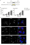
Figure 1
Figure 1
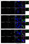
Fluorescent protein-fused GP retained its function and intracellular distribution. (A) Schematic representation of fluorescent protein-fused GP. MLD, Mucin-like domain; FP, fluorescent protein; GS; glycine-serine linker. Amino acid positions for the MLD are shown. (B) Neutralization assay of the VSV pseudotyped with fluorescent protein-fused GP. VSV pseudotyped with the indicated GPs were incubated with monoclonal antibodies, ZGP133/3.16 or ZGP226/8.1, followed by inoculation into Vero E6 cells. The relative infectivity is shown by setting the IU in the absence of neutralizing antibody. The mean and standard deviation of three independent experiments are shown. (C) Intracellular distribution of wild-type, mCherry-, or Venus-fused GP. HEK293 cells were transfected with expression plasmids for wtGP (top), mCherry-GP (middle), or Venus-GP (bottom). The cells were harvested at 24 hpt and distribution of individual GP derivatives (left, magenta in the merged images) and TGN46 (middle, green in the merged images) were analyzed by immunofluorescence staining. The nuclei were counterstained with DAPI (blue in the merged images). Scale bars: 10 μm.
Figure 2
Figure 2
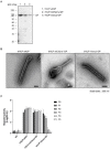
Morphological properties of Ebola VLPs containing fluorescent protein-fused GPs. (A) Validation of incorporation of fluorescent protein-fused GPs into viral particles. Expi293F cells were transfected with the expression plasmids for wt and two GP derivatives, NP and VP40. Ebola VLPs released into culture supernatants were purified by ultracentrifugation and expression of GP (left), NP (middle), and VP40 (right) in purified VLPs was analyzed by western blot analysis. (B) Analysis of morphological features of VLPs containing fluorescent protein-fused GPs. Purified VLPs spiked with wtGP (left), mCherry-GP (middle) and Venus-GP (right) were analyzed by negative staining with electron microscopy. Scale bars: 1 μm.
Figure 3
Figure 3
Establishment of EBOV trVLPs encoding the fluorescent protein-fused GP (A) Incorporation of the fluorescent protein-fused GP into the tetracistronic trVLPs. trVLP-wtGP, -mCherry-GP, or -Venus-GP released into culture supernatant of transfected-HEK293 cells (P0) were purified by ultracentrifugation. Incorporation of GP derivatives into purified trVLP was determined by western blot analysis. (B) Confirmation of morphology of tetracistronic trVLPs encoding the fluorescent protein-fused GP. Purified trVLP-wtGP (left), trVLP-mCherry-GP (middle), or trVLP-Venus-GP (right) were visualized by negative staining. Scale bars: 200 nm. (C) Reporter activities of trVLPs possessing a tetracistronic minigenome that encodes fluorescent protein-fused GP throughout multiple generations. Huh7 cells (P1) pre-transfected with expression plasmids encoding the RNP proteins were inoculated with culture supernatant of transfected HEK293 cells (P0). Seventy-two hpi, reporter activities were measured using the Dual-Luciferase Reporter Assay System. This infection was repeated every 72 h for 5 passages (P5). As a negative control, naïve HEK293 cells are shown. The means and standard deviations from 3 independent experiments are shown.
Figure 4
Figure 4
Visualization of entry process of trVLP-mCherry-GP into recipient cells. trVLP-mCherry-GP was inoculated into Vero E6 cells expressing eGFP-Rab7 and incubated for 30 min on ice. After adsorption, cells were incubated for 0 h (top) or 2 h (bottom) at 37°C. The distribution of mCherry signals (left) on the cell surface (0 h, top), in the cytoplasm (2 h, bottom), and in eGFP-Rab7 (middle) was analyzed by confocal laser scanning microscopy. mCherry-GP and eGFP-Rab7 are shown in magenta and green, respectively, in the merged image. The insets show boxed areas. The white arrows represent colocalized signals. The nuclei were counterstained with DAPI (blue in the merged images). Scale bars: 10 μm.
Figure 5
Figure 5
Visualization of intracellular traffic of mCherry-GP in the trVLP-infected cells. (A) Distribution of mCherry-GP in HEK293 (P0 cells). HEK293 cells were co-transfected with the plasmid for minigenome and the expression plasmids for T7 and RNP proteins. At 48 hpt, the cells were harvested and intracellular distributions of wtGP (top, magenta in the merged images) or mCherry-GP (bottom, magenta in the merged images) and TGN46 (middle, green in the merged images) were analyzed by immunofluorescence staining with antibodies against GP (for wtGP) or TGN46, respectively. (B) Distribution of mCherry-GP in Vero-E6 (P1 cells). Vero-E6 cells pre-transfected with the expression plasmids for the RNP proteins were incubated with trVLP-wtGP (top) or trVLP-mCherry-GP (bottom). Forty-eight hours after inoculation, the cells were harvested and intracellular distributions of wtGP (top) or mCherry-GP (bottom) and TGN46 (middle) were analyzed as described above. (C) Live-cell imaging of mCherry-GP in P0 and P1 cells. HEK293 (P0 cells, top) or Vero-E6 cells (P1 cells, bottom) were pre-transfected with an expression plasmid for GFP-Golgi. At 48 hpt, distribution of mCherry-GP was analyzed by confocal laser scanning microscopy. Nuclei (blue) were counterstained with DAPI (A,B) or Hoechst 33342 (C). Insets show the enlarged images of boxed areas. Arrows represent co-localized signals. Scale bars: 10 μm.

Similar articles

References

    1. Adu-Gyamfi E., Digman M. A., Gratton E., Stahelin R. V. (2012). Investigation of Ebola VP40 assembly and oligomerization in live cells using number and brightness analysis. Biophys. J. 102, 2517–2525. doi: 10.1016/j.bpj.2012.04.022, PMID: - DOI - PMC - PubMed
    1. Baseler L., Chertow D. S., Johnson K. M., Feldmann H., Morens D. M. (2017). The pathogenesis of Ebola virus disease. Annu. Rev. Pathol. 12, 387–418. doi: 10.1146/annurev-pathol-052016-100506 - DOI - PubMed
    1. Bhatia B., Furuyama W., Hoenen T., Feldmann H., Marzi A. (2021). Ebola virus glycoprotein domains associated with protective efficacy. Vaccines 9:9. doi: 10.3390/vaccines9060630 - DOI - PMC - PubMed
    1. Bhattacharyya S., Hope T. J. (2011). Full-length Ebola glycoprotein accumulates in the endoplasmic reticulum. Virol. J. 8:11. doi: 10.1186/1743-422X-8-11, PMID: - DOI - PMC - PubMed
    1. Biedenkopf N., Hoenen T. (2017). Modeling the ebolavirus life cycle with transcription and replication-competent Viruslike particle assays. Methods Mol. Biol. 1628, 119–131. doi: 10.1007/978-1-4939-7116-9_9, PMID: - DOI - PubMed

LinkOut - more resources