Review
. 2022 Nov 3:13:1035100.
doi: 10.3389/fmicb.2022.1035100. eCollection 2022.

An old confusion: Entomophthoromycosis versus mucormycosis and their main differences

Affiliations
Review

An old confusion: Entomophthoromycosis versus mucormycosis and their main differences

Jaime David Acosta-España et al. Front Microbiol. .

Abstract

Fungal diseases were underestimated for many years. And the global burden of fungal infections is substantial and has increased in recent years. Invasive fungal infections have been linked to several risk factors in humans which basically depend on the individual homeostasis of the patients. However, many fungi can infect even apparently healthy people. Knowledge of these pathogens is critical in reducing or stopping morbidity and/or mortality statistics due to fungal pathogens. Successful therapeutic strategies rely on rapid diagnosis of the causative fungal agent and the underlying disease. However, the terminology of the diseases was updated to existing phylogenetic classifications and led to confusion in the definition of mucormycosis, conidiobolomycosis, and basidiobolomycosis, which were previously grouped under the now-uncommon term zygomycosis. Therefore, the ecological, taxonomic, clinical, and diagnostic differences are addressed to optimize the understanding and definition of these diseases. The term "coenocytic hyphomycosis" is proposed to summarize all fungal infections caused by Mucorales and species of Basidiobolus and Conidiobolus.

Keywords: basidiobolomycosis; conidiobolomycosis; entomophthoramycosis; mucoralomycosis; phycomycosis; zygomycosis.


Conflict of interest statement

The authors declare that the research was conducted in the absence of any commercial or financial relationships that could be construed as a potential conflict of interest.

Figures

FIGURE 1
FIGURE 1
Main differences between basidiobolomycosis, conidiobolomycosis and mucormycosis. On the left, the main differences of the main forms of mucormycosis (red), conidiobolomycosis (blue) and basidiobolomycosis (green) are explained. This image was created with Biorender.com.
FIGURE 2
FIGURE 2
Taxonomy and terms used in mucormycosis and entomophthoromycosis. This image shows the old nomenclature for zygomycosis in the light purple section. The current taxonomic classification and terminology is indicated in the light green section. This image was created with Biorender.com.
FIGURE 3
FIGURE 3
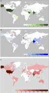
Global prevalence of infections by Mucorales, Conidiobolus spp. and Basidiobolus spp. based on a systematic review of case reports. (A) A systematic review of case reports with the term basidiobolomycosis and Basidiobolus from 1987 to 8/29/2022 was performed in PubMed. Cases with confirmed Basidiobolus spp. infections that allowed identification of the reporting country were included. Detailed information can be found in supplement A. (B) A systematic review of case reports with the term conidiobolomycosis and Conidiobolus spp. from 1978 to 8/29/2022 was performed in PubMed. Cases with confirmed Conidiobolus spp. infections that allowed identification of the reporting country were included. Detailed information can be found in supplement B. (C) A systematic review of case reports with the term mucormycosis between from 2000 to 8-22-2022 was performed in PubMed. Cases with confirmed Mucorales infections that allowed the identification of the reporting country were included. Detailed information can be found in supplement C. Systematic reviews were performed following the PRISMA 2020 criteria (Page et al., 2021).

References

    1. Acosta-España J. D., Voigt K. (2022). Mini Review: Risk Assessment, Clinical Manifestation, Prediction, and Prognosis of Mucormycosis: Implications for Pathogen- and Human-Derived Biomarkers. Front. Microbiol. 13:895989. 10.3389/FMICB.2022.895989 - DOI - PMC - PubMed
    1. Afshar P., Larijani L. V., Rouhanizadeh H. (2018). A comparison of conventional rapid methods in diagnosis of superficial and cutaneous mycoses based on KOH, Chicago sky blue 6B and calcofluor white stains. Iran. J. Microbiol. 10:433. - PMC - PubMed
    1. Agrawal R., Yeldandi A., Savas H., Parekh N. D., Lombardi P. J., Hart E. M. (2020). Pulmonary mucormycosis: Risk factors, radiologic findings, and pathologic correlation. Radiographics 40 656–666. 10.1148/RG.2020190156 - DOI - PubMed
    1. al Jarie A., al Azraki T., al Mohsen I., al Jumaah S., Almutawa A., Mohd Fahim Y., et al. (2011). Basidiobolomycosis: Case series. J. Mycol. Med. 21 37–45. 10.1016/J.MYCMED.2010.11.002 - DOI - PubMed
    1. Alanio A., Garcia-Hermoso D., Mercier-Delarue S., Lanternier F., Gits-Muselli M., Menotti J., et al. (2015). Molecular identification of Mucorales in human tissues: Contribution of PCR electrospray-ionization mass spectrometry. Clin. Microbiol. Infect. 21 594.e1–5. 10.1016/J.CMI.2015.01.017 - DOI - PubMed

LinkOut - more resources