

Visualization of a Juvenile Australopithecus afarensis Specimen: Implications for Functional Foot Anatomy
- PMID: 36406634
- PMCID: PMC9138551
- DOI: 10.5210/jbc.v43i2.10229
Visualization of a Juvenile Australopithecus afarensis Specimen: Implications for Functional Foot Anatomy
Abstract
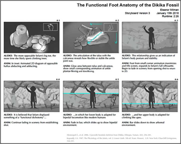
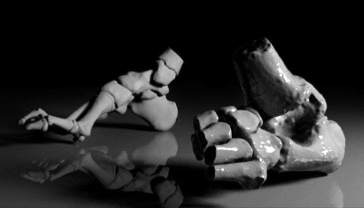
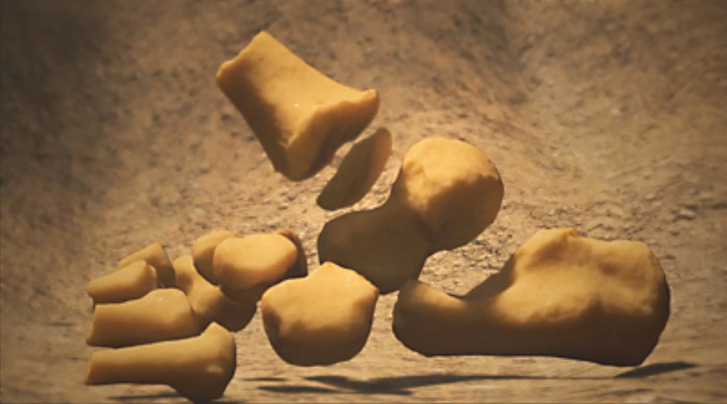
Since it was named in 1978, analyses of Australopithecus afarensis have culminated in several dominant theories on how humans acquired many of their unique adaptations. Because bipedal locomotion is one of the earliest characteristics of human functional anatomy to appear in the fossil record, its associated anatomy in early hominins has significant implications for human evolution (Stern 2000). The skeleton and overall morphological characteristics of the foot in Australopithecus afarensis provide important clues about the origins of upright bipedal locomotion. Popularly known as "Selam," the 3.3 million-year-old DIK-1-1 fossil was discovered in Dikika, Ethiopia by Dr. Zeresenay Alemseged and his team in 2000. Selam was an australopithecine who died at three years old, making her the youngest early hominin specimen known today (Alemseged et al. 2006). This discovery allows researchers to investigate not only locomotor patterns of A. afarensis within the context of human evolution, but also to examine what child development may have looked like during this pivotal time. The purpose of this project is to create a 3D animation that accurately reconstructs the anatomy and taphonomy of the Dikika foot. By segmenting CT data, 3D modelling, and animating, this investigation aims to contribute to the breadth of fossil reconstruction techniques in the field of biomedical visualization. This method provides a robust means of communication within, and beyond, the paleoanthropological community about new discoveries and how to visualize them.
Keywords: 3D animation; Australopithecus afarensis; fossil; human evolution; paleoanthropology; segmentation.
Figures


Similar articles
-
A nearly complete foot from Dikika, Ethiopia and its implications for the ontogeny and function of Australopithecus afarensis.Sci Adv. 2018 Jul 4;4(7):eaar7723. doi: 10.1126/sciadv.aar7723. eCollection 2018 Jul. Sci Adv. 2018. PMID: 29978043 Free PMC article.
-
A juvenile early hominin skeleton from Dikika, Ethiopia.Nature. 2006 Sep 21;443(7109):296-301. doi: 10.1038/nature05047. Nature. 2006. PMID: 16988704
-
Thoracic vertebral count and thoracolumbar transition in Australopithecus afarensis.Proc Natl Acad Sci U S A. 2017 Jun 6;114(23):6000-6004. doi: 10.1073/pnas.1702229114. Epub 2017 May 22. Proc Natl Acad Sci U S A. 2017. PMID: 28533391 Free PMC article.
-
Challenges and perspectives on functional interpretations of australopith postcrania and the reconstruction of hominin locomotion.J Hum Evol. 2023 Feb;175:103304. doi: 10.1016/j.jhevol.2022.103304. Epub 2022 Dec 21. J Hum Evol. 2023. PMID: 36563461 Review.
-
Interpreting the posture and locomotion of Australopithecus afarensis: where do we stand?Am J Phys Anthropol. 2002;Suppl 35:185-215. doi: 10.1002/ajpa.10185. Am J Phys Anthropol. 2002. PMID: 12653313 Review.
References
-
- Boucher EM, Artz T. (2013, Feb. & March). Digital paleoart: reconstruction and restoration from laser-scanned fossils (Doctoral dissertation).
LinkOut - more resources
Full Text Sources