. 2022 Nov 14:17:5265-5286.
doi: 10.2147/IJN.S369761. eCollection 2022.

Novel Curcumin Derivative-Decorated Ultralong-Circulating Paclitaxel Nanoparticles: A Novel Delivery System with Superior Anticancer Efficacy and Safety

Affiliations

Novel Curcumin Derivative-Decorated Ultralong-Circulating Paclitaxel Nanoparticles: A Novel Delivery System with Superior Anticancer Efficacy and Safety

Yumeng Wei et al. Int J Nanomedicine. .

Abstract

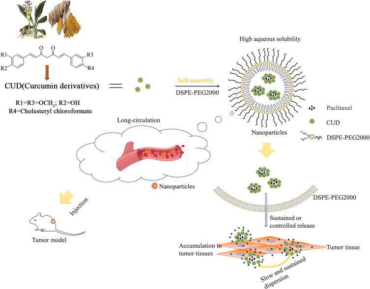
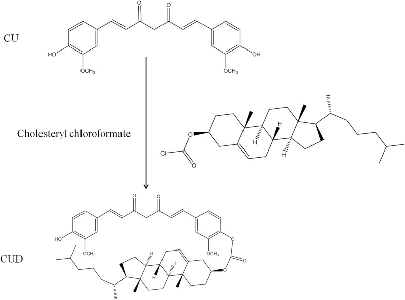
Purpose: Paclitaxel (PTX) has been widely utilized for the treatment of breast cancer. However, drawbacks, such as poor aqueous solubility, rapid blood clearance and severe toxicity, greatly reduce its efficacy and safety. Herein, a novel self-developed curcumin derivative (CUD) was chosen as the carrier to develop a long-acting PTX nano-delivery system (PTX-Sln@CUD) in order to improve its pharmacokinetic behavior, anti-breast cancer efficacy and safety.

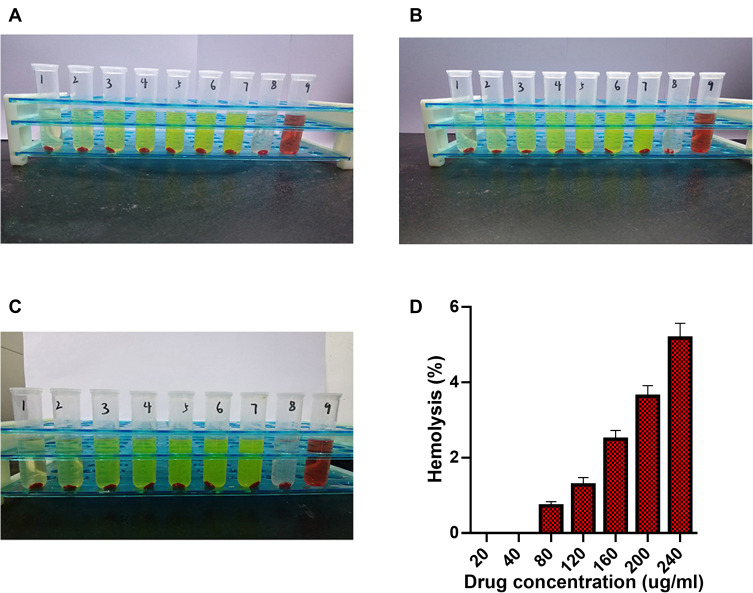
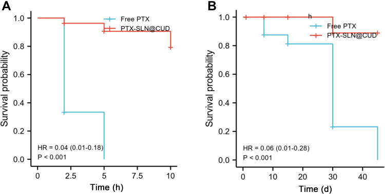
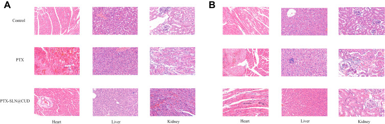
Methods: PTX-Sln@CUD was prepared using solid dispersion and ultrasonic technology. Relevant physical and chemical properties, including stability and release behavior, were characterized. The clearance of PTX-Sln@CUD in vivo was studied by pharmacokinetic experiments. The anti-tumor activity of PTX-Sln@CUD was investigated in vitro and in vivo. Hemolysis experiments, acute toxicity and cumulative toxicity studies were performed in mice to determine the safety of PTX-Sln@CUD.

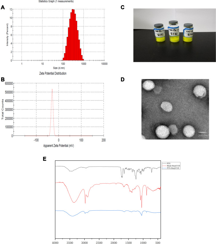
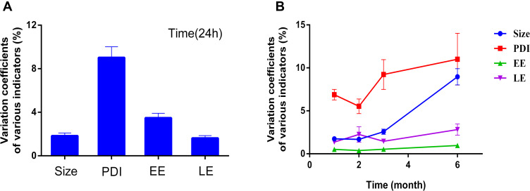
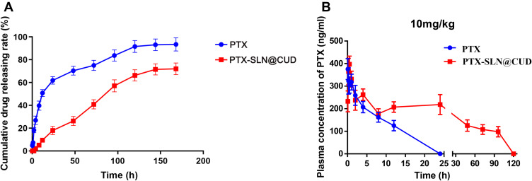
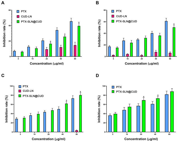
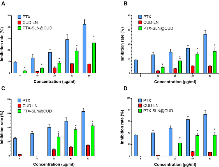
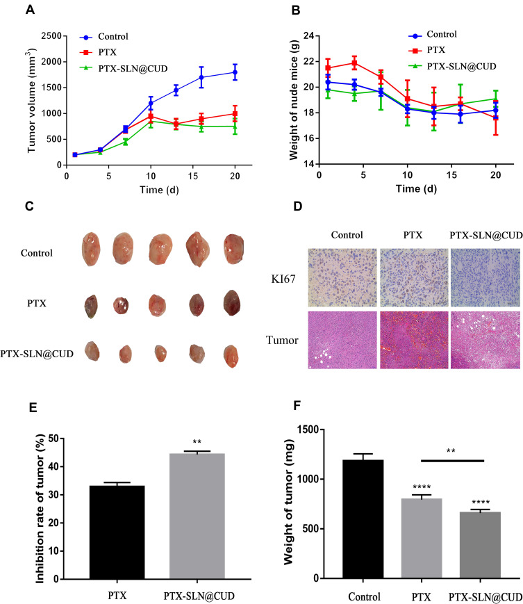
Results: The average particle size, PDI, Zeta potential, encapsulation efficiency and loading efficiency of the PTX-Sln@CUD were 238.5 ± 4.79 nm, 0.225 ± 0.011, -33.8 ± 1.26 mV, 94.20 ± 0.49% and 10.98 ± 0.31%, respectively. PTX-Sln@CUD was found to be stable at room temperature for half a year. The cumulative release rates of PTX-Sln@CUD at 24, 96 and 168 h were 17.98 ± 2.60, 57.09 ± 2.32 and 72.66 ± 4.16%, respectively, which were adherent to zero-order kinetics. T1/2, MRT (0-t) and AUC (0-t) of the PTX-Sln@CUD group were 4.03-fold (44.293 h), 7.78-fold (38.444 h) and 6.18-fold (14.716 mg/L*h) of the PTX group, respectively. PTX-Sln@CUD group demonstrated stronger anti-breast cancer activity than the PTX group. Importantly, the PTX-Sln@CUD group was safer compared to the PTX group both in vitro and in vivo.

Conclusion: PTX-Sln@CUD was verified as promising therapeutic nanoparticles for breast cancer and provided a novel strategy to solve the problem of low efficacy and poor safety of clinical chemotherapy drugs.

Keywords: breast cancer; curcumin derivative; long-acting; paclitaxel nanoparticles.


Conflict of interest statement

The authors declare that they have no competing interests in this work.

Figures

None
Graphical abstract
Figure 1
Figure 1
The structure of CUD ((3S,8R,9S,10S,13R,14S,17R)-10,13-dimethyl-17-((R)-6-methylheptan-2-yl) hexadecahydro-1H-cyclopenta[a]phenanthren-3-yl (4-((1E,6E)-7-(4-hydroxy-3-methoxyphenyl)-3,5-dioxohepta-1,6-dien-1-yl)-2-methoxyphenyl) carbonate).
Figure 2
Figure 2
Size and Zeta potential of optimized formulation of PTX-SLN@CUD (A) Size and PDI; (B) Zeta potential n=3 per group). (C) Imaging of PTX-SLN@CUD after freeze-drying. (D) TEM image of PTX-SLN@CUD. (E) FTIR patterns of PTX, PTX-SLN@CUD, and Blank-SLN@CUD.
Figure 3
Figure 3
PTX nanoparticle stability. Variation coefficients of various indicators of PTX-SLN@CUD at 24 h(A). Coefficient of variation curve of PTX-SLN@CUD lyophilized powder preparation(B), and the nanoparticles were collected at 1, 2, 3 and 6 months. Each data point represents the mean ± SEM (n=3).
Figure 4
Figure 4
(A) Release profile of free PTX and PTX-SLN@CUD at different time points over 168 h. Each data point represents the mean ± SEM (n=3). (B) Plasma drug concentrations vs time profiles following injection administration of PTX-SLN@CUD and PTX over 120 h. Each data point represents the mean ± SEM (n=5).
Figure 5
Figure 5
The inhibition rate (%) of PTX, CUD-LN, and PTX-SLN@CUD on MCF-7 cells was measured at 24 h (A), 48 h (B), 72 h (C) and 96 h (D), respectively (formula image, n=3). *P < 0.05, there was a significant difference compared with PTX.
Figure 6
Figure 6
The inhibition rate (%) of PTX, CUD-LN, and PTX-SLN@CUD on L02 cells was measured at 24 h (A), 48 h (B), 72 h (C) and 96 h (D), respectively (formula image, n=3). *P < 0.05, there was a significant difference compared with PTX.
Figure 7
Figure 7
In vivo activity of PTX-SLN@CUD. (A) Variety of tumor volume in vivo anticancer trials. (B) Changes in mouse body weight (n=6 per group). (C) After 20 days of drug administration, the mice in each group were sacrificed, and tumor tissues were collected and imaged. (D) Ki-67 immunohistochemical staining of tumors excised from animals in the control, PTX, and CU-PTX-LN groups, respectively. (E) Tumor inhibitory rates (%) of MCF-7-bearing nude mice treated with free PTX and PTX-SLN@CUD on day 20, **p < 0.01 compared with PTX. (F) Tumor weights (mg) of MCF-7-bearing nude mice treated with PTX and PTX-SLN@CUD on day 20, after which the mice were humanely sacrificed, **p < 0.01, ****p < 0.0001, administration group vs control.
Figure 8
Figure 8
The phenomenon of hemolysis of PTX-SLN@CUD with different time points ((A) 1 h; (B) 3 h; (C) 5 h). The hemolysis rates of different concentrations of PTX-SLN@CUD (D).
Figure 9
Figure 9
Survival curves of mice in toxicity assays. (A) Survival curves of mice in free PTX and PTX-SLN@CUD groups over 10 days after a single 40 mg/kg dose administration. (B) Survival curves of mice in free PTX and PTX-SLN@CUD groups after continuous administration at a dose of 10 mg/kg for 2 months.
Figure 10
Figure 10
Pathological assessments of H&E-stained organs excised from mice subjected to normal saline, PTX, and PTX-SLN@CUD. (A) The dose was 40 mg/kg and injected intraperitoneally once, (B) The dose was 10 mg/kg, injected intraperitoneally every three days for 60 days.

Similar articles

Cited by

References

    1. Chong Y, Jin W. A physical mechanism and global quantification of breast cancer. PLoS One. 2016;11(7):422–426. - PMC - PubMed
    1. Ghoncheh M, Pournamdar Z, Salehiniya H. Incidence and mortality and epidemiology of breast cancer in the world. Asian Pac J Cancer Prev. 2016;17(S3):43–46. doi:10.7314/APJCP.2016.17.S3.43 - DOI - PubMed
    1. Abu Samaan TM, Samec M, Liskova A, Kubatka P, Büsselberg D. Paclitaxel’s mechanistic and clinical effects on breast cancer. Biomolecules. 2019;9(12):789–810. doi:10.3390/biom9120789 - DOI - PMC - PubMed
    1. Au YC, Co NN, Tsuruga T, et al. Exosomal transfer of stroma-derived miR21 confers paclitaxel resistance in ovarian cancer cells through targeting APAF1. Nat Commun. 2016;7:150–162. - PMC - PubMed
    1. Della Corte L, Barra F, Foreste V, et al. Advances in paclitaxel combinations for treating cervical cancer. Expert Opin Pharmacother. 2020;21(6):663–677. doi:10.1080/14656566.2020.1724284 - DOI - PubMed