. 2022 Nov 16:10:tkac044.
doi: 10.1093/burnst/tkac044. eCollection 2022.

Neutrophil extracellular traps contribute to myofibroblast differentiation and scar hyperplasia through the Toll-like receptor 9/nuclear factor Kappa-B/interleukin-6 pathway

Affiliations

Neutrophil extracellular traps contribute to myofibroblast differentiation and scar hyperplasia through the Toll-like receptor 9/nuclear factor Kappa-B/interleukin-6 pathway

Yiming Shao et al. Burns Trauma. .

Abstract

Background: Inflammation is an important factor in pathological scarring. The role of neutrophils, one of the most important inflammatory cells, in scar hyperplasia remains unclear. The purpose of this article is to study the correlation between neutrophil extracellular traps (NETs) and scar hyperplasia and identify a new target for inhibiting scar hyperplasia.

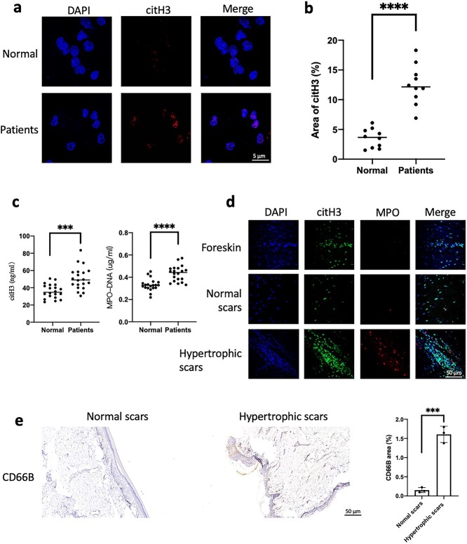
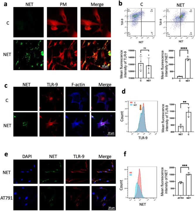
Methods: Neutrophils were isolated from human peripheral blood by magnetic-bead sorting. NETs in plasma and scars were detected by enzyme-linked immunosorbent assays (ELISAs), immunofluorescence and flow cytometry. Immunohistochemistry was used to assess neutrophil (CD66B) infiltration in hypertrophic scars. To observe the entry of NETs into fibroblasts we used immunofluorescence and flow cytometry.

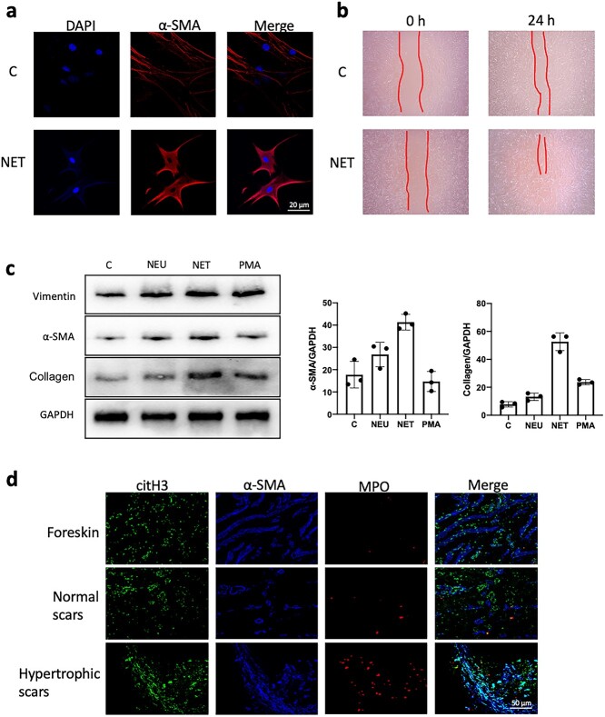
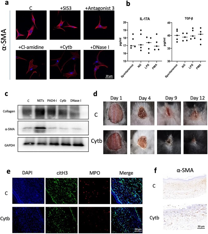
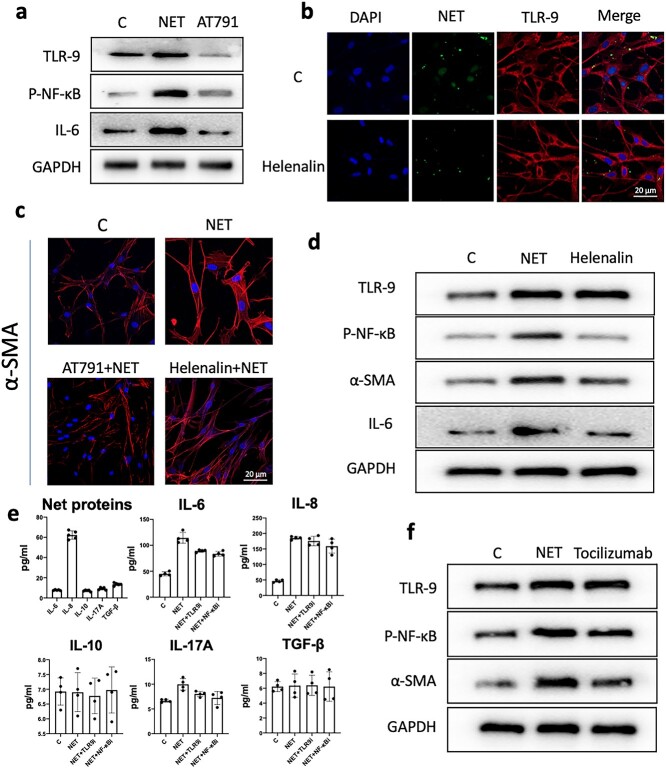
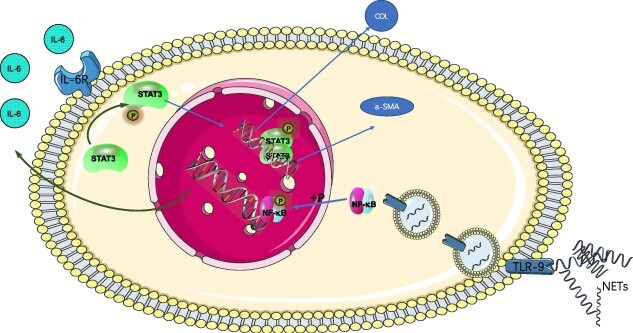
Results: We found that peripheral blood neutrophils in patients with hypertrophic scars were more likely to form NETs (p < 0.05). Hypertrophic scars showed greater infiltration with neutrophils and NETs (p < 0.05). NETs activate fibroblasts in vitro to promote their differentiation and migration. Inhibition of NETs with cytochalasin in wounds reduced the hyperplasia of scars in mice. We induced neutrophils to generate NETs with different stimuli in vitro and detected the proteins carried by NETs. We did not find an increase in the expression of common scarring factors [interleukin (IL)-17 and transforming growth factor-β (TGF-β), p > 0.05]. However, inhibiting the production of NETs or degrading DNA reduced the differentiation of fibroblasts into myofibroblasts. In vitro, NETs were found to be mediated by Toll-like receptor 9 (TLR-9) in fibroblasts and further phosphorylated nuclear factor Kappa-B (NF-κB). We found that IL-6, which is downstream of NF-κB, was increased in fibroblasts. Additionally, IL-6 uses autocrine and paracrine signaling to promote differentiation and secretion.

Conclusions: Our experiments found that NETs activate fibroblasts through the TLR-9/NF-κB/IL-6 pathway, thereby providing a new target for regulating hypertrophic scars.

Keywords: Fibroblast, Inflammation, Differentiation, Nuclear factor Kappa-B, Interleukin-6; Hypertrophic scar; Neutrophil extracellular traps; Toll-like receptor 9.


Figures

Figure 1.
Figure 1.
Increased production of NETs in patients with hypertrophic scarring. Peripheral blood was collected from 20 patients with scar hyperplasia and healthy volunteers (Normal group). (a, b) After purification of the neutrophils of the two groups, immunofluorescence staining of neutrophil citrullinated histones (red) was performed in the two groups (scale bar: 5 μm). The citH3 level in the neutrophil nuclei of the patient group was significantly higher than that of the healthy volunteer group (Normal group) (****p < 0.0001). (c) ELISAs detected citH3 and NETs (MPO–DNA) in plasma, and the levels of the patient group was significantly higher than those of the healthy volunteer group (Normal group) (***p < 0.001, ****p < 0.0001). (d) Foreskin, normal scar and hypertrophic scar tissues were sliced and stained, and the NETs (citH3 + MPO) in the hypertrophic scar increased significantly compared with those in the other two groups (scale bar: 50 μm). (e) Immunohistochemical staining of normal scars and hypertrophic scar tissue showed that neutrophil infiltration (CD66B) in hypertrophic scars was significantly increased compared with that in normal scars (scale bar: 50 μm, ***p < 0.001). C the blank control, NETsneutrophil extracellular traps, citH3 citrullinated histone H3, MPO myeloperoxidase, ELISAs enzyme-linked immunosorbent assays
Figure 2.
Figure 2.
NETs stimulate fibroblast differentiation and secretion. (a) Fibroblasts were stimulated with NETs for 24 h in vitro. NETs led to a significant increase in the expression of α-SMA in fibroblasts compared with that in the control group (scale bar: 20 μm). (b) NET stimulation of fibroblasts for 24 h resulted in accelerated migration of fibroblasts compared to the controls. (c) Fibroblasts were stimulatedwith neutrophils (NEU), PMA and NET in vitro, and compared to no stimulation (C). WB detection showed that compared with other treatments, stimulation with NETs led to increased expression of α-SMA and collagen in fibroblasts. (GAPDH, glyceraldehyde-3-phosohate dehydrogenase) (d) Immunofluorescence staining of foreskin, normal scars and hyperplastic scar tissue showed increased expression and more disorganized arrangement of α-SMA (blue) within the hyperplastic scars compared with the other two groups (scale bar: 50 μm). C the blank control, NETs neutrophil extracellular traps, α-SMA α-smooth muscle actin,  PMA phorbol-12-Myristate-13-Acetate, WB western blot, GAPDH glyceraldehyde-3-phosohate dehydrogenase
Figure 3.
Figure 3.
Reducing NET formation can inhibit scar hyperplasia. (a) Preincubation of fibroblasts with a TGF-β inhibitor (SIS3) and an IL-17A inhibitor (antagonist 3) in vitro did not inhibit the expression of α-SMA in fibroblasts. Inhibition or degradation of NETs with PAD4 inhibitor (Cl-amidine), cytochalasin (Cytb) or DNase I reduced the expression of a-SMA in fibroblasts (scale bar: 20 μm). (b) ELISA detection of NET-carrying proteins stimulated by PMA, LPS and NG on the surface of neutrophils. The amount of IL-17A and TGF-β carried on the surface was low and there was no significant difference compared with spontaneously imagined NETs. (c) WB analysis showed that PAD4 inhibitor (Cl-amidine), cytochalasin (Cytb) and DNase I could inhibit the expression of α-SMA and collagen in fibroblasts induced by NETs. (d) A skin lesion with a diameter of 1.5 cm was made on the back of the mouse and the dressings were changed with normal saline and Cytb. The wound scars of the Cytb group mice were smaller when the wound healing time was the same in the two groups of mice. (e) Immunofluorescence staining was performed on the back healing wound tissue of the two groups of mice and Cytb effectively inhibited the infiltration of NETs (MPO + DNA) (scale bar: 50 μm). (f) Immunohistochemical detection of the back healing wound tissue of the two groups of mice showed that Cytb could effectively reduce the expression of α-SMA in the tissue (scale bar: 50 μm). Cthe blank control, NETs neutrophil extracellular traps,TGF-β transforming growth factor-beta, IL interleukin, α-SMA α-smooth muscle actin, PAD4 peptidylarginine deiminase 4, PMA phorbol-12-Myristate-13-Acetate, LPS Lipopolysaccharide, MPO myeloperoxidase, GAPDH glyceraldehyde-3-phosohate dehydrogenase
Figure 4.
Figure 4.
TLR-9 receptor mediates NET entry into fibroblasts. (a) The TLR-9 receptor mediates NET entry into fibroblasts. Fibroblasts were stimulated with stained NETs (green) for 24 h in vitro and some NETs entered fibroblasts (scale bar: 20 μm). (b) Fibroblasts were stimulated with stained NETs (SYTOX) for 24 h in vitro and the expression of total TLR-9 receptor and NETs in fibroblasts was detected by flow cytometry. Compared with that of the control group, the total TLR-9 receptors of fibroblasts did not change significantly (ns: p > 0.05) and the NETs entering the cells increased (****p < 0.0001). (c) Fibroblasts were stimulated with stained NETs (green) for 24 h in vitro and the intracellular TLR-9 expression receptor was significantly increased and colocalized with the NETs that entered the cells (scale bar: 20 μm). (d) Flow cytometry detection of the TLR-9 receptor in fibroblast membranes. NETs stimulated for 24 h led to decreased expression of the TLR-9 receptor in fibroblast membranes (**p < 0.01). (e) After preincubation of fibroblasts with a TLR-9 receptor inhibitor (AT791) in vitro, AT791 effectively reduced the entry of NETs into fibroblasts (scale bar: 20 μm). (f) NETs entered fibroblasts, as detected by flow cytometry. AT791 effectively reduced the entry of NETs into fibroblasts (***p < 0.001). C the blank control, TLR-9 Toll-like receptors 9, NETs neutrophil extracellular traps, ns not statistically, DAPI4',6-diamidino-2-phenylindole
Figure 5.
Figure 5.
The NETs/TLR-9/NF-κB pathway mediates fibroblast IL-6 secretion and leads to scar hyperplasia. (a) Fibroblasts were preincubated with a TLR-9 receptor inhibitor (AT791) in vitro and WB detection showed that AT791 could reduce the expression of P-NF-κB and IL-6 induced by NET-stimulated fibroblasts. (b) Fibroblasts were preincubated with an NF-κB phosphorylation inhibitor (helenalin) in vitro and immunofluorescence showed that helenalin did not affect the expression of the TLR-9 receptor or the entry of NETs into fibroblasts (scale bar: 20 μm). (c) In vitro preincubation of fibroblasts with TLR-AT791 or helenalin inhibited the expression of α-SMA in fibroblasts induced by NETs (scale bar: 20 μm). (d) Fibroblasts were preincubated with an NF-κB phosphorylation inhibitor (helenalin) in vitro and WB analysis showed that helenalin did not affect the expression of the TLR-9 receptor but could reduce P-NF-κB and IL-6 in fibroblasts induced by NET-SMA expression. (e) PMA stimulates neutrophils to form NETs. The protein carried by NETs was purified and several cytokines were detected by ELISAs. IL-8 had the highest expression, while the expression of other cytokines was very low. After NET stimulation of fibroblasts for 24 h, the corresponding cytokines in the culture supernatant were detected and it was found that only the expression levels of IL-6 and IL-8 were increased, and IL-6 could be inhibited by AT791(TLR9i) or helenalin(NF-κBi). (f) Fibroblasts were preincubated with an IL-6 receptor inhibitor (tocilizumab) in vitro. Tocilizumab did not affect the expression of the TLR-9 receptor or P-NF-κB induced by NET stimulation but reduced the expression of α-SMA. the blank control, TLR-9 Toll-like receptors 9, NETs neutrophil extracellular traps, IL interleukin, α-SMA  α-smooth muscle actin, PMA phorbol-12-Myristate-13-Acetate, P-NF-κB Phosphorylation nuclear factor kappa-B, WB western blot, GAPDH glyceraldehyde-3-phosohate dehydrogenase, ELISAs enzyme-linked immunosorbent assays, TGF-β transforming growth factor-β
Figure 6.
Figure 6.
Schematic diagram of the TLR-9/NF-κB/IL-6 pathway.  NETs translocate into fibroblasts through TLR-9 receptor interactions and lead to an increase in nuclear P-NF-κB, which in turn initiates the expression and increase of IL-6 in fibroblasts. The secretion of IL-6 promotes the expression of α-SMA and the production of collagen in fibroblasts through autocrine and paracrine signaling, resulting in scar hyperplasia. TLR-9 Toll-like receptors 9, NETs neutrophil extracellular traps, IL interleukin, α-SMA  α-smooth muscle actin, PMA phorbol-12-Myristate-13-Acetate

Similar articles

Cited by

References

    1. Monstrey S, Middelkoop E, Vranckx JJ, Bassetto F, Ziegler UE, Meaume S, et al. Updated scar management practical guidelines: non-invasive and invasive measures. J Plast Reconstr Aesthet Surg. 2014;67:1017–25. - PubMed
    1. Elsaie ML. Update on management of keloid and hypertrophic scars: a systemic review. J Cosmet Dermatol. 2021;20:2729–38. - PubMed
    1. Ogawa R. Keloid and hypertrophic scars are the result of chronic inflammation in the reticular dermis. Int J Mol Sci. 2017;18:606. 10.3390/ijms18030606. - DOI - PMC - PubMed
    1. Wang ZC, Zhao WY, Cao Y, Liu YQ, Sun Q, Shi P, et al. The roles of inflammation in keloid and hypertrophic scars. Front Immunol. 2020;11:603187. - PMC - PubMed
    1. Fischer A, Wannemacher J, Christ S, Koopmans T, Kadri S, Zhao J, et al. Neutrophils direct preexisting matrix to initiate repair in damaged tissues. Nat Immunol. 2022;23:518–31. - PMC - PubMed