. 2022 Nov 3:10:1058895.
doi: 10.3389/fcell.2022.1058895. eCollection 2022.

Tissue-specific requirement of sodium channel and clathrin linker 1 (Sclt1) for ciliogenesis during limb development

Affiliations

Tissue-specific requirement of sodium channel and clathrin linker 1 (Sclt1) for ciliogenesis during limb development

Hankyu Lee et al. Front Cell Dev Biol. .

Abstract

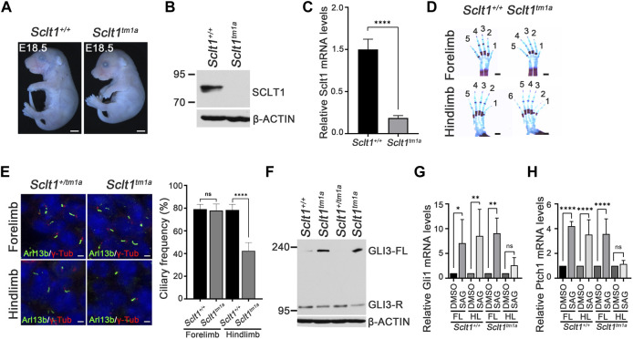
Primary cilia have essential roles as signaling centers during development and adult homeostasis. Disruption of ciliary structure or function causes congenital human disorders called ciliopathies. Centriolar distal appendage (DAP) proteins are important for anchoring cilia to the membrane. However, the exact functions of DAP during in vivo ciliogenesis and animal development remain poorly understood. Here, we showed that the DAP component sodium channel and clathrin linker 1 (Sclt1) mutant mice had abnormal craniofacial and limb development with postnatal lethality. In mutant embryos, most of the affected tissues had defects in DAP recruitment to the basal body and docking to the membrane that resulted in reduced ciliogenesis and disrupted hedgehog (Hh) signaling in limb bud mesenchymal cells. However, limb digit formation and ciliogenesis in Sclt1 mutant mice were differentially affected between the fore- and hindlimb buds. The forelimbs developed normally in Sclt1 mutants, but the hindlimbs had preaxial polydactyly. Heterozygous loss of Cep83, another core DAP component, in Sclt1 mutant mice, caused forelimb and hindlimb polydactyly. These findings revealed the tissue-specific differential requirement of DAPs. Taken together, these results indicated that during limb development the ciliary base components, DAPs, play an essential role in ciliogenesis and Hh signaling in vivo in a position-dependent manner.

Keywords: SCLT1; ciliogenesis; ciliopathy; distal appendage; limb development; primary cilia.


Conflict of interest statement

The authors declare that the research was conducted in the absence of any commercial or financial relationships that could be construed as a potential conflict of interest.

Figures

FIGURE 1
FIGURE 1
Sclt1 mutant mice display hindlimb-specific preaxial polydactyly caused by disruption of ciliogenesis and Hh signaling (A) Gross morphological analysis of Sclt1 tm1a and wildtype control littermates at stage E18.5. Scale bars = 1 mm (B) Western blot analysis to detect levels of SCLT1 from E10.5 whole embryo extracts. Signal specific to anti-SCLT1 antibody is undetectable in Sclt1 mutant embryos, compared with wildtype control (C) Quantitative RT-PCR (qRT-PCR) analyses with RNAs from whole embryos at E10.5 confirmed that expression levels of properly spliced full-length transcripts were lower than those of wildtype transcripts (D) Alcian blue/alizarin red staining of limbs in E18.5 embryos. Hindlimbs of Sclt1 mutant had abnormal digit patterning such as preaxial polydactyly (E) Left panel, representative immunofluorescence image of co-staining with ciliary marker, Arl13b (Arl13b, green), and basal body marker, gamma-tubulin (γ-Tub, red). Right panel, quantitative analysis of ciliogenesis from immunofluorescent stained Sclt1 mutant and control limb buds. Error bars represent SEM and asterisk denotes statistical significance, based on Student′s t test results (***p <0.001) (F) GLI3 processing was analyzed using western blotting with anti-GLI3 antibody. Whole embryo extracts from E10.5 wildtype and Sclt1 mutant embryos were obtained and then examined to detect the full-length (GLI3-FL) and processed (GLI3-R) forms of GLI3 protein with anti-GLI3 antibody. Two independent embryos for each genotype were used and β-actin levels were used as a control (G) To analyze the response to Hh activation in cultured primary mesenchymal cells of forelimb and hindlimb buds, 100 nM Smoothened agonist (SAG) were treated, and then qRT-PCR analysis was performed to measure mRNA levels of Hh target genes, Gli1 and Ptch1. Error bars represent SEM and asterisk denotes statistical significance, based on Student′s t test results (*p <0.05; **p <0.01; ****p <0.0001).
FIGURE 2
FIGURE 2
Sclt1 is involved in hierarchical DAP assembly during ciliogenesis of the hindlimb.To confirm SCLT1 mediated DAP assembly in mesenchymal cells obtained from E11.5 limb buds, we analyzed the localization of the DAP proteins such as CEP83, CEP89, CEP164, and FBF1 using immunofluorescent staining with indicated antibodies (green), and γ-Tub antibody (red) for basal body. Disruption of Sclt1 in mesenchymal cells prevents CEP164 and FBF1 recruitment to the basal body. Scale bars = 1 µm.
FIGURE 3
FIGURE 3
Sclt1 is required for initiation of ciliogenesis by anchoring ciliary vesicle through DAPs and subsequent tau-tubulin kinase-2 (TTBK2) recruitment (A) Transmission electron microscopy analysis of developing limb buds at E11.5 was carried out to identify the early stage of ciliogenesis, ciliary vesicle docking to the mother centriole. Loss of Sclt1 resulted in reduced ciliary vesicle formation in the distal end of the centriole. Quantitative analysis (right panel) was conducted to compare the extent of ciliary vesicle docking in limb buds from Sclt1 mutant and wildtype control embryos. Error bars represent SEM (n = 3 independent samples; **p <0.01, Student′s t tests) (B) To examine the effects of Sclt1 on TTBK2 and CP110 localization in cultured limb mesenchymal cells, immunofluorescent images were taken from cells co-stained with anti-TTBK2 (green) or with CP110 antibodies (green), and with γ-Tub antibody (red). Representative images were shown in the top panel and quantitative results are presented in the bottom panel. Error bars represent SEM values and asterisks denote statistical significance according to Student′s t tests (****p <0.0001).
FIGURE 4
FIGURE 4
Heterozygosity of Cep83 in Sclt1 mutant induces fore- and hindlimb polydactyly (A) Photomicroscopic image to compare the gross morphology of the Sclt1 single mutant, Sctl1 KO , and heterozygous Cep83 combined with Sclt1 null mutant, Cep83 +/KO ;Sctl1 KO with control embryos at E18.5. Scale bars = 2 mm (B) To evaluate the genetic interaction of Cep83 with Sclt1 in limb digit specification, we examined the digit patterning in the Sclt1 single mutant, Cep83 +/+ ;Sclt1 KO ; Cep83 and Sclt1 double mutant,Cep83 +/KO ;Sclt1 KO ; and control littermates, Cep83 +/+ ;Sclt1 +/+ , by staining limbs with alcian blue/alizarin red. Heterozygous loss of Cep83 gene with Sclt1 homozygote mutant affects forelimb and hindlimb digit specification at E18.5. Scale bars = 500 µm (C) Immunofluorescent images were taken from cryosectioned E11.5 embryo limbs and analyzed with anti-Arl13b (green) and γ-Tub antibodies (red). Representative images were shown in the left panel and quantitative results presented in the right panel. Error bars represent SEM and asterisks denote statistical significance according to Student′s t tests (****p <0.0001).
FIGURE 5
FIGURE 5
Heterozygosity of Cep83 in Sclt1 mutant disrupts complete DAP formation in the forelimb Sclt1 mutant embryos had further disrupted forelimb digit patterning by heterozygous loss of Cep83. To confirm the role of Cep83 in complete DAP assembly in Sclt1 mutant mice, limb mesenchymal cells were analyzed with DAP proteins specific antibodies to determine their proper localization in basal bodies. Scale bars = 1 µm. Quantitative analyses were done with more than 150 cells counted and statistical analysis with Student′s t tests were used (*p < 0.05; **p < 0.01).

Similar articles

Cited by

References

    1. Adly N., Alhashem A., Ammari A., Alkuraya F. S. (2014). Ciliary genes TBC1D32/C6orf170 and SCLT1 are mutated in patients with OFD type IX. Hum. Mutat. 35 (1), 36–40. 10.1002/humu.22477 - DOI - PubMed
    1. Badano J. L., Mitsuma N., Beales P. L., Katsanis N. (2006). The ciliopathies: an emerging class of human genetic disorders. Annu. Rev. Genomics Hum. Genet. 7, 125–148. 10.1146/annurev.genom.7.080505.115610 - DOI - PubMed
    1. Blanco-Ameijeiras J., Lozano-Fernández P., Martí E. (2022). Centrosome maturation - in tune with the cell cycle. J. Cell Sci. 135 (2), jcs259395. 10.1242/jcs.259395 - DOI - PubMed
    1. Bowler M., Kong D., Sun S., Nanjundappa R., Evans L., Farmer V., et al. (2019). High-resolution characterization of centriole distal appendage morphology and dynamics by correlative STORM and electron microscopy. Nat. Commun. 10 (1), 993. 10.1038/s41467-018-08216-4 - DOI - PMC - PubMed
    1. Čajánek L., Nigg E. A. (2014). Cep164 triggers ciliogenesis by recruiting Tau tubulin kinase 2 to the mother centriole. Proc. Natl. Acad. Sci. U. S. A. 111 (28), E2841–E2850. 10.1073/pnas.1401777111 - DOI - PMC - PubMed

LinkOut - more resources