Case Reports
. 2022 Oct 18;14(10):e30415.
doi: 10.7759/cureus.30415. eCollection 2022 Oct.

Histopathological Features of Gerhardt Syndrome in a Patient With Multiple System Atrophy: An Autopsy Case Report

Affiliations
Case Reports

Histopathological Features of Gerhardt Syndrome in a Patient With Multiple System Atrophy: An Autopsy Case Report

Keiichi Nakahara et al. Cureus. .

Abstract

Multiple system atrophy (MSA) is a progressive neurodegenerative disease characterized by autonomic failure, parkinsonism, and cerebellar ataxia. Gerhardt syndrome, which is inspiratory dyspnea with laryngeal stridor associated with dysfunction of the vocal folds, is a frequent and fatal complication of MSA. A 59-year-old man with a six-year history of MSA presented with ataxia and dysarthria. He also had dyspnea and stridor, which had worsened in the last three months, and died from respiratory distress. Autopsy revealed neurogenic group atrophy of the posterior cricoarytenoid (PCA) muscle, which suggested that laryngeal nerve damage caused abductor vocal fold paralysis in addition to cerebellar and brainstem atrophy with glial cytoplasmic inclusions. Our histopathological findings suggest that Gerhardt syndrome may be associated with neurogenic atrophy of the laryngeal abductor muscle (PCA muscle) of the vocal folds.

Keywords: gerhardt syndrome; group atrophy; multiple system atrophy; pathological autopsy; posterior cricoarytenoid muscle.


Conflict of interest statement

The authors have declared that no competing interests exist.

Figures

Figure 1
Figure 1. Brain MRI findings
Brain MRI shows atrophy of cerebellar and middle cerebellar peduncles (MCP), enlargement of the fourth ventricle, hyperintensity in the MCP (MCP-sign) (white arrow), and hot cross bun sign (black arrow). A: Axial T1-weighted MRI, B: Axial T2-weighted MRI, C: Fluid attenuated inversion recovery (FLAIR)
Figure 2
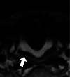
Figure 2. Laryngeal MRI finding
Axial T2-weighted MRI on initial admission shows no atrophy of the posterior cricoarytenoid (PCA) muscle (arrow).
Figure 3
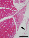
Figure 3. Neurogenic group atrophy of the posterior cricoarytenoid muscle in an autopsy case with multiple system atrophy (MSA)
Neurogenic groups atrophy of the posterior cricoarytenoid muscle (arrow). Hematoxylin and eosin, scale bar 200 µm.

Similar articles

References

    1. The Movement Disorder Society criteria for the diagnosis of multiple system atrophy. Wenning GK, Stankovic I, Vignatelli L, et al. Mov Disord. 2022;37:1131–1148. - PMC - PubMed
    1. Stridor in multiple system atrophy: consensus statement on diagnosis, prognosis, and treatment. Cortelli P, Calandra-Buonaura G, Benarroch EE, et al. Neurology. 2019;93:630–639. - PMC - PubMed
    1. Laryngeal movement disorders in multiple system atrophy: a diagnostic biomarker? Gandor F, Vogel A, Claus I, et al. Mov Disord. 2020;35:2174–2183. - PMC - PubMed
    1. Laryngeal stridor in multiple system atrophy: clinicopathological features and causal hypotheses. Ozawa T, Sekiya K, Aizawa N, Terajima K, Nishizawa M. J Neurol Sci. 2016;361:243–249. - PubMed
    1. Stridor and death in multiple system atrophy. Silber MH, Levine S. Mov Disord. 2000;15:699–704. - PubMed

Publication types

LinkOut - more resources