Tattoo Pigment in an Intramammary Lymph Node Mimicking Breast Malignancy
- PMID: 36407269
- PMCID: PMC9663708
- DOI: 10.7759/cureus.30336
Tattoo Pigment in an Intramammary Lymph Node Mimicking Breast Malignancy
Abstract
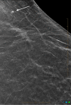
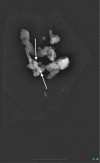
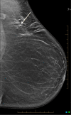
There are many patterns of microcalcification in mammography. Distinguishing between these patterns can be challenging. A malignant cause needs to be assessed through further diagnostic workup. We present a case of a 36-year-old BRCA1 mutation carrier, presenting with a small mass containing calcification on her screening mammogram. A vacuum-assisted biopsy under tomosynthesis guidance was performed and demonstrated an intramammary lymph node showing prominent extracellular black pigment. To our knowledge, this is the first case report of tattoo pigment mimicking breast malignancy on mammography.
Keywords: breast calcification; breast malignancy; case report; intramammary lymph node; tattoo pigment.
Copyright © 2022, Jung et al.
Conflict of interest statement
The authors have declared that no competing interests exist.
Figures


Similar articles
-
Think of ink: Tattoo pigment masquerading as a breast mass.J Med Imaging Radiat Oncol. 2024 Jun;68(4):424-426. doi: 10.1111/1754-9485.13659. Epub 2024 Apr 17. J Med Imaging Radiat Oncol. 2024. PMID: 38632859
-
Tattoo pigment mimicking axillary lymph node calcifications on mammography.Radiol Case Rep. 2020 Jun 9;15(8):1194-1196. doi: 10.1016/j.radcr.2020.05.014. eCollection 2020 Aug. Radiol Case Rep. 2020. PMID: 32550957 Free PMC article.
-
Tattoo pigment in axillary lymph node mimicking calcification of breast cancer.BMJ Case Rep. 2013 Aug 8;2013:bcr2013200284. doi: 10.1136/bcr-2013-200284. BMJ Case Rep. 2013. PMID: 23929611 Free PMC article.
-
Ovarian cancer initially presenting as intramammary metastases and mimicking a primary breast carcinoma: a case report and literature review.J Womens Health (Larchmt). 2010 Jan;19(1):169-74. doi: 10.1089/jwh.2009.1465. J Womens Health (Larchmt). 2010. PMID: 20088673 Review.
-
Digital breast tomosynthesis of gynecomastia and associated findings-a pictorial review.Clin Imaging. 2014 Sep-Oct;38(5):565-70. doi: 10.1016/j.clinimag.2014.04.005. Epub 2014 Apr 21. Clin Imaging. 2014. PMID: 24852677 Review.
Cited by
-
Generalized lymphadenopathy mimicking lymphoma caused by tattoo pigment in a cosmetic worker.Intern Emerg Med. 2024 Nov;19(8):2363-2364. doi: 10.1007/s11739-024-03706-4. Epub 2024 Jul 11. Intern Emerg Med. 2024. PMID: 38990499 No abstract available.
References
-
- BI-RADS descriptors for mammographically detected microcalcifications verified by histopathology after needle-localized open breast biopsy. Shin HJ, Kim HH, Ko MS, Kim HJ, Moon JH, Son BH, Ahn SH. Am J Roentgenol. 2010;195:1466–1471. - PubMed
-
- Insights into worldwide interest in tattoos using Google trends. Kluger N. Dermatology. 2019;235:240–242. - PubMed
-
- Epidemiology of tattoos in industrialized countries. Kluger N. Curr Probl Dermatol. 2015;48:6–20. - PubMed
-
- A class act? Social background and body modifications in Australia. Tranter B, Grant R. J Sociol . 2018;54:412–428.
-
- Fell Fell, A. (2020. Tattoos in Australia; Perceptions, trends and regrets. [ Jul; 2022 ]. 2020. https://mccrindle.com.au/article/blog/tattoos-in-australia-perceptions-t... https://mccrindle.com.au/article/blog/tattoos-in-australia-perceptions-t...
Publication types
LinkOut - more resources
Full Text Sources
Miscellaneous
