. 2022 Nov 3:9:994080.
doi: 10.3389/fcvm.2022.994080. eCollection 2022.

Role of miR-199a-5p in the post-transcriptional regulation of ABCA1 in response to hypoxia in peritoneal macrophages

Affiliations

Role of miR-199a-5p in the post-transcriptional regulation of ABCA1 in response to hypoxia in peritoneal macrophages

Juan Francisco Aranda et al. Front Cardiovasc Med. .

Abstract

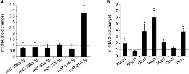
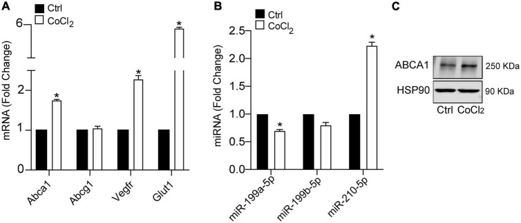
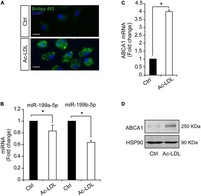
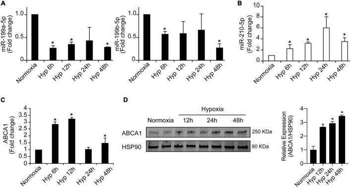
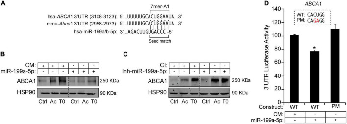
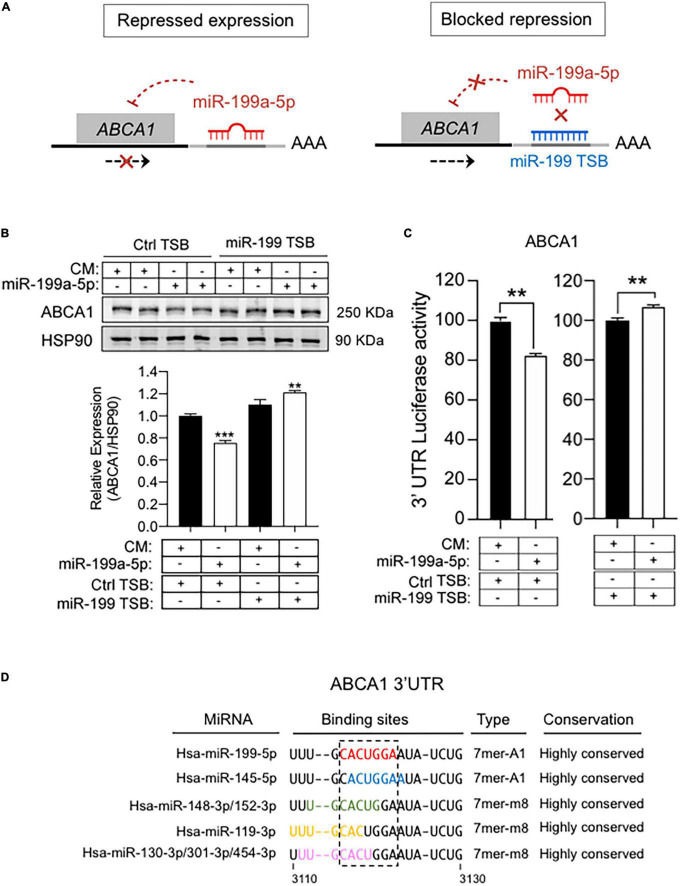
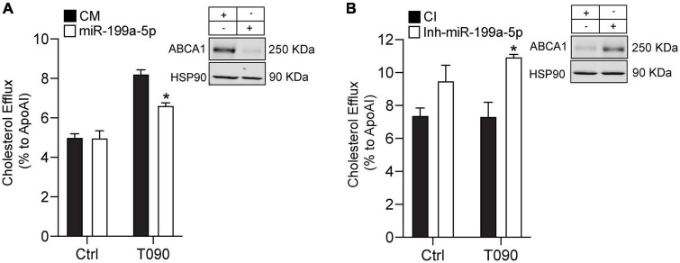
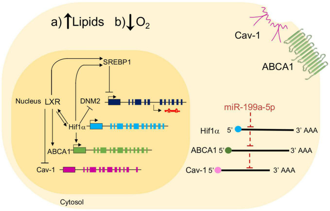
Hypoxia is a crucial factor contributing to maintenance of atherosclerotic lesions. The ability of ABCA1 to stimulate the efflux of cholesterol from cells in the periphery, particularly foam cells in atherosclerotic plaques, is an important anti-atherosclerotic mechanism. The posttranscriptional regulation by miRNAs represents a key regulatory mechanism of a number of signaling pathways involved in atherosclerosis. Previously, miR-199a-5p has been shown to be implicated in the endocytic and retrograde intracellular transport. Although the regulation of miR-199a-5p and ABCA1 by hypoxia has been already reported independently, the role of miR-199a-5p in macrophages and its possible role in atherogenic processes such us regulation of lipid homeostasis through ABCA1 has not been yet investigated. Here, we demonstrate that both ABCA1 and miR-199a-5p show an inverse regulation by hypoxia and Ac-LDL in primary macrophages. Moreover, we demonstrated that miR-199a-5p regulates ABCA1 mRNA and protein levels by directly binding to its 3'UTR. As a result, manipulation of cellular miR-199a-5p levels alters ABCA1 expression and cholesterol efflux in primary mouse macrophages. Taken together, these results indicate that the correlation between ABCA1-miR-199a-5p could be exploited to control macrophage cholesterol efflux during the onset of atherosclerosis, where cholesterol alterations and hypoxia play a pathogenic role.

Keywords: ABCA1; atherosclerosis; cholesterol efflux; hypoxia; macrophage; miRNAs.


Conflict of interest statement

The authors declare that the research was conducted in the absence of any commercial or financial relationships that could be construed as a potential conflict of interest.

Figures

FIGURE 1
FIGURE 1
Analysis of miR-199a-5p targets involved in Atherosclerosis. (A) Representation of miR-199a-5p expression in human tissue using Gtex Portal. Expression values are shown in TPM (Transcripts Per Million), calculated from a gene model with isoforms collapsed to a single gene. No other normalization steps have been applied. Box plots are shown as median and 25th and 75th percentiles and points are displayed as outliers if they are above or below 1.5 times the interquartile range. (B) Bioinformatic analysis of prominent miR-199a-5p target genes obtained thought TargetScan 6.2. involved in atherosclerosis using GeneMania. Blue lines indicate pathway co-localization, yellow lines indicate predicted and purple lines indicate co-expression. (C) Table of human predicted targets genes of miR-199a-5p involved in atherosclerosis obtained using TargetScan 6.2., showing the number and type of the putative binding sites and the conservation in mouse and human.
FIGURE 2
FIGURE 2
Hypoxia regulates miRNAs and cholesterol transporter ABCA1. (A) qRT-PCR analysis of miR-199a-5pa, miR-199b-5p, miR-33a-5p, miR-758-5p, and miR-144-5p in peritoneal macrophages exposed to hypoxic conditions. HypoxamiR miR-210-5p was used as a positive control. (B) qRT-PCR analysis of Abca1, Abcg1, Glut-1, Vegfr, Mcp1, Cox-2, and iNos expression in mouse peritoneal macrophages during hypoxia conditions. Data are expressed as relative expression levels and correspond to the means ± SEM from three independent experiments performed in triplicate. *P < 0.05, significantly different from control normoxic cells.
FIGURE 3
FIGURE 3
Hypoxia mimicking condition dowregulates miR-199a-5p expression. (A) qRT-PCR analysis of miR-199a-5p, miR-199b-5p, and miR-210-5p mouse peritoneal macrophages stimulated with CoCl2 (100 μM) that mimics hypoxia conditions. (B) qRT-PCR analysis Abca1, Hifα, Vegfr, and Glut-1 expression levels in was used as a positive control for hypoxia. (C) Representative Western Blot analysis of ABCA1 in mouse peritoneal macrophages stimulated with CoCl2 (100 μM) for 24 h. HSP90 was used as a loading control. Data are expressed as relative expression levels and correspond to the means ± SEM from three independent experiments performed in triplicate. *P < 0.05, significantly different from control (Ctrl) non-treated cells.
FIGURE 4
FIGURE 4
Cholesterol loading in peritoneal macrophages downregulates miR-199a-5p and upregulates ABCA1. (A) Representative images showing lipid droplets in mouse peritoneal macrophages treated with Ac-LDL for 24 h and stained with Bodipy 468 (green). Nuclei were stained with DAPI (blue). These experiment were performed 3 independent times. Scalebar: 10 μm. (B,C) qRT-PCR analysis of miR-199a-5p, miR-199b-5p (B) and Abca1 (C), expression levels in mouse peritoneal macrophages stimulated with Ac-LDL (120 μg/mL) for 24 h. Data are expressed as relative expression levels and correspond to the means ± SEM from three independent experiments performed in triplicate. *P < 0.05, significantly different from control (Ctrl) cells. (D) Representative Western blot of ABCA1 in mouse peritoneal macrophages stimulated with Ac-LDL. HSP90 was used as a loading control.
FIGURE 5
FIGURE 5
Inverse regulation of miR-199a-5p and ABCA1 during hypoxia. (A) qRT-PCR analysis of miR-199a-5p, and miR-199b-5p expression levels in mouse peritoneal macrophages stimulated with hypoxia at the indicated time points. (B) miR-210-5p was used as a positive control of hypoxia. (C) qRT-PCR analysis of ABCA1 in mouse peritoneal macrophages subjected to hypoxia at the indicated time points. (D) Representative Western blot of ABCA1 in mouse peritoneal macrophages under hypoxia at the indicated time points. HSP90 was used as a loading control. Right panel shows relative ABCA1 protein expression normalized to HSP90 (n = 3). Data are expressed as relative expression and correspond to the means ± SEM from three independent experiments. *P < 0.05, significantly different from cells in normoxia.
FIGURE 6
FIGURE 6
MiR-199a-5p suppresses ABCA1 expression. (A) Human and mouse ABCA1 3′UTRs sequences containing target sites for miR-199a/b-5p. Highlighted in bold are the sequences for miR-199a/b-5p-binding site and seed match. (B,C) Representative Western blot analysis of ABCA1 in mouse peritoneal macrophages transfected with CM and miR-199a-5p or Ctrl-Inh or Inh-miR-199a-5p and stimulated with T090 3 μM for 12 h and Ac-LDL for 24 h. HSP90 was used as a loading control. Dashed lines separates different transfection conditions within the same Western blot. Data are expressed as relative expression levels and correspond to the means ± SEM from three independent experiments performed in triplicate. *P < 0.05, significantly different from CM or CI without treatment. (D) Luciferase reporter activity in COS-7 cells transfected with CM or miR-199a-5p mimic and the ABCA1 3’UTR [wild type (WT)] or the constructs containing the indicated point mutations (PM). Nucleotides highlighted in red indicate the point mutations in the miR-199a-5p-binding sites. Data are expressed as relative luciferase activity compared to the activity in control samples cotransfected with an equal concentration of CM and correspond to the means ± SEM of three experiments performed in triplicate. *P < 0.05, significantly different from cells cotransfected with CM and the WT or PM 3’UTR.
FIGURE 7
FIGURE 7
MiR-199 target site blockers and ABCA1 expression. (A) Schematic overview of LNA-enhanced target site blocker (TSB) mode of action. Without miR-199 TSB or with scramble TSB, miR-199/RISC complex binds to the 3’UTR of ABCA1 mRNA, thereby attenuating its expression. By contrast, addition of miR-199 TSB blocks the binding of miR-199 and derepress ABCA1. (B) Representative Western blot of ABCA1 in mouse peritoneal macrophages transfected as indicated. transfected with synthetic miR-199a-5p mimic in the presence of scramble TSBs or specific miR-199 TSBs for 48 h. Lower panel shows relative ABCA1 protein expression normalized to HSP90 (n = 6). *P < 0.05, significantly different from cells cotransfected with CM and Ctrl TSB. (C) Luciferase reporter activity in COS-7 cells transfected with CM or miR-199a-5p mimic and the ABCA1 3’UTR in the presence of Ctrl TSB or miR-199a-5p TSB. Data are expressed as relative luciferase and correspond to the means ± SEM of three experiments performed in triplicate. **P < 0.01, significantly different from cells transfected with CM (left panel) or Ctrl TSB (right panel) ***P > 0.001. (D) Representation of the human 3’UTR region of ABCA1 where miR-199-5p binding site is located (red), and the overlapping with binding sites for the indicated miRNAs.
FIGURE 8
FIGURE 8
MiR-199a-5p inhibits cholesterol efflux. (A,B) Cholesterol efflux analysis to ApoA1 performed in mouse peritoneal macrophages transfected with Ctrl-miR (CM) and miR-199a-5p or Ctrl-Inh (CI) or Inh-miR-199a-5p and stimulated with or without T0901317 (T090) 3 μm for 12 h. Data are the means ± SEM of three independent experiments in triplicate. *P < 0.05 Ctrl-miR-199a-5p compared with miR-199a-5p or Ctrl-Inh-199 compared with Inh-miR-199a-5p. (A,B) Representative Western blots of ABCA1 in mouse peritoneal macrophages transfected with CM and miR-199a-5p or CI or Inh-miR-199a-5p. HSP90 was used as a loading control.
FIGURE 9
FIGURE 9
Schematic showing the effect of miR-199a-5p on ABCA1 and potential interplay mechanisms with key genes involved in atherosclerosis. Schematic overview about a possible regulation of miR-199a-5p in the relation between ABCA1, Cav-1, LXR in hypoxia conditions in mouse peritoneal macrophages. Black arrows represent activation processes and inhibition processes. LXR activates several genes involve in atherosclerosis such as ABCA1, SREBP1 and HIF1α, but inhibits Cav-1. On the other hand, some of these genes are inhibited by miR-199a-5p and present a possible direct binding for this miRNA on its predicted site in the 3’UTR.

Similar articles

Cited by

References

    1. Thomas H, Diamond J, Vieco A, Chaudhuri S, Shinnar E, Cromer S, et al. Global atlas of cardiovascular disease 2000-2016: the path to prevention and control. Glob Heart. (2018) 13:143–63. 10.1016/J.GHEART.2018.09.511 - DOI - PubMed
    1. Glass CK, Witztum JL. Atherosclerosis: the road ahead. Cell. (2001) 104:503–16. 10.1016/S0092-8674(01)00238-0 - DOI - PubMed
    1. Takahashi K, Takeya M, Sakashita N. Multifunctional roles of macrophages in the development and progression of atherosclerosis in humans and experimental animals. Med Electron Microsc. (2002) 35:179–203. 10.1007/S007950200023 - DOI - PubMed
    1. Parathath S, Yang Y, Mick S, Fisher EA. Hypoxia in murine atherosclerotic plaques and its adverse effects on macrophages. Trends Cardiovasc Med. (2013) 23:80–4. - PMC - PubMed
    1. Sluimer JC, Gasc JM, van Wanroij JL, Kisters N, Groeneweg M, Sollewijn Gelpke MD, et al. Hypoxia, hypoxia-inducible transcription factor, and macrophages in human atherosclerotic plaques are correlated with intraplaque angiogenesis. J Am Coll Cardiol. (2008) 51:1258–65. 10.1016/J.JACC.2007.12.025 - DOI - PubMed