

HOXA10 enhances cell proliferation and suppresses apoptosis in esophageal cancer via activating p38/ERK signaling pathway
- PMID: 36407869
- PMCID: PMC9635270
- DOI: 10.1515/med-2022-0558
HOXA10 enhances cell proliferation and suppresses apoptosis in esophageal cancer via activating p38/ERK signaling pathway
Abstract
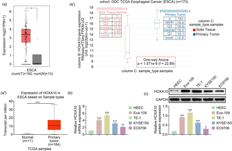
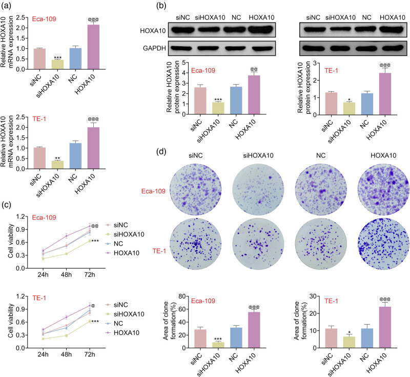
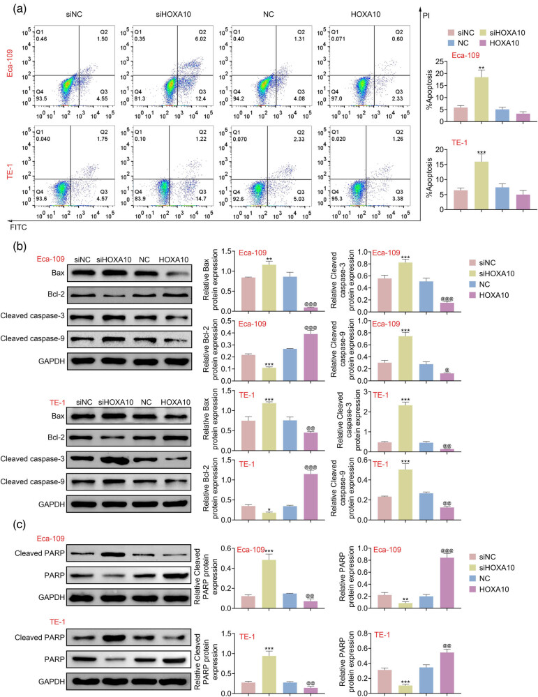
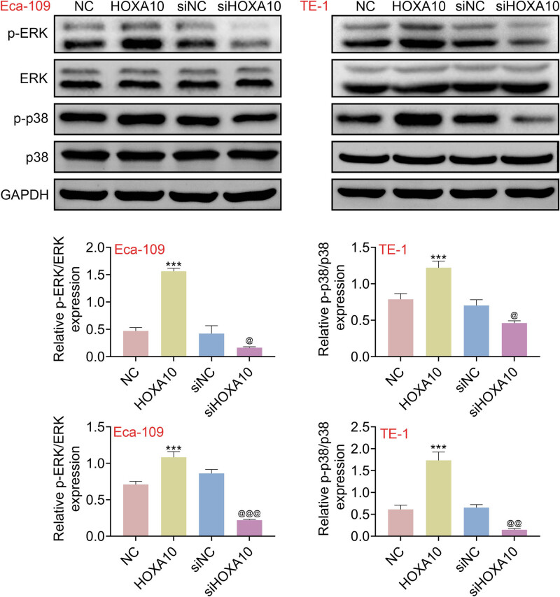
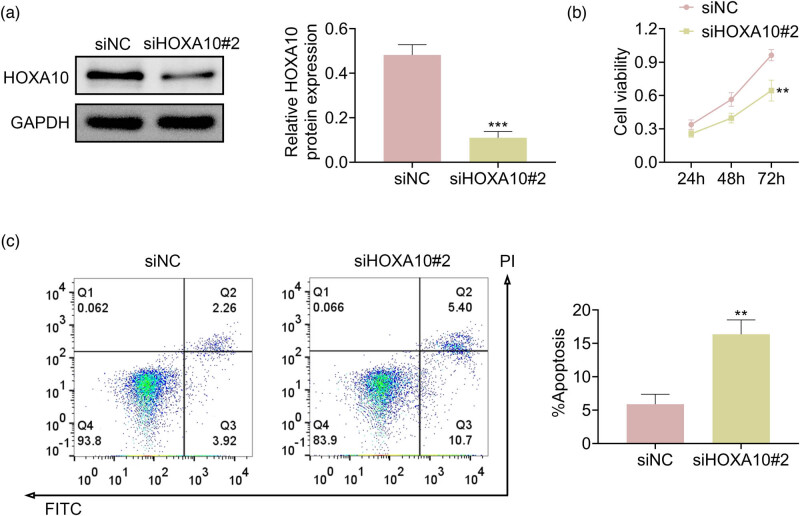
Esophageal cancer (EC) is an extremely aggressive malignant tumor. Homeobox A10 (HOXA10) is highly expressed and plays an important role in a variety of tumors. However, the function of HOXA10 in EC remains unclear. In this study, HOXA10 was observed to highly express in EC tissues and cells. Interestingly, the CCK-8 assay, flow cytometry, and colony formation assay confirmed that overexpression of HOXA10 promoted proliferation and suppressed cell apoptosis in EC cells. More importantly, the western blot assay indicated that the phosphorylation levels of ERK and p38 were elevated in EC cells overexpressed HOXA10, indicating that overexpression of HOXA10 activated p38/ERK signaling pathway in EC cells. These findings concluded that HOXA10 aggravated EC progression via activating p38/ERK signaling pathway, providing a potential therapeutic target for EC.
Keywords: ERK signaling pathway; HOXA10; apoptosis; esophageal cancer; p38 signaling pathway; proliferation.
© 2022 Lifeng Jiang and Qixian Yang, published by De Gruyter.
Conflict of interest statement
Conflict of interest: Authors state no conflict of interest.
Figures
Similar articles
-
HOXA10 promotes cell invasion and MMP-3 expression via TGFβ2-mediated activation of the p38 MAPK pathway in pancreatic cancer cells.Dig Dis Sci. 2014 Jul;59(7):1442-51. doi: 10.1007/s10620-014-3033-6. Epub 2014 Jan 25. Dig Dis Sci. 2014. PMID: 24464212
-
HoxA10 Facilitates SHP-1-Catalyzed Dephosphorylation of p38 MAPK/STAT3 To Repress Hepatitis B Virus Replication by a Feedback Regulatory Mechanism.J Virol. 2019 Mar 21;93(7):e01607-18. doi: 10.1128/JVI.01607-18. Print 2019 Apr 1. J Virol. 2019. PMID: 30674631 Free PMC article.
-
HOXA10 knockdown inhibits proliferation, induces cell cycle arrest and apoptosis in hepatocellular carcinoma cells through HDAC1.Cancer Manag Res. 2019 Jul 26;11:7065-7076. doi: 10.2147/CMAR.S199239. eCollection 2019. Cancer Manag Res. 2019. PMID: 31440094 Free PMC article.
-
Circ_0010235 Regulates HOXA10 Expression to Promote Malignant Phenotypes and Radioresistance in Non-small Cell Lung Cancer Cells Via Decoying miR-588.Balkan Med J. 2022 Jul 22;39(4):255-266. doi: 10.4274/balkanmedj.galenos.2022.2022-2-50. Balkan Med J. 2022. PMID: 35872625 Free PMC article.
-
MiRNA-128 and MiRNA-142 Regulate Tumorigenesis and EMT in Oral Squamous Cell Carcinoma Through HOXA10.Cancer Manag Res. 2020 Oct 12;12:9987-9997. doi: 10.2147/CMAR.S250093. eCollection 2020. Cancer Manag Res. 2020. PMID: 33116855 Free PMC article.
Cited by
-
EIF4A3 induced circGRIK2 promotes the malignancy of glioma by regulating the miR-1303/HOXA10 axis.Am J Cancer Res. 2023 Dec 15;13(12):5868-5886. eCollection 2023. Am J Cancer Res. 2023. PMID: 38187044 Free PMC article.
-
Natural Antisense Transcript-Mediated Regulation of HOXA10-AS in Oral Squamous Cell Carcinoma.J Oral Pathol Med. 2025 Apr;54(4):217-231. doi: 10.1111/jop.13613. Epub 2025 Mar 4. J Oral Pathol Med. 2025. PMID: 40038044 Free PMC article.
-
Leveraging epigenetic aberrations in the pathogenesis of endometriosis: from DNA methylation to non-coding RNAs.Front Genet. 2025 Jul 28;16:1597287. doi: 10.3389/fgene.2025.1597287. eCollection 2025. Front Genet. 2025. PMID: 40792071 Free PMC article. Review.
-
HOXA10 DNA Methylation Level in the Endometrium Women with Endometriosis: A Systematic Review.Biology (Basel). 2023 Mar 20;12(3):474. doi: 10.3390/biology12030474. Biology (Basel). 2023. PMID: 36979165 Free PMC article. Review.
References
-
- Hongsuphan N, Faikongngeon S, Wongtanasarasin W. Cardiac tamponade as an unusual initial presentation of squamous cell carcinoma of the esophagus. Signa Vitae. 2021:1–4. 10.22514/sv.2021.120. - DOI
LinkOut - more resources
Full Text Sources
Research Materials
Miscellaneous