. 2022 Nov 3:16:1013712.
doi: 10.3389/fnins.2022.1013712. eCollection 2022.

Upregulation of neuropeptide Y in cardiac sympathetic nerves induces stress (Takotsubo) cardiomyopathy

Affiliations

Upregulation of neuropeptide Y in cardiac sympathetic nerves induces stress (Takotsubo) cardiomyopathy

Takahide Arai et al. Front Neurosci. .

Abstract

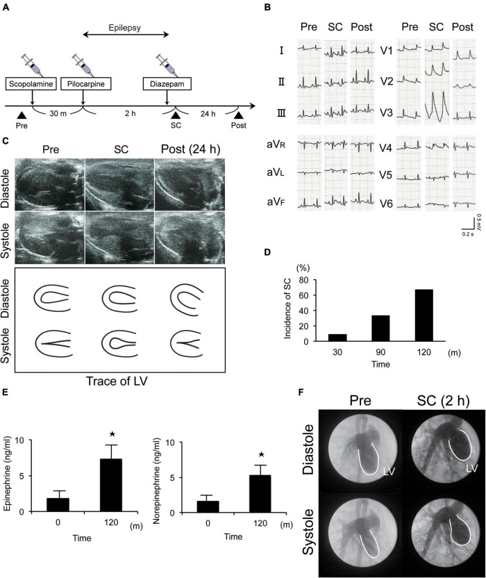
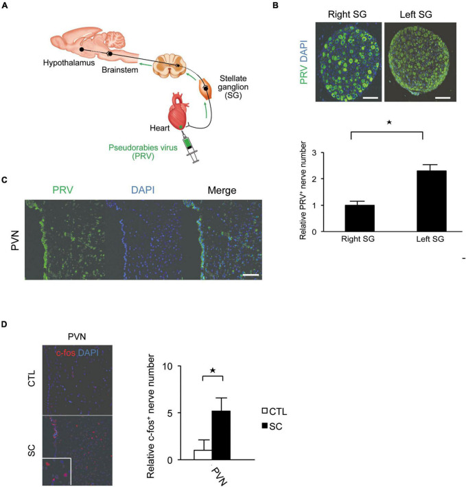
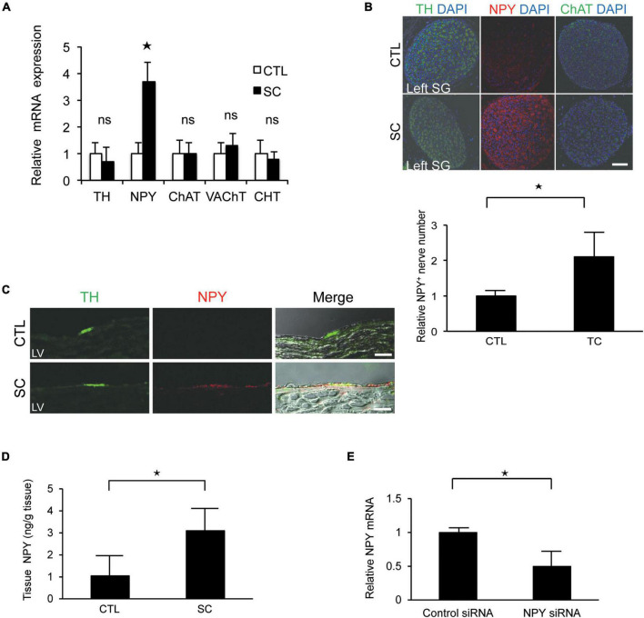
Substantial emotional or physical stress may lead to an imbalance in the brain, resulting in stress cardiomyopathy (SC) and transient left ventricular (LV) apical ballooning. Even though these conditions are severe, their precise underlying mechanisms remain unclear. Appropriate animal models are needed to elucidate the precise mechanisms. In this study, we established a new animal model of epilepsy-induced SC. The SC model showed an increased expression of the acute phase reaction protein, c-Fos, in the paraventricular hypothalamic nucleus (PVN), which is the sympathetic nerve center of the brain. Furthermore, we observed a significant upregulation of neuropeptide Y (NPY) expression in the left stellate ganglion (SG) and cardiac sympathetic nerves. NPY showed neither positive nor negative inotropic and chronotropic effects. On the contrary, NPY could interrupt β-adrenergic signaling in cardiomyocytes when exposure to NPY precedes exposure to noradrenaline. Moreover, its elimination in the left SG via siRNA treatment tended to reduce the incidence of SC. Thus, our results indicated that upstream sympathetic activation induced significant upregulation of NPY in the left SG and cardiac sympathetic nerves, resulting in cardiac dysfunctions like SC.

Keywords: Takotsubo cardiomyopathy; cardiac sympathetic nervous; neuropeptide Y; stellate ganglion; stress.


Conflict of interest statement

The authors declare that the research was conducted in the absence of any commercial or financial relationships that could be construed as a potential conflict of interest.

Figures

FIGURE 1
FIGURE 1
New animal model of epilepsy-induced stress cardiomyopathy (SC). (A) Protocol for the induction of SC in Wistar rats using pilocarpine. (B) Electrocardiograms revealing typical ST elevation during SC. Pre, pre-inducing epilepsy; Post, post-inducing epilepsy; (C,F) Echocardiography (c) and left ventriculography (F) revealed that wall motion of the left ventricle (LV) apical segment was akinetic or dyskinetic during SC. (D) Incidence of epilepsy-induced SC (n = 9). (E) Upregulated serum epinephrine, norepinephrine, and dopamine levels 2 h after the induction of epilepsy (n = 4). Where appropriate, data are provided as the mean ± SD. *P < 0.05 compared with 0 min (E).
FIGURE 2
FIGURE 2
Pseudorabies virus (PRV) staining of the central portions of sympathetic nerve fibers innervating the apex of the left ventricle. (A) Schema of retrograde labeling by PRV. SG, stellate ganglion. (B) Neurons were labeled by PRV. More neurons were labeled by PRV in the left SG than in the right SG. DAPI, 4′,6′-diamidino-2-phenylindole. (C) PRV staining revealed that the central portion of the cardiac sympathetic nerve originated mainly in the paraventricular hypothalamic nucleus (PVN). Scale bars: 50 μm (B); 100 μm (C). (D) Immunofluorescence staining for c-fos in the PVN of the control and SC groups. Quantitative analysis of c-fos-positive cells is shown (n = 5). *p < 0.05.
FIGURE 3
FIGURE 3
Upregulation of neuropeptide Y (NPY) expression in the left stellate ganglion (SG) and cardiac sympathetic nerves. (A) mRNA expression of various neuronal markers in the left SG in the control (CTL) and stress cardiomyopathy (SC) groups. SGs in SC model were collected 2 h after the commencement of epilepsy. NPY expression was significantly increased in the SC model, whereas there was no increase in the expression of the choline transporter (CHT), choline acetyltransferase (ChAT), vesicular acetylcholine transporter (VAchT), or tyrosine hydroxylase (TH; n = 5). (B,C) Triple-immunohistochemistry staining of the left SG and left ventricle (LV) for TH (green), NPY (red), and 4′,6′-diamidino-2-phenylindole (DAPI; nuclei; blue). NPY upregulation was observed in the SG and cardiac sympathetic nerve of the LV in the SC model. Scale bars: 50 μm (B) and 100 μm (C). (D) NPY content of the apical part of the LV in control and SC rats (n = 6). (E) Expression level of NPY mRNA significantly reduced following the injection of NPY siRNA into left SG compared to control siRNA (n = 6). Where appropriate, data are provided as the mean ± SD. *P < 0.05 relative to the control.
FIGURE 4
FIGURE 4
(A) Investigation of the effects of neuropeptide Y (NPY) on left ventricle (LV) contraction using the Langendorff procedure. Pretreatment with NPY completely blocked NA-induced increase in heart rate and LV developed pressure (n = 4 for each group). (B,C) The measurement of beat frequency and contraction length using primary cultured cardiomyocytes revealed that when hearts were exposed noradrenaline (NA), beat frequency and contraction length significantly increased (n = 4). Conversely, when hearts were first exposed NPY, beat frequency and contraction length did not increase after NA administration (n = 4). (D–F) Effects of NPY on ventricular cardiomyocytes isolated from adult rats based on the measurement of the frequency of Ca2+ transients and the peak to transients. The frequency of Ca2+ transients and the peak to transients in isolated ventricular cardiomyocytes increased significantly when cells were exposed to NA only, but increases were not observed when cells were first exposed to NPY and then administered with NA (n = 4 for each group). *p < 0.05, ns: not significant.

Similar articles

Cited by

References

    1. Akashi Y. J., Nef H. M., Möllmann H., Ueyama T. (2010). Stress cardiomyopathy. Annu. Rev. Med. 61 271–286. 10.1146/annurev.med.041908.191750 - DOI - PubMed
    1. Al-Rafiah A. R., Mehdar K. M. (2021). Histopathological and biochemical assessment of neuroprotective effects of sodium valproate and lutein on the pilocarpine albino rat model of epilepsy. Behav. Neurol. 2021:5549638. 10.1155/2021/5549638 - DOI - PMC - PubMed
    1. Anderson J. L., Adams C. D., Antman E. M., Bridges C. R., Califf R. M., Casey D. E., Jr., et al. (2007). ACC/AHA 2007 guidelines for the management of patients with unstable angina/non-ST-elevation myocardial infarction: A report of the American college of cardiology/American heart association task force on practice guidelines (Writing committee to revise the 2002 guidelines for the management of patients with unstable angina/non-ST-elevation myocardial infarction) developed in collaboration with the American college of emergency physicians, the society for cardiovascular angiography and interventions, and the society of thoracic surgeons endorsed by the American association of cardiovascular and pulmonary rehabilitation and the society for academic emergency medicine. J. Am. Coll. Cardiol. 50 e1–e157. 10.1016/j.jacc.2007.02.013 - DOI - PubMed
    1. Aston-Jones G., Card J. P. (2000). Use of pseudorabies virus to delineate multisynaptic circuits in brain: Opportunities and limitations. J. Neurosci. Methods 103 51–61. 10.1016/s0165-0270(00)00295-8 - DOI - PubMed
    1. Callanan E. Y., Lee E. W., Tilan J. U., Winaver J., Haramati A., Mulroney S. E., et al. (2007). Renal and cardiac neuropeptide Y and NPY receptors in a rat model of congestive heart failure. Am. J. Physiol. Renal Physiol. 293 F1811–F1817. 10.1152/ajprenal.00191.2007 - DOI - PubMed