. 2022 Apr:18:17448069221142523.
doi: 10.1177/17448069221142523.

Adverse effects of methylene blue in peripheral neurons: An in vitro electrophysiology and cell culture study

Affiliations

Adverse effects of methylene blue in peripheral neurons: An in vitro electrophysiology and cell culture study

Megan L Uhelski et al. Mol Pain. 2022 Apr.

Abstract

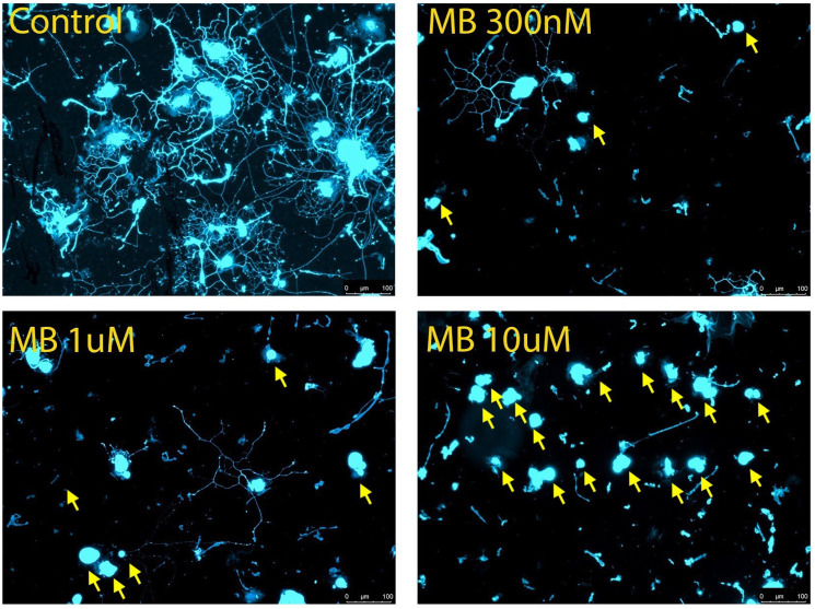
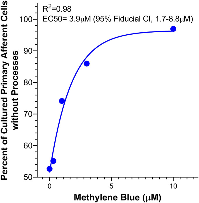
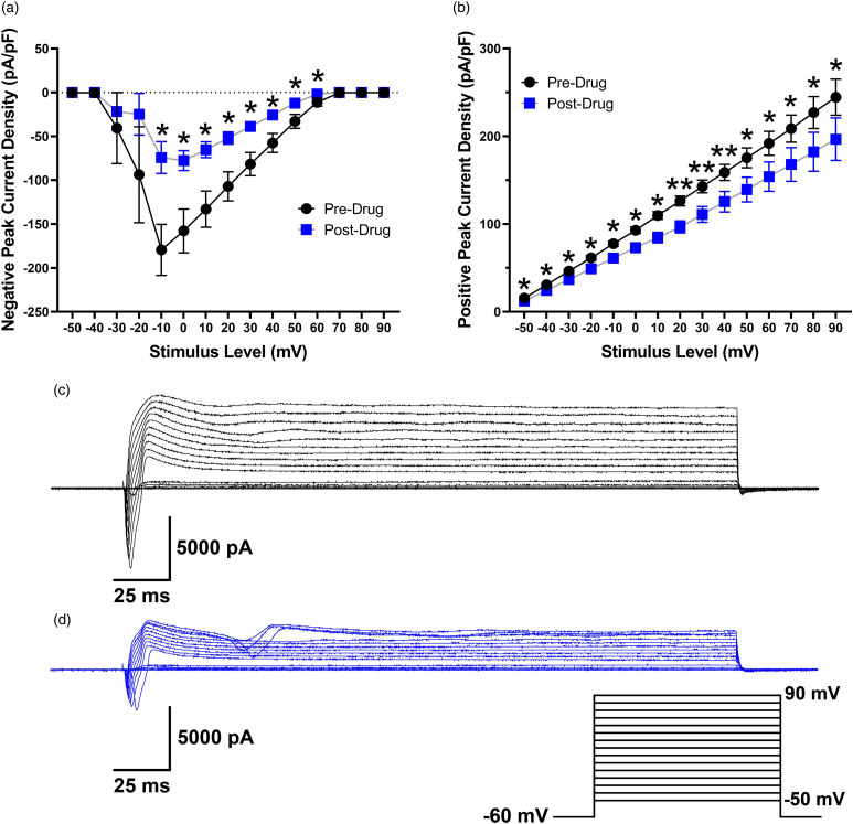
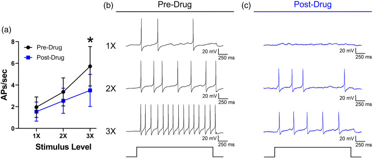
Methylene blue (MB) is an effective treatment for methemoglobinemia, ifosfamide-induced encephalopathy, cyanide poisoning, and refractory vasoplegia. However, clinical case reports and preclinical studies indicate potentially neurotoxic activity of MB at certain concentrations. The exact mechanisms of MB neurotoxicity are not known, and while the effects of MB on neuronal tissue from different brain regions and myenteric ganglia have been examined, its effects on primary afferent neurons from dorsal root ganglia (DRG) have not been studied. Mouse DRG were exposed to MB (0.3-10 μM) in vitro to assess neurite outgrowth. Increasing concentrations of MB (0.3-10 μM) were associated with neurotoxicity as shown by a substantial loss of cells with neurite formation, particularly at 10 μM. In parallel experiments, cultured rat DRG neurons were treated with MB (100 μM) to examine how MB affects electrical membrane properties of small-diameter sensory neurons. MB decreased peak inward and outward current densities, decreased action potential amplitude, overshoot, afterhyperpolarization, increased action potential rise time, and decreased action potential firing in response to current stimulation. MB induced dose-dependent toxicity in peripheral neurons, in vitro. These findings are consistent with studies in brain and myenteric ganglion neurons showing increased neuronal loss and altered membrane electrical properties after MB application. Further research is needed to parse out the toxicity profile for MB to minimize damage to neuronal structures and reduce side effects in clinical settings.

Keywords: dorsal root ganglion; immunocytochemistry; methylene blue; neurite; neurotoxicity.


Conflict of interest statement

The author(s) declared no potential conflicts of interest with respect to the research, authorship, and/or publication of this article.

Figures

Figure 1.
Figure 1.
Cytotoxicity of methylene blue in cultured mouse dorsal root ganglion (DRG) neurons. Approximately 1.1 × 10³ DRG cells were seeded in each well of an 8 well slide along with methylene blue at different concentrations for 16 h. Immunocytochemistry was performed using anti-PGP 9.5 and neurite outgrowth in each well viewed under fluorescence microscope. Representative images for each concentration except 3 μM are shown: (A) control, 0 μM; (B) MB 0.3 μM; (C) MB 1 μM; (D) MB 10 μM. Neurons without neurite extensions are indicated by yellow arrows. Scale bar represents 100 μm.
Figure 2.
Figure 2.
Concentration response curve for the proportion of DRG cells without neurite processes used to calculate the EC50 concentration of methylene blue.
Figure 3.
Figure 3.
Peak negative (A) and positive (B) current density (pA/pF) in rat DRG neurons before and after a 10 min application of MB (n = 7). Traces represent current evoked by command voltage step from −60 mV (holding potential) to test potentials between −50 and 90 mV in 10-mV increments as shown in the inset for a rat DRG neuron before (C) and after (D) application of MB. *p < 0.05, **p < 0.01.
Figure 4.
Figure 4.
Action potential (AP) characteristics in rat DRG neurons (n = 10) before and after a 10 min application of methylene blue (100 μM). Overlapping raw traces from two neurons, one with a shoulder on the falling phase of the action potential (A) and one without (B).
Figure 5.
Figure 5.
(A) Mean responses of rat DRG neurons (n = 11) to current stimulation expressed as the number of APs per second at 1X, 2X, and 3X rheobase before and after a 10 min application of methylene blue (100 μM). Traces represent responses to 1X, 2X, and 3X rheobase current stimulation before (B) and after (C) methylene blue application. *p < 0.05.

Similar articles

References

    1. Oz M, Lorke DE, Hasan M, Petroianu GA. Cellular and molecular actions of Methylene Blue in the nervous system. Med Research Reviews 2011; 31: 93–117. - PMC - PubMed
    1. Pelgrims J, De Vos F, Van den Brande J, Schrijvers D, Prové A, Vermorken J. Methylene blue in the treatment and prevention of ifosfamide-induced encephalopathy: report of 12 cases and a review of the literature. Br Journal Cancer 2000; 82: 291–294. - PMC - PubMed
    1. Habib AM, Elsherbeny AG, Almehizia RA. Methylene blue for vasoplegic syndrome postcardiac surgery. Indian J Crit Care Med 2018; 22: 168–173. - PMC - PubMed
    1. Hanzlik P. Methylene blue as antidote for cyanide poisoning. JAMA: J Am Med Assoc 1933; 100: 357–357. - PMC - PubMed
    1. Li J, Chen X, Qi M, Li Y. Sentinel lymph node biopsy mapped with methylene blue dye alone in patients with breast cancer: a systematic review and meta-analysis. PLoS One 2018; 13: e0204364. - PMC - PubMed

Publication types

LinkOut - more resources