. 2023 Feb 14;227(4):543-553.
doi: 10.1093/infdis/jiac452.

Human Cytomegalovirus Infection of Epithelial Cells Increases SARS-CoV-2 Superinfection by Upregulating the ACE2 Receptor

Collaborators, Affiliations

Human Cytomegalovirus Infection of Epithelial Cells Increases SARS-CoV-2 Superinfection by Upregulating the ACE2 Receptor

Marianne R Perera et al. J Infect Dis. .

Abstract

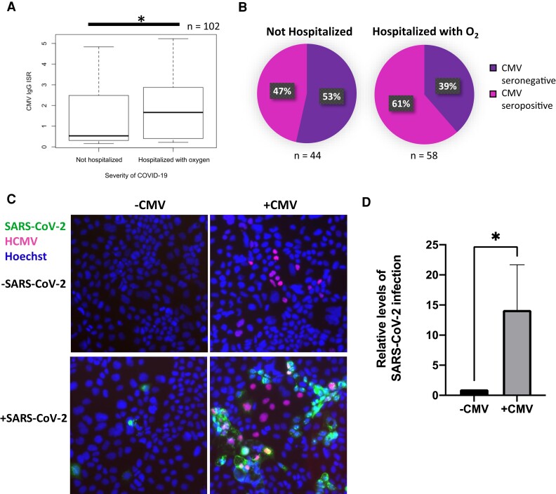
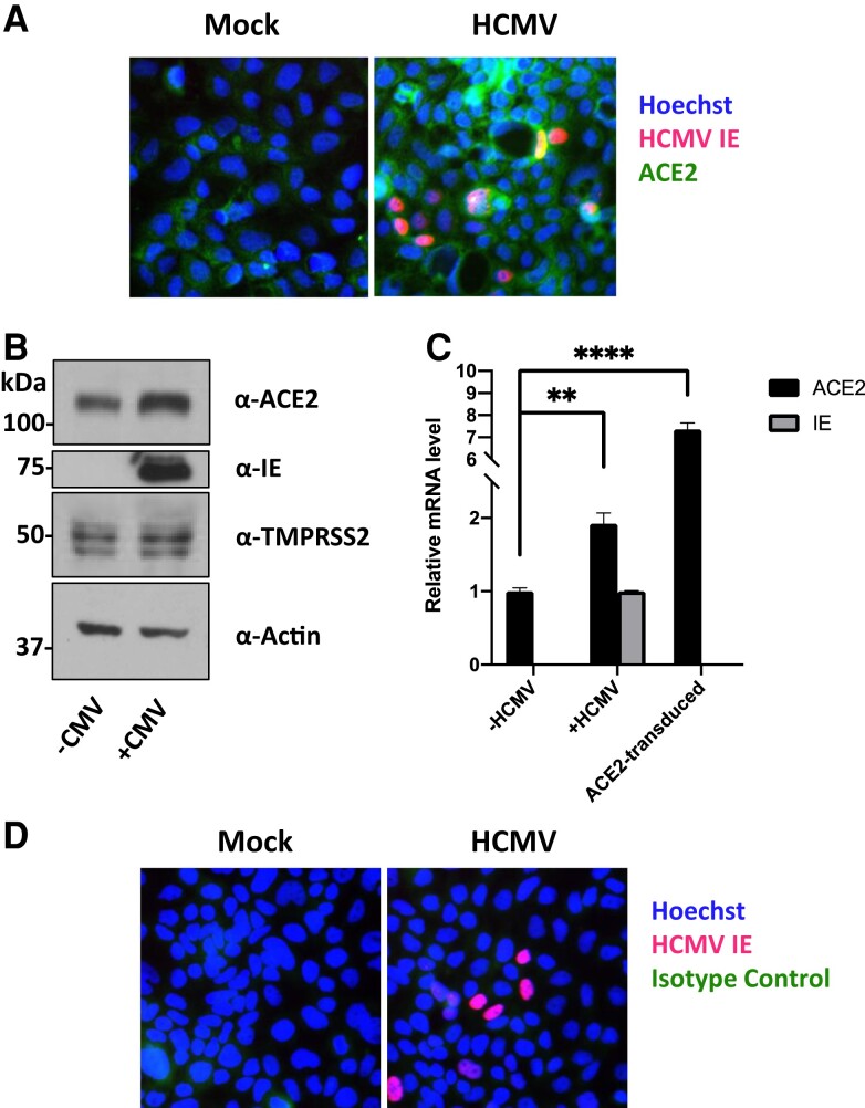
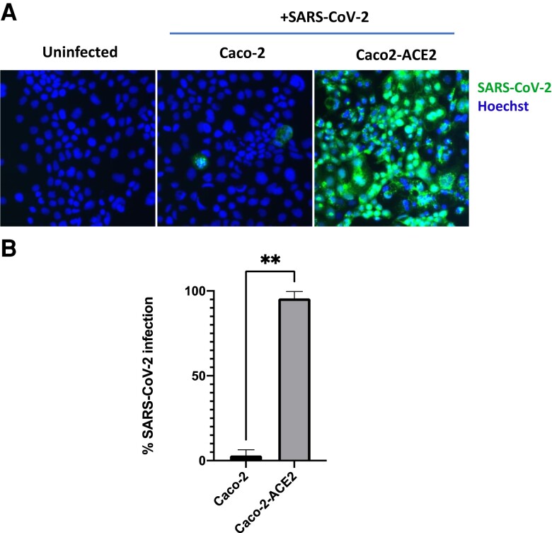
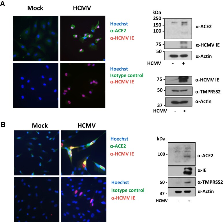
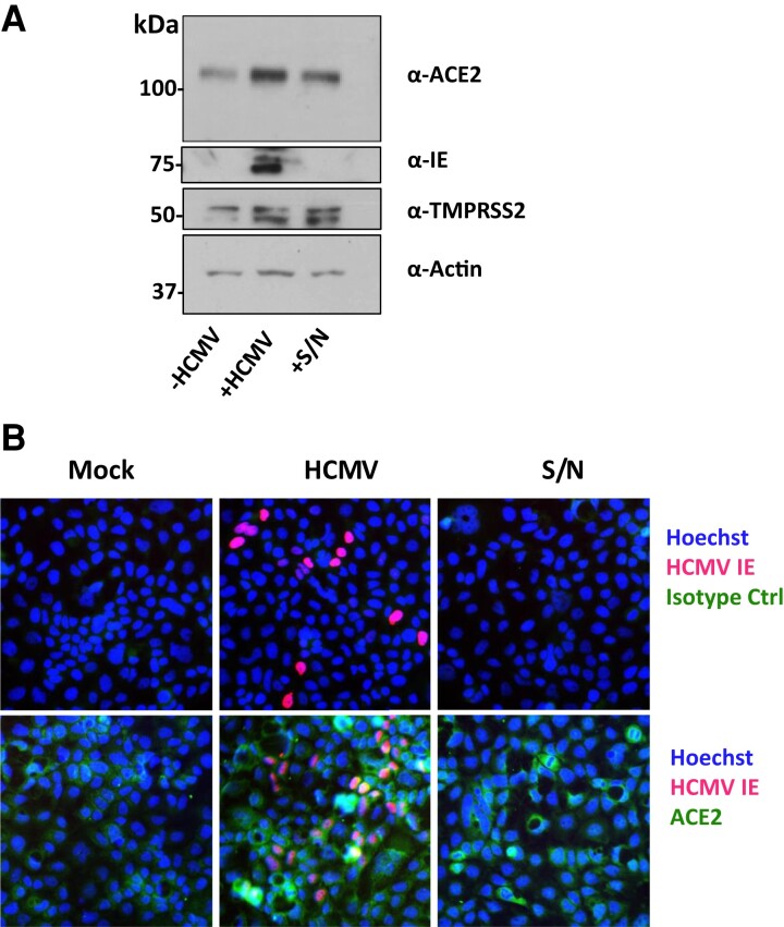
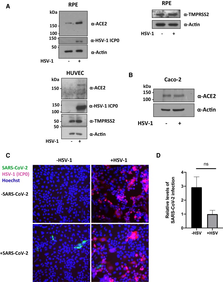
Severe acute respiratory syndrome coronavirus 2 (SARS-CoV-2), the causative agent of coronavirus disease 2019 (COVID-19), has caused widespread morbidity and mortality since its onset in late 2019. Here, we demonstrate that prior infection with human cytomegalovirus (HCMV) substantially increases infection with SARS-CoV-2 in vitro. HCMV is a common herpesvirus carried by 40%-100% of the population, which can reactivate in the lung under inflammatory conditions, such as those resulting from SARS-CoV-2 infection. We show in both endothelial and epithelial cell types that HCMV infection upregulates ACE2, the SARS-CoV-2 cell entry receptor. These observations suggest that HCMV reactivation events in the lung of healthy HCMV carriers could exacerbate SARS-CoV-2 infection and subsequent COVID-19 symptoms. This effect could contribute to the disparity of disease severity seen in ethnic minorities and those with lower socioeconomic status, due to their higher CMV seroprevalence. Our results warrant further clinical investigation as to whether HCMV infection influences the pathogenesis of SARS-CoV-2.

Keywords: ACE2; COVID-19; HCMV; SARS-CoV-2; coinfection; human cytomegalovirus.


Conflict of interest statement

Potential conflicts of interest. All authors: No reported conflicts of interest. All authors have submitted the ICMJE Form for Disclosure of Potential Conflicts of Interest. Conflicts that the editors consider relevant to the content of the manuscript have been disclosed.

Figures

Figure 1.
Figure 1.
Prior infection with HCMV increases SARS-CoV-2 superinfection in vitro. A, Levels of HCMV IgG in patients with PCR-confirmed SARS-CoV-2 infection were measured by ELISA, and the ISR determined (which reflects the levels of HCMV IgG). Patients were stratified into not hospitalized (asymptomatic or mild infection) or hospitalized with O2 (hospitalized with supplemental O2 or hospitalized with assisted ventilation). Box and whisker plots show ISR distributions for these 2 groups; n = 102. Significant difference was determined using a two-tailed Mann Whitney U test. *P < .05. B, Immune response ratio from (A) was used to class patients as CMV seropositive or seronegative, and the proportion of patients who were not hospitalized or hospitalized with oxygen that were CMV seropositive or CMV seronegative is illustrated in a pie chart. C and D, Caco-2 epithelial cells were infected with HCMV for 4 days before being infected with ZsGreen-tagged SARS-CoV-2 at a multiplicity of infection of 0.01. C, Two days postinfection with SARS-CoV-2, cells were fixed, stained for HCMV IE1/2 (red), and stained with Hoechst (blue) and imaged on a fluorescence microscope. D, Graph shows mean percent SARS-CoV-2 infection from 3 randomly chosen images for each of 4 replicates over 2 independent repeats. Error bars show standard deviation. Statistical significance was determined using a 2-tailed Mann-Whitney U test, *P < .05. Abbreviations: ELISA, enzyme-linked immunosorbent assay; HCMV, human cytomegalovirus; IgG, immunoglobulin G; ISR, immune status ratio; PCR, polymerase chain reaction; SARS-CoV-2, polymerase chain reaction.
Figure 2.
Figure 2.
HCMV infection upregulates ACE2 on Caco-2 epithelial cells. Caco-2 cells were infected with HCMV at a multiplicity of infection of 0.1. A, Four dpi, cells were fixed and stained with an anti-HCMV IE1/2 antibody (red), Hoechst stain (blue), and α-ACE2 antibody (green). B, Four dpi, protein lysates from cells were harvested and assessed for levels of ACE2, TMPRSS2, HCMV IE1/2, and the loading control actin by immunoblot. C, Four dpi, RNA was harvested from cells and levels of ACE2, HCMV IE, and the housekeeping RNA, 18S, were measured by RTqPCR. As a positive control, RNA was also harvested from HeLa cells transduced to overexpress ACE2. Significance was determined using a 1-way ANOVA with Dunnett post hoc testing. Error bars show standard deviation. D, As in (A), but cells were stained with an isotype control antibody (green). Abbreviations: ACE2, angiotensin converting enzyme 2; dpi, days postinfection; HCMV, human cytomegalovirus; IE, immediate early protein; RTqPCR, quantitative real-time polymerase chain reaction.
Figure 3.
Figure 3.
Overexpression of ACE2 increases SARS-CoV-2 infection rate in Caco-2 cells. Untransduced Caco-2 cells or Caco-2 cells transduced with an ACE2-overexpression lentivirus were infected with ZsGreen-tagged SARS-CoV-2 virus at a low multiplicity of infection (0.01). A, Two days postinfection, cells were fixed, Hoechst stained, and imaged with a fluorescence microscope. B, Quantification of results shown in (A), counted from 6 random fields of view over 2 independent repeats. Error bars show standard deviation. Statistical significance was determined by a 2-tailed Mann-Whitney U test. **P < .01. Abbreviations: ACE2, angiotensin converting enzyme 2; SARS-CoV-2, polymerase chain reaction.
Figure 4.
Figure 4.
HCMV infection upregulates ACE2 on epithelial and endothelial cells. A, Retinal pigment epithelial cells or (B) human umbilical vein endothelial cells were infected with HCMV at a multiplicity of infection of 1 or 0.1, respectively. A and B, (left) Four dpi, cells were fixed and stained with an anti-HCMV IE1/2 antibody (red), Hoechst stain (blue), and either an α-ACE2 antibody or its isotype control (green). A and B, (right) Four dpi, protein lysates from cells were harvested and assessed for levels of ACE2 or TMPRSS2, HCMV IE1/2, and the loading control actin by immunoblot. Abbreviations: ACE2, angiotensin converting enzyme 2; dpi, days postinfection; HCMV, human cytomegalovirus; IE, immediate early protein.
Figure 5.
Figure 5.
Supernatant from HCMV-infected cells upregulates ACE2 on uninfected Caco-2 cells. Caco-2 cells were mock infected (−HCMV) or infected (+HCMV) with HCMV at a multiplicity of infection of 0.1. Every 24 hours, supernatant from HCMV infected cells was transferred to another well of uninfected Caco-2 cells (+S/N). A, Protein lysates from these cells were then immunoblotted for ACE2, TMPRSS2, HCMV IE1/2, and the loading control actin. B, Four days postinfection, cells were fixed and stained with anti-HCMV IE1/2 antibody (red), Hoechst stain (blue), and either α-ACE2 antibody or its isotype control (green). Abbreviations: ACE2, angiotensin converting enzyme 2; HCMV, human cytomegalovirus; IE, immediate early protein; S/N, supernatant.
Figure 6.
Figure 6.
Exogenous IFNs do not increase levels of ACE2 in Caco-2 cells. Caco-2 cells were treated with increasing concentrations of (A) IFN-α or IFN-β or (B) IFN-γ. Four days posttreatment, protein was harvested form these cells and assayed for levels of ACE2, the loading control actin, or the positive control ISG15. Abbreviations: ACE2, angiotensin converting enzyme 2; IFN, interferon; ISG15, interferon-stimulated gene 15.
Figure 7.
Figure 7.
Prior HSV-1 infection does not increase SARS-CoV-2 infection in Caco-2 epithelial cells, although it can upregulate ACE2 in other cell types. A, RPE (above) or HUVEC (below) were infected with wild-type YFP-HSV-1 at an MOI of 1. At 24 hpi, protein lysates were harvested from cells and immunoblotted for ACE2, TMPRSS2, and the loading control, actin. B, Caco-2 cells were infected with wild-type YFP-HSV-1. At 24 hpi, protein was harvested and blotted for ACE2 levels. C and D, Caco-2 epithelial cells were infected with ΔgE HSV-1 for 24 hours before being infected with ZsGreen-tagged SARS-CoV-2 at an MOI of 0.01. C, Two days postinfection with SARS-CoV-2, cells were fixed, stained for HSV-1 ICP0 (red) and stained with Hoechst (blue), and imaged on a fluorescence microscope. D, Graph shows mean percent SARS-CoV-2 infection from randomly chosen images from 3 replicates. Error bars show standard error. Statistical significance was determined using a 2-tailed Mann-Whitney U test. Abbreviations: ACE2, angiotensin converting enzyme 2; hpi, hours postinfection; HSV, herpes simplex virus-1; HUVEC, human umbilical venous endothelial cells; MOI, multiplicity of infection; ns, not significant; RPE, retinal pigment epithelial cells; SARS-CoV-2, polymerase chain reaction.

References

    1. Berlin DA, Gulick RM, Martinez FJ. Severe COVID-19. N Engl J Med 2020; 383:2451–60. - PubMed
    1. Musuuza JS, Watson L, Parmasad V, Putman-Buehler N, Christensen L, Safdar N. Prevalence and outcomes of co-infection and superinfection with SARS-CoV-2 and other pathogens: a systematic review and meta-analysis. PLoS One 2021; 16:e0251170. - PMC - PubMed
    1. Bai L, Zhao Y, Dong J, et al. . Coinfection with influenza A virus enhances SARS-CoV-2 infectivity. Cell Res 2021; 31:395–403. - PMC - PubMed
    1. Kadambari S, Klenerman P, Pollard AJ. Why the elderly appear to be more severely affected by COVID-19: the potential role of immunosenescence and CMV. Rev Med Virol 2020; 30:e2144. - PMC - PubMed
    1. Söderberg-Nauclér C. Does reactivation of cytomegalovirus contribute to severe COVID-19 disease? Immun Ageing 2021; 18:12. - PMC - PubMed

Publication types

Substances