

Quantitative myelin imaging with MRI and PET: an overview of techniques and their validation status
- PMID: 36408715
- PMCID: PMC10115240
- DOI: 10.1093/brain/awac436
Quantitative myelin imaging with MRI and PET: an overview of techniques and their validation status
Abstract
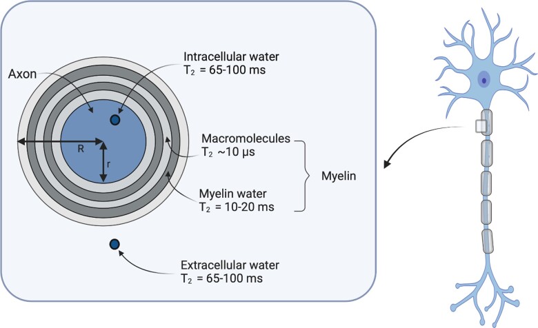
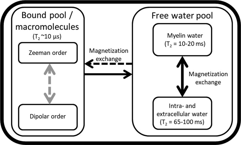
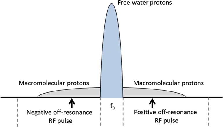
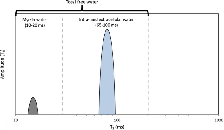
Myelin is the protective sheath wrapped around axons, consisting of a phospholipid bilayer with water between the wraps. The measurement of damage to the myelin sheaths, the evaluation of the efficacy of therapies aiming to promote remyelination and monitoring the degree of brain maturation in children all require non-invasive quantitative myelin imaging methods. To date, various myelin imaging techniques have been developed. Five different MRI approaches can be distinguished based on their biophysical principles: (i) imaging of the water between the lipid bilayers directly (e.g. myelin water imaging); (ii) imaging the non-aqueous protons of the phospholipid bilayer directly with ultra-short echo-time techniques; (iii) indirect imaging of the macromolecular content (e.g. magnetization transfer; inhomogeneous magnetization transfer); (iv) mapping of the effects of the myelin sheath's magnetic susceptibility on the MRI signal (e.g. quantitative susceptibility mapping); and (v) mapping of the effects of the myelin sheath on water diffusion. Myelin imaging with PET uses radioactive molecules with high affinity to specific myelin components, in particular myelin basic protein. This review aims to give an overview of the various myelin imaging techniques, their biophysical principles, image acquisition, data analysis and their validation status.
Keywords: MRI; PET; brain maturation; demyelination; myelin imaging.
© The Author(s) 2022. Published by Oxford University Press on behalf of the Guarantors of Brain.
Conflict of interest statement
The authors report no competing interests.
Figures
Similar articles
-
Inhomogeneous Magnetization Transfer (ihMT) imaging in the acute cuprizone mouse model of demyelination/remyelination.Neuroimage. 2023 Jan;265:119785. doi: 10.1016/j.neuroimage.2022.119785. Epub 2022 Dec 1. Neuroimage. 2023. PMID: 36464096
-
Predicting PET-derived myelin content from multisequence MRI for individual longitudinal analysis in multiple sclerosis.Neuroimage. 2020 Dec;223:117308. doi: 10.1016/j.neuroimage.2020.117308. Epub 2020 Sep 2. Neuroimage. 2020. PMID: 32889117
-
White matter intercompartmental water exchange rates determined from detailed modeling of the myelin sheath.Magn Reson Med. 2019 Jan;81(1):628-638. doi: 10.1002/mrm.27398. Epub 2018 Sep 19. Magn Reson Med. 2019. PMID: 30230605 Free PMC article.
-
Probing myelin content of the human brain with MRI: A review.Magn Reson Med. 2021 Feb;85(2):627-652. doi: 10.1002/mrm.28509. Epub 2020 Sep 16. Magn Reson Med. 2021. PMID: 32936494 Review.
-
Ultrahigh field imaging of myelin disease models: Toward specific markers of myelin integrity?J Comp Neurol. 2019 Sep 1;527(13):2179-2189. doi: 10.1002/cne.24598. Epub 2019 Jan 4. J Comp Neurol. 2019. PMID: 30520034 Review.
Cited by
-
Unsupervised Pattern Analysis to Differentiate Multiple Sclerosis Phenotypes Using Principal Component Analysis on Various MRI Sequences.J Clin Med. 2024 Sep 4;13(17):5234. doi: 10.3390/jcm13175234. J Clin Med. 2024. PMID: 39274448 Free PMC article.
-
The value of synthetic MRI in detecting the brain changes and hearing impairment of children with sensorineural hearing loss.Front Neurosci. 2024 Jun 11;18:1365141. doi: 10.3389/fnins.2024.1365141. eCollection 2024. Front Neurosci. 2024. PMID: 38919907 Free PMC article.
-
Measuring Pathology in Patients with Multiple Sclerosis Using Positron Emission Tomography.Curr Neurol Neurosci Rep. 2023 Sep;23(9):479-488. doi: 10.1007/s11910-023-01285-z. Epub 2023 Jul 7. Curr Neurol Neurosci Rep. 2023. PMID: 37418219 Review.
-
Iron accumulation/overload and Alzheimer's disease risk factors in the precuneus region: A comprehensive narrative review.Aging Med (Milton). 2024 Oct 22;7(5):649-667. doi: 10.1002/agm2.12363. eCollection 2024 Oct. Aging Med (Milton). 2024. PMID: 39507230 Free PMC article. Review.
-
Myelin Imaging of the Spinal Cord in Animal Models and Patients with Multiple Sclerosis Using [11C]MeDAS PET: A Translational Study.J Nucl Med. 2025 Jan 3;66(1):136-141. doi: 10.2967/jnumed.123.266896. J Nucl Med. 2025. PMID: 39638431 Free PMC article.
References
-
- Stadelmann C, Timmler S, Barrantes-Freer A, Simons M. Myelin in the central nervous system: Structure, function, and pathology. Physiol Rev. 2019;99:1381–1431. - PubMed
-
- Baumann N, Pham-Dinh D. Biology of oligodendrocyte and myelin in the mammalian central nervous system. Physiol Rev. 2001;81:871–927. - PubMed
-
- Nave K-A, Werner HB. Myelination of the nervous system: Mechanisms and functions. Annu Rev Cell Dev Biol. 2014;30:503–533. - PubMed
-
- van der Knaap MS, Valk J. Magnetic resonance of myelination and myelin disorders. Padiatr Prax. 2006;68:452.
