

Conbercept improves macular microcirculation and retinal blood supply in the treatment of nonischemic branch retinal vein occlusion macular edema
- PMID: 36408725
- PMCID: PMC9757001
- DOI: 10.1002/jcla.24774
Conbercept improves macular microcirculation and retinal blood supply in the treatment of nonischemic branch retinal vein occlusion macular edema
Abstract
Purpose: To investigate the effect of conbercept on macular microvascular system and retinal blood supply in the treatment of nonischemic branch retinal vein occlusion macular edema (BRVO-ME).
Methods: Patients were divided into three groups: group A (containing 12 nonischemic BRVO-ME eyes), group B (containing contralateral 12 healthy eyes), and group C (containing 30 cataract eyes to obtain normal aqueous humor cytokine levels). Group A received monthly intravitreal injections of conbercept for 3 months. General data and best-corrected visual acuity (BCVA) were compared among the three groups. Optical coherence tomography angiography (OCTA) results (including central macular thickness [CMT], retinal vascular density and perfusion, and foveal avascular zone [FAZ]) at baseline were compared among groups A and B. Aqueous humor cytokine levels (including VEGF, IL-8, PDGF-AA, TNF-α, and ANGPTL-4) at baseline were compared between groups A and C. Moreover, BCVA, OCTA results, and aqueous humor cytokine levels of group A before and after conbercept treatment were compared.
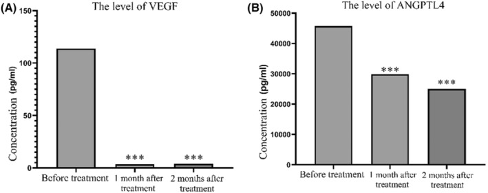
Result: At baseline, group A had a significantly worse BCVA, lower retinal vascular density and perfusion, and numerically larger CMT and FAZ area comparing to the group B, and had a higher aqueous cytokine level (IL-8, VEGF, and ANGPTL-4) comparing to the group C (all ps < 0.05). After the injection of conbercept, group A presented a better BCVA (at initial diagnosis vs. after three conbercept injections: 1.16 ± 0.51 vs. 0.81 ± 0.30, logMAR, p < 0.05), higher retinal vascular density (11.56 ± 4.73 vs. 15.88 ± 2.31, mm-1 , p < 0.05) and perfusion (0.28 ± 0.12 vs. 0.39 ± 0.06, mm2 , p < 0.05), smaller CMT (504.92 ± 184.11 vs. 219.83 ± 46.63, mm2 , p < 0.05), as well as a lower levels of VEGF (before first injection vs. before third injection: 113.84 [70.81, 235.4] vs. 3.94 [3.56, 8.07], pg/ml, p < 0.05) and ANGPTL-4 (45,761 [7327.5, 81,402.5] vs. 25,015.5 [6690, 43,396], pg/ml, p < 0.05). However, the average FAZ area of group A expanded (at initial diagnosis vs. after three conbercept injections: 0.41 ± 0.14 vs. 0.62 ± 0.36, mm2 , p < 0.05).
Conclusion: This study demonstrated that intraocular injection of conbercept could effectively improve macular microcirculation and increase retinal blood supply in the treatment of nonischemic BRVO-ME based on the combination of visual acuity, OCTA parameters, and aqueous humor cytokine assay results. However, further study with a larger sample size and longer observation period is still needed in the future.
Keywords: ANGPTL-4; OCTA; branch retinal vein occlusions; conbercept; macular edema.
© 2022 The Authors. Journal of Clinical Laboratory Analysis published by Wiley Periodicals LLC.
Conflict of interest statement
All authors report no conflicts of interest. The authors alone are responsible for the content and writing of this article.
Figures
Similar articles
-
Evaluation of Microvascular Structure Changes after Conbercept Treatment on Macular Edema Secondary to Retinal Vein Occlusion.Biomed Res Int. 2020 Jun 19;2020:9046781. doi: 10.1155/2020/9046781. eCollection 2020. Biomed Res Int. 2020. PMID: 32685542 Free PMC article. Clinical Trial.
-
Quantitative Analysis of Retinal Microvascular Changes after Conbercept Therapy in Branch Retinal Vein Occlusion Using Optical Coherence Tomography Angiography.Ophthalmologica. 2019;242(2):69-80. doi: 10.1159/000499608. Epub 2019 May 21. Ophthalmologica. 2019. PMID: 31112969
-
Optical coherence tomography angiography for macular microvessels in ischemic branch retinal vein occlusion treated with conbercept: predictive factors for the prognosis.Int J Ophthalmol. 2023 Dec 18;16(12):2049-2055. doi: 10.18240/ijo.2023.12.18. eCollection 2023. Int J Ophthalmol. 2023. PMID: 38111937 Free PMC article.
-
A systematic review and meta-analysis to compare the efficacy of conbercept with ranibizumab in patients with macular edema secondary to retinal vein occlusion.Medicine (Baltimore). 2020 May 22;99(21):e20222. doi: 10.1097/MD.0000000000020222. Medicine (Baltimore). 2020. PMID: 32481293 Free PMC article.
-
Comparison between Ozurdex and intravitreal anti-vascular endothelial growth factor treatment for retinal vein occlusion-related macular edema: A systematic review and meta-analysis of randomized controlled trials.Indian J Ophthalmol. 2019 Nov;67(11):1800-1809. doi: 10.4103/ijo.IJO_382_19. Indian J Ophthalmol. 2019. PMID: 31638037 Free PMC article.
Cited by
-
New Markers for the Assessment of Microvascular Complications in Patients with Metabolic Syndrome.Metabolites. 2025 Mar 10;15(3):184. doi: 10.3390/metabo15030184. Metabolites. 2025. PMID: 40137149 Free PMC article. Review.
-
Effect of multiple intravitreal injections of conbercept on the cornea in patients with branch retinal vein occlusion-induced macular edema.Front Med (Lausanne). 2025 Jun 25;12:1595543. doi: 10.3389/fmed.2025.1595543. eCollection 2025. Front Med (Lausanne). 2025. PMID: 40636373 Free PMC article.
References
-
- Jaulim A, Ahmed B, Khanam T, Chatziralli IP. Branch retinal vein occlusion: epidemiology, pathogenesis, risk factors, clinical features, diagnosis, and complications. An update of the literature. Retina. 2013;33(5):901‐910. - PubMed
-
- Hayreh SS. Photocoagulation for retinal vein occlusion. Prog Retin Eye Res. 2021;85:100964. - PubMed
-
- Heier JS, Bressler NM, Avery RL, et al. Comparison of aflibercept, bevacizumab, and ranibizumab for treatment of diabetic macular edema: extrapolation of data to clinical practice. JAMA Ophthalmol. 2016;134(1):95‐99. - PubMed
MeSH terms
Substances
Grants and funding
LinkOut - more resources
Full Text Sources
