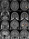
Temporal encephaloceles and coexisting epileptogenic lesions
- PMID: 36408781
- PMCID: PMC9977755
- DOI: 10.1002/epi4.12674
Temporal encephaloceles and coexisting epileptogenic lesions
Abstract
Objective: This study was performed to identify coexisting structural lesions in patients with epilepsy and known temporal encephaloceles (TEs).
Methods: Forty-seven structural magnetic resonance imaging (MRI) scans of patients with epilepsy and radiologically diagnosed TEs were retrospectively reviewed visually and using an automated postprocessing software, the Morphometric Analysis Program v2018 (MAP18), to depict additional subtle, potentially epileptogenic lesions in the 3D T1-weighted MRI data. All imaging findings were evaluated in the context of clinical and electroencephalographical findings.
Results: The study population consisted of 47 epilepsy patients (38.3% female, n = 18). The median age at the time of the scan was 40 years (range 12-81 years). Twenty-one out of 47 MRI scans (44.7%) showed coexisting lesions in the initial MRI evaluation; in 38.3% (n = 18) of patients, those lesions were considered probably epileptogenic. After postprocessing, probable epileptogenic lesions were identified in 53.2% (n = 25) of patients. Malformations of cortical development had initially been reported in 17.0% (n = 8) of patients with TEs, which increased to 38.3% (n = 18) after postprocessing. TEs and other epileptogenic lesions were considered equally epileptogenic in 21.3% (n = 10) of the cases in the initial MR reports and 25.5% (n = 12) of the cases after postprocessing.
Significance: Temporal encephaloceles are a potential cause of MRI-negative temporal lobe epilepsy. According to our data, TEs can occur with other lesions, suggesting that increased awareness is also required in patients with lesional epilepsy. TEs may not always be epileptogenic; hence, their occurrence with other structural pathologies may influence the presurgical evaluation and surgical approach. Finally, TEs can be associated with malformations of cortical development, which may indicate a common developmental etiology of those lesions.
Keywords: epilepsy surgery; focal cortical dysplasia; structural epilepsy; temporal encephaloceles; temporal lobe epilepsy.
© 2022 The Authors. Epilepsia Open published by Wiley Periodicals LLC on behalf of International League Against Epilepsy.
Conflict of interest statement
Christopher Nimsky is a scientific consultant for Brainlab. Adam Strzelczyk reports personal fees and grants from Angelini Pharma/Arvelle Therapeutics, Desitin Arzneimittel, Eisai, GW Pharmaceuticals, Marinus Pharma, Medtronic, UCB, UNEEG Medical, and Zogenix. All other authors have no conflict of interest to disclose with respect to this study.
Figures


References
-
- Siffel C, Wong LYC, Olney RS, Correa A. Survival of infants diagnosed with encephalocele in Atlanta, 1979‐98. Paediatr Perinat Epidemiol. 2003;17(1):40–8. - PubMed
-
- Urbach H, Jamneala G, Mader I, Egger K, Yang S, Altenmüller D. Temporal lobe epilepsy due to meningoencephaloceles into the greater sphenoid wing: a consequence of idiopathic intracranial hypertension? Neuroradiology. 2018;60(1):51–60. - PubMed
-
- Wind JJ, Caputy AJ, Roberti F. Spontaneous encephaloceles of the temporal lobe. Neurosurg Focus. 2008;25(6):E11. - PubMed

