Review
. 2022 Dec 6;23(12):e55420.
doi: 10.15252/embr.202255420. Epub 2022 Nov 21.

Composition, organization and mechanisms of the transition zone, a gate for the cilium

Affiliations
Review

Composition, organization and mechanisms of the transition zone, a gate for the cilium

Kwangjin Park et al. EMBO Rep. .

Abstract

The cilium evolved to provide the ancestral eukaryote with the ability to move and sense its environment. Acquiring these functions required the compartmentalization of a dynein-based motility apparatus and signaling proteins within a discrete subcellular organelle contiguous with the cytosol. Here, we explore the potential molecular mechanisms for how the proximal-most region of the cilium, termed transition zone (TZ), acts as a diffusion barrier for both membrane and soluble proteins and helps to ensure ciliary autonomy and homeostasis. These include a unique complement and spatial organization of proteins that span from the microtubule-based axoneme to the ciliary membrane; a protein picket fence; a specialized lipid microdomain; differential membrane curvature and thickness; and lastly, a size-selective molecular sieve. In addition, the TZ must be permissive for, and functionally integrates with, ciliary trafficking systems (including intraflagellar transport) that cross the barrier and make the ciliary compartment dynamic. The quest to understand the TZ continues and promises to not only illuminate essential aspects of human cell signaling, physiology, and development, but also to unravel how TZ dysfunction contributes to ciliopathies that affect multiple organ systems, including eyes, kidney, and brain.

Keywords: cilia; ciliary gate; ciliary trafficking; ciliopathies; transition zone.


Figures

Figure 1
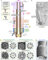
Figure 1. Overview of evolutionarily conserved components and compartments of cilia, including the transition zone “ciliary gate”
(A) All cilia possess a microtubule‐based axoneme that stems from a basal body foundation anchored to the plasma membrane via transition fibers. To build the cilium and make it dynamic, an intraflagellar transport (IFT) machinery powered by molecular motors (IFT‐Kinesin and IFT‐Dynein) docks at transition fibers and uses three functional modules (IFT‐A, IFT‐B and BBS protein complex or BBSome) to traffic ciliary proteins (cargoes) into and out of the organelle. Immediately distal to the basal body is a transition zone (TZ) region, widely believed to form a ciliary gate or diffusion barrier for membrane and soluble proteins. The TZ harbors typically Y‐shaped structures that span from the axoneme to the overlying membrane. In metazoans, the proximal segment includes a so‐called Inversin compartment that functionally interacts with the TZ. Most cilia also possess a distal segment with singlet microtubules. (i) TZ‐associated Y‐links are shown with arrowheads in cross‐section electron microscope images of motile cilia TZs from Chlamydomonas (modified from Craige et al, 2010) and rabbit oviduct (Anderson, 1974), as well as nonmotile cilia TZs from C. elegans (Jensen et al, 2018) and rat photoreceptor (Besharse et al, ; “Copyright 1985 Society for Neuroscience”). (ii) The ends of the TZ Y‐links, which connect to the membrane, likely form ciliary necklace “beads.” Shown is an electron micrograph of freeze‐fractured hamster respiratory airway cilia showing the ciliary necklace region, which contains 7–8 strands of membrane particles (arrowheads) and may form a spiral (arrows show start/end; modified from Heller & Gordon, 1986). Scale bars, 100 nm. (B) Rhodopsin (arrowheads) is highly concentrated in the distal ciliary region of the cilium (outer segment), and virtually absent from the connecting cilium (TZ) and near the basal body. Modified from ref. (Liu et al, ; “Copyright 1999 Society for Neuroscience”). Scale bar, 0.5 μm.
Figure 2
Figure 2. Genetic, physical, and functional interaction network of transition zone proteins
Interactions within the ring include among the most evolutionarily conserved transition zone (TZ) proteins, and interactions with other selected TZ proteins are shown outside of the ring in gray dotted boxes. Genetic interactions between two genes encoding TZ proteins (circles) and/or TZ protein localization dependency (arrows point to dependent protein) are shown on gray connecting lines and colored according to the model organism (Chlamydomonas, purple; Drosophila, blue, C. elegans, green, and vertebrates, red). Direct physical interactions and co‐precipitation of proteins are represented by yellow dotted and solid connections, respectively (wine‐colored dotted lines depict both types of interactions). Tentative grouping of proteins into MKS, NPHP or core scaffolding modules are colored purple, blue and green, respectively; uncertain grouping is shown as gray. Proteins are shown alongside their ciliopathy associations (MKS, Meckel syndrome; JBTS, Joubert syndrome; BBS, Bardet–Biedl syndrome; NPHP, Nephronophthisis; OFD, Orofaciodigital syndrome; COACH, COACH syndrome; SLSN, Senior–Løken syndrome; LCA, Leber congenital amaurosis) as well as domain structures (C2 and related B9 domains; TM, transmembrane; CC, coiled coil; β‐prop, β‐propeller; SH3, SRC homology 3; CYS, cysteine‐rich; MSP, major sperm protein). See text for additional details. All interaction data are presented in Table EV1.
Figure 3
Figure 3. Spatial organization of evolutionarily conserved transition zone proteins
Tentative spatial organization of evolutionarily conserved transition zone (TZ) proteins based on their ciliary subcellular localization in mammalian cells and other species (see text for details). The longitudinal cutout view shows one slice of the ninefold symmetrical microtubule (MT) basal body triplets and TZ doublets (brown layer), together with Y‐link structures that connect the axoneme to the ciliary membrane (blue layer). Since the exact relationship between the TZ proteins and the Y‐link is unclear, the Y‐shaped structure is simply drawn as a projection. In this model, CEP290 and RPGRIP1L (shaped as cuboids; green layer) are interacting, core scaffolding proteins found close to the axoneme MT doublets, with CEP290 being more proximal (closer to the basal body) compared with other TZ proteins in some cilia. RPGRIP1L is required for the localization of all TZ proteins, including NPHP module (triangular prisms) and MKS module (cylinders) components. CEP290 is most closely affiliated with the MKS module. MKS and NPHP module proteins are situated either at or within the membrane (blue layer) or in an intermediate position between the membrane and axoneme (red layer). TF, transition fiber.
Figure 4
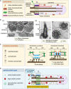
Figure 4. Proposed mechanisms for transition zone function as a diffusion barrier for membrane and soluble proteins
At least four potential, nonmutually exclusive properties of the transition zone (TZ) may explain its role as a ciliary gate. (A) A protein picket fence or “physical diffusion barrier,” consisting of septins and/or additional proteins, may be present within the TZ region, potentially in association with Y‐links and the ciliary necklace. This barrier may prevent membrane‐associated proteins and lipids from outside or inside of the cilium from freely diffusing across the barrier. (B) A condensed, lipid‐ordered microdomain at the TZ may restrict the diffusion of lipids and membrane‐associated proteins. (i) The TZ, which harbors TZ necklaces, is devoid of filipin‐sterol complexes (protrusions) relative to more distal regions of the cilium (where some are circled). These freeze‐fracture views of tracheal cells incubated with filipin suggest the lack of or inaccessibility of free cholesterol in the TZ. Modified from ref. (Montesano, 1979). (ii) The TZ has a detergent‐resistant membrane. Detergent extraction of photoreceptors removes plasma and ciliary membranes, but leaves continuous or interrupted membrane patches at the TZ (white arrows), visible in longitudinal (left) and transverse (right) electron micrographs. The positions of some Y‐links are shown with arrowheads. Modified from ref. (Anderson, 1974). (C) Distinct lipid bilayer properties of the TZ, such as curvature and thickness, may be conferred by different lipid species with different aliphatic and head groups (cylindrical, conical, inverse conical, long‐tailed, short‐tailed), the Y‐links, as well as specific lipid‐binding and membrane‐shaping proteins (red). These properties may influence the diffusion of lipids and membrane‐associated proteins across the TZ. PM, plasma membrane; CM, ciliary membrane; TF, transition fiber. (D) A mesh‐like protein gate, consisting of nucleoporins and potentially other proteins, restricts the movement of larger soluble proteins into and out of the ciliary compartment. Although this sieve‐type gate (green mesh) depends on the TZ, several studies suggest that nucleoporins are localized at the basal body, making its exact site of action unclear (see text for details). The transport of certain soluble proteins (including tubulins) across the gate may benefit from trafficking systems such as IFT (shown at the basal body before entering the cilium).
Figure 5
Figure 5. Trafficking systems that cross the transition zone ciliary gate create a dynamic signaling compartment
Intraflagellar transport (IFT) and other transport systems such as lipidated protein intraflagellar targeting (LIFT) cross the TZ and therefore make the cilium a dynamic compartment. The LIFT system, which includes the lipidated protein‐binding chaperones UNC119 and PDE6D, facilitates the transport of lipid‐modified proteins such as INPP5E and NPHP3 into cilia. Cargo release is mediated by effectors, including ARL3 and RP2. At least for some ciliary cargos, including GPCR proteins, the IFT system (including IFT‐A complex‐associated TULP3) makes use of a PIP2‐PI4P lipid gradient established by INPP5E to bind cargo outside the cilium (high PIP2) and release it inside the cilium (low PIP2). Functional interactions between IFT and LIFT (shown as question mark) have been documented but require further investigation (see text for details).

Similar articles

Cited by

References

    1. AFZELIUS BA (1961) The fine structure of the cilia from ctenophore swimming‐plates. J Biophys Biochem Cytol 9: 383–394 - PMC - PubMed
    1. Andersen OS, Koeppe RE (2007) Bilayer thickness and membrane protein function: an energetic perspective. Annu Rev Biophys Biomol Struct 36: 107–130 - PubMed
    1. Anderson RG (1974) Isolation of ciliated or unciliated basal bodies from the rabbit oviduct. J Cell Biol 60: 393–404 - PMC - PubMed
    1. Anvarian Z, Mykytyn K, Mukhopadhyay S, Pedersen LB, Christensen ST (2019) Cellular signalling by primary cilia in development, organ function and disease. Nat Rev Nephrol 15: 199–219 - PMC - PubMed
    1. Arima T, Shibata Y, Yamamoto T (1984) A deep‐etching study of the guinea pig tracheal cilium with special reference to the ciliary transitional region. J Ultrastruct Res 89: 34–41 - PubMed