. 2022 Nov 15;149(22):dev201017.
doi: 10.1242/dev.201017. Epub 2022 Nov 21.

Cranium growth, patterning and homeostasis

Affiliations

Cranium growth, patterning and homeostasis

Phillip S Ang et al. Development. .

Abstract

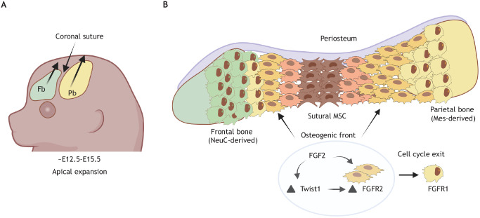
Craniofacial development requires precise spatiotemporal regulation of multiple signaling pathways that crosstalk to coordinate the growth and patterning of the skull with surrounding tissues. Recent insights into these signaling pathways and previously uncharacterized progenitor cell populations have refined our understanding of skull patterning, bone mineralization and tissue homeostasis. Here, we touch upon classical studies and recent advances with an emphasis on developmental and signaling mechanisms that regulate the osteoblast lineage for the calvaria, which forms the roof of the skull. We highlight studies that illustrate the roles of osteoprogenitor cells and cranial suture-derived stem cells for proper calvarial growth and homeostasis. We also discuss genes and signaling pathways that control suture patency and highlight how perturbing the molecular regulation of these pathways leads to craniosynostosis. Finally, we discuss the recently discovered tissue and signaling interactions that integrate skull and cerebrovascular development, and the potential implications for both cerebrospinal fluid hydrodynamics and brain waste clearance in craniosynostosis.

Keywords: Craniofacial development; Craniosynostosis; Osteogenic front; Osteoprogenitor cell; Supraorbital mesenchyme; Sutural stem cells.


Conflict of interest statement

Competing interests The authors declare no competing or financial interests.

Figures

Fig. 1.
Fig. 1.
Comparative human and mouse skull anatomy. The major calvarial bones and intervening sutures are labeled. CS, coronal suture; Fb, frontal bone; FS, frontal suture; Ipb, interparietal bone; LS, lambdoid suture; MS, metopic suture; Nb, nasal bone; Ob, occipital bone; Pb, parietal bone; SS, sagittal suture; SqS, squamosal suture.
Fig. 2.
Fig. 2.
Osteoblast differentiation in the supraorbital mesenchyme. (A) Molecular regulation of the osteoblast lineage. Mesenchymal stem cells (MSCs) give rise to osteoprogenitor cells (OPCs) and then to committed preosteoblasts, which differentiate into postmitotic osteoblasts. (B) Following migration into the supraorbital ridge, neural crest (NeuC, green)-derived and mesoderm (Mes, yellow)-derived OPCs form and condense in the supraorbital mesenchyme (SOM) between ∼E10.5 and E12.5. (C,D) Morphogen signaling drives differentiation of the osteoblast lineage. Wnt signaling (C) from the surface ectoderm is necessary for the specification of OPCs from uncommitted MSCs. β-Catenin activates Twist1 in OPCs, which prevents chondrogenesis by inhibiting Sox9 activity, while also maintaining OPC responsiveness to autocrine Wnt signaling. BMP signaling (D) activates Runx2, which drives Osx1 expression in committed preosteoblasts. Once Twist1 levels decline (arrowhead), the brake on Runx2 function is relieved (dashed lines), allowing upregulation of Osx1 expression. Osx1 is required for differentiation towards mature osteoblasts.
Fig. 3.
Fig. 3.
Apical expansion and growth of the calvarium. (A) Between E12.5 and E15.5, the frontal bone (Fb) and parietal bone (Pb) primordia form as condensations in the supraorbital mesenchyme expand towards the apex of the head. (B) Apical expansion is influenced by the proliferation of preosteoblasts at the osteogenic front. Proliferating preosteoblasts require FGF2 signaling, which also serves to upregulate FGFR2 expression via Twist1. As cells differentiate, one daughter cell continues to divide at the leading edge, whereas the other exits the cell cycle to differentiate into a FGFR1+ osteoblast. Note that a small portion of the frontal bone contains coronal suture-derived osteoblasts, as shown in this example, whereas the coronal suture makes a major contribution to the parietal bone. Also, the periosteum is thought to be a source of osteoblasts. MSC, mesenchymal stem cell.
Fig. 4.
Fig. 4.
Suture morphology and pathogenesis of craniosynostosis. (A) The frontal bone (Fb), which is predominately derived from the neural crest (NeuC), and the coronal suture, which is derived from the mesoderm (Mes), form a natural neural crest-mesoderm boundary (NeuCMesB) in the skull. Note that a small portion of the frontal bone contains osteoblasts derived from the coronal suture, as shown in this example, so the boundary is unidirectional. Within the suture, a cell-fate border also exists that delineates uncommitted mesenchymal stem cells (MSCs) from cells undergoing specification into OPCs via exposure to morphogen signaling. Examples of various mechanisms postulated to cause suture fusion are shown. (B) Craniosynostosis. (Ba) Twist1 maintains the boundary between the NeuC and the Mes in the coronal suture by mediating repulsive EphA-ephrin A signaling. When these repulsive interactions are lost, the NeuC invades the OF into the sutural mesenchyme, causing ossification at the midline. (Bb) Twist1 haploinsufficiency can also cause premature differentiation of MSCs into OPCs at the sutural midline, depleting the pool of MSCs and leading to ossification. (Bc) Gain-of-function mutations in FGFR2 increase the proliferation of preosteoblasts at the osteogenic OF, causing the OF to encroach on the midline sutural mesenchyme. CS, coronal suture; Pb, parietal bone; OB, osteoblast; OF, osteogenic front.
Fig. 5.
Fig. 5.
Integrated development of the skull and dural venous sinuses. (A) Growth and remodeling of the dural venous sinuses requires paracrine BMP signaling from preosteoblasts and the dura. This process is dependent on Twist1 for proper generation of OPCs and expansion of the developing dura. (B) The transverse sinus develops from the primitive head vein and the associated anterior and middle capillary plexi, which are present at E11.75 in mouse (left). By E12.75 (middle), the middle plexus grows into the anterior plexus, which is located in the dura immediately adjacent to the coronal suture (CS) and the parietal bone. This forms the transverse sinus and a new drainage route, causing the primitive head vein to regress as it is no longer the primary vessel draining blood from the brain. Knocking out Twist1 activity in the dura, sutural and supraorbital mesenchyme (top right panel) stunts parietal bone development and the formation of the coronal suture. The loss of preosteoblasts and hypoplastic dura diminishes paracrine BMP signaling, leading to delayed remodeling and poorly developed transverse sinuses, one of which regresses, leading to unilateral absence of the vessel. By E13.75 (B′), the sigmoid sinus remodels in response to temporal bone (Tb) development. Both the sigmoid and transverse sinuses often fail to properly form in craniosynostosis. Fb, frontal bone; Pb, parietal bone.

Similar articles

Cited by

References

    1. Alaqeel, S., Hinton, R. and Opperman, L. (2006). Cellular response to force application at craniofacial sutures. Orthod. Craniofac. Res. 9, 111-122. 10.1111/j.1601-6343.2006.00371.x - DOI - PubMed
    1. Alfonso-Durrruty, M. P., Giles, B. T., Misarti, N., San Roman, M. and Morello, F. (2015). Antiquity and geographic distribution of cranial modification among the prehistoric groups of Fuego-Patagonia, Chile: Cranial Modification in Fuego-Patagonia. Am. J. Phys. Anthropol. 158, 607-623. 10.1002/ajpa.22832 - DOI - PubMed
    1. Ambrosetti, D., Holmes, G., Mansukhani, A. and Basilico, C. (2008). Fibroblast growth factor signaling uses multiple mechanisms to inhibit wnt-induced transcription in osteoblasts. Mol. Cell. Biol. 28, 4759-4771. 10.1128/MCB.01849-07 - DOI - PMC - PubMed
    1. Anderson, J. (1998). Apert syndrome mutations in fibroblast growth factor receptor 2 exhibit increased affinity for FGF ligand. Hum. Mol. Genet. 7, 1475-1483. 10.1093/hmg/7.9.1475 - DOI - PubMed
    1. Ang, P. S., Matrongolo, M. J. and Tischfield, M. A. (2022). The growth and expansion of meningeal lymphatic networks are affected in craniosynostosis. Development 149, dev200065. 10.1242/dev.200065 - DOI - PubMed

Publication types