. 2022 Dec 20;13(6):e0281922.
doi: 10.1128/mbio.02819-22. Epub 2022 Nov 21.

The ESX-1 Substrate PPE68 Has a Key Function in ESX-1-Mediated Secretion in Mycobacterium marinum

Affiliations

The ESX-1 Substrate PPE68 Has a Key Function in ESX-1-Mediated Secretion in Mycobacterium marinum

Merel P M Damen et al. mBio. .

Abstract

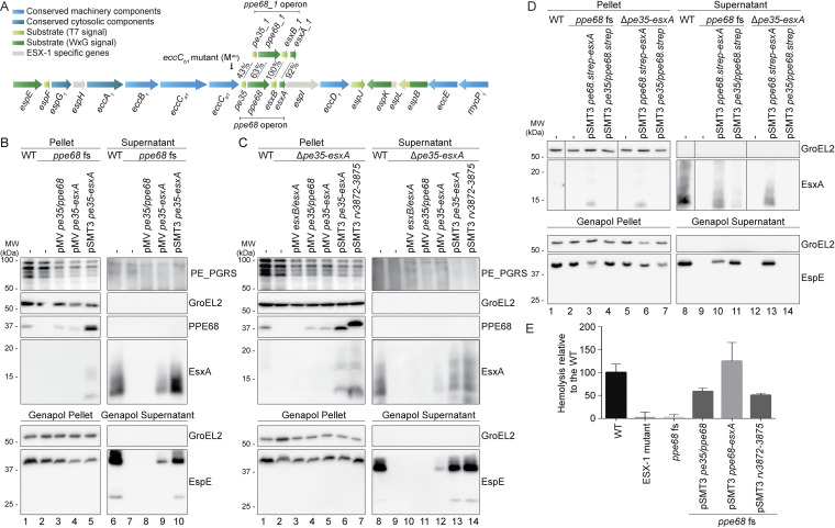
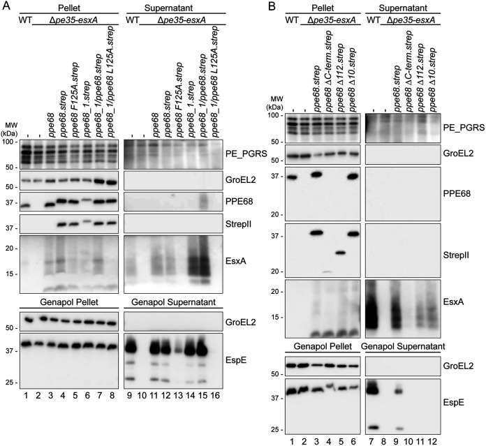
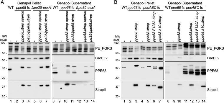
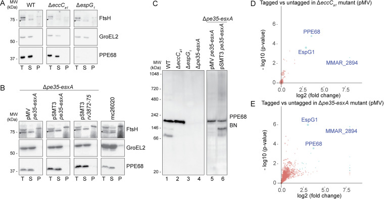
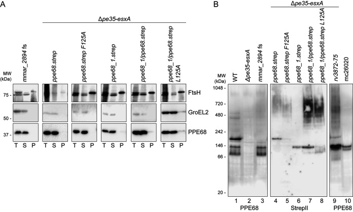
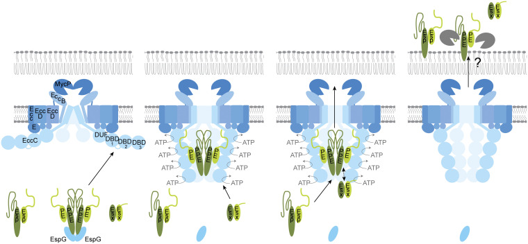
Mycobacteria use specialized type VII secretion systems (T7SSs) to secrete proteins across their diderm cell envelope. One of the T7SS subtypes, named ESX-1, is a major virulence determinant in pathogenic species such as Mycobacterium tuberculosis and the fish pathogen Mycobacterium marinum. ESX-1 secretes a variety of substrates, called Esx, PE, PPE, and Esp proteins, at least some of which are folded heterodimers. Investigation into the functions of these substrates is problematic, because of the intricate network of codependent secretion between several ESX-1 substrates. Here, we describe the ESX-1 substrate PPE68 as essential for secretion of the highly immunogenic substrates EsxA and EspE via the ESX-1 system in M. marinum. While secreted PPE68 is processed on the cell surface, the majority of cell-associated PPE68 of M. marinum and M. tuberculosis is present in a cytosolic complex with its PE partner and the EspG1 chaperone. Interfering with the binding of EspG1 to PPE68 blocked its export and the secretion of EsxA and EspE. In contrast, esxA was not required for the secretion of PPE68, revealing a hierarchy in codependent secretion. Remarkably, the final 10 residues of PPE68, a negatively charged domain, seem essential for EspE secretion, but not for the secretion of EsxA and of PPE68 itself. This indicates that distinctive domains of PPE68 are involved in secretion of the different ESX-1 substrates. Based on these findings, we propose a mechanistic model for the central role of PPE68 in ESX-1-mediated secretion and substrate codependence. IMPORTANCE Pathogenic mycobacteria, such Mycobacterium tuberculosis and Mycobacterium marinum, use a type VII secretion system (T7SS) subtype, called ESX-1, to mediate intracellular survival via phagosomal rupture and subsequent translocation of the mycobacterium to the host cytosol. Identifying the ESX-1 substrate that is responsible for this process is problematic because of the intricate network of codependent secretion between ESX-1 substrates. Here, we show the central role of the ESX-1 substrate PPE68 for the secretion of ESX-1 substrates in Mycobacterium marinum. Unravelling the mechanism of codependent secretion will aid the functional understanding of T7SSs and will allow the analysis of the individual roles of ESX-1 substrates in the virulence caused by the significant human pathogen Mycobacterium tuberculosis.

Keywords: ESX-1; EsxA; Mycobacterium; PPE; chaperones; protein transport; protein-protein interactions; tuberculosis; type VII secretion.


Conflict of interest statement

The authors declare no conflict of interest.

Figures

FIG 1
FIG 1
pe35/ppe68 is involved in the secretion of ESX-1 substrates EsxA and EspE, and is required for ESX-1 mediated hemolysis. A) Schematic overview of the esx-1 gene cluster and its paralogous region. Genes are color-coded to their subcellular localization (key color). Notably, the pe gene upstream of ppe68_1 was originally named pe35, while the paralogous gene upstream of ppe68 in the esx-1 locus was named mmar_5447. In this study, we renamed these two genes pe35_1 and pe35, respectively. B, C, D) SDS-PAGE and immunostaining of the cell pellet, culture supernatant, Genapol pellet and Genapol supernatant fractions of M. marinum M wild type (WT) and the ppe68 fs mutant without and with various complementation constructs (B, D) or WT and the ppe68 operon mutant (Δpe35-esxA) with and without various complementation constructs (C, D). Genes were expressed from an integrative pMV vector or a multicopy pSMT3 vector under control of the hsp60 promotor, and for D) with a Strep-tag on the C-terminus of PPE68. In B and D) the WT, ppe68 fs mutant and the ppe68 fs mutant containing a pSMT3 vector also contained a pMV TdTomato plasmid. Proteins were visualized with anti-EsxA, anti-EspE, and anti-PPE68 antibodies (ESX-1 substrates). A processed band of 25 kDa was detected by the anti-EspE antibody in the Genapol supernatant fraction of the WT, which has been reported before (46). As a loading and lysis control, blots were incubated with antibodies directed against the cytosolic GroEL2 protein and secreted PE_PGRS proteins (ESX-5 substrates). In all blots, equivalent OD units were loaded: 0.2 OD for pellet and Genapol pellet and 0.4 OD for supernatant and Genapol supernatant fractions (B, C) or 0.2 OD for supernatant fractions and 2 OD for Genapol supernatant fractions (D). Data shown are representative of three independent experiments. E) Hemoglobin release after incubation of sheep red blood cells with M. marinum WT, an ESX-1 mutant (eccCb1 mutant; MVU), the ppe68 fs mutant, and the ppe68 fs mutant complemented with either the entire ppe68 operon of M. marinum, with only pe35/ppe68, or with the ppe68 operon of M. tuberculosis (rv3872-rv3875). Hemoglobin release was quantified by determining the OD450 of the medium after incubation. Data shown are from two independent experiments including 3 technical replicates each.
FIG 2
FIG 2
Conserved features of PPE68 are important for its central role in ESX-1 secretion. (A) Role of the EspG1-binding domain of PPE68. SDS-PAGE and immunostaining of the cell pellet, culture supernatant, Genapol pellet, and Genapol supernatant fractions of M. marinum WT, the ppe68 operon mutant (Δpe35-esxA), and the ppe68 operon mutant complemented with either the ppe68 operon, the ppe68_1 operon, or a hybrid operon containing pe35_1, the N-terminal domain of ppe68_1, or the C-terminal domain of ppe68, esxB, and esxA. Single amino acid substitutions were made in the helical tip of PPE68 and the PPE68_1/PPE68 hybrid to investigate the role of the EspG1-binding domain. (B) The role of the C-terminal domain of PPE68. SDS-PAGE and immunostaining of the cell pellet, culture supernatant, Genapol pellet, and Genapol supernatant fractions of M. marinum WT, the ppe68 operon mutant (Δpe35-esxA), and the ppe68 operon mutant complemented with either the WT or ppe68 operon variants, encoding PPE68 lacking either the entire (PPE68ΔC-term) or partial, i.e., the final 112 (PPE68Δ112) or 10 (PPE68Δ10) amino acids, of the C-terminal domain. In both panels A and B, the operons were expressed from a multicopy pSMT3 vector under control by the hsp60 promoter and with a Strep tag on the C terminus of PPE68 and its variants. Proteins were visualized with anti-EsxA, anti-EspE, anti-StrepII (PPE68), and anti-PPE68 antibodies (ESX-1 substrates). A processed band of 25 kDa was detected by the anti-EspE antibody in the Genapol supernatant fraction of the WT, which has been reported before (Phan et al., 2018 [46]). As a loading and lysis control, blots were incubated with antibodies directed against the cytosolic GroEL2 protein and secreted PE_PGRS proteins (ESX-5 substrates). In all blots, equivalent OD units were loaded: 0.2 OD for pellet and Genapol pellet and 0.4 OD for supernatant and Genapol supernatant fractions. Data shown in each panel are representative of three independent experiments.
FIG 3
FIG 3
PPE68 is secreted independently of esxB and esxA and processed on the cell surface of M. marinum. (A) SDS-PAGE and immunostaining on Genapol pellet and concentrated Genapol supernatant fractions of M. marinum WT, the ppe68 fs mutant, the ppe68 operon mutant, or both mutants complemented with pe35/ppe68 or the entire ppe68 operon. (B) Additional SDS-PAGE and immunostaining of Genapol pellet and concentrated Genapol supernatant fractions of the pecABC fs mutant overexpressing the WT ppe68 operon (ppe68.strep), the operon producing either the PPE68F125A with an amino acid substitution in the helical tip, PPE68Δ10 with a C-terminal truncation of the last 10 amino acids. Operons were expressed from a multicopy pSMT3 vector controlled by the hsp60 promoter and with a Strep tag on the C terminus of PPE68 and the variants. Proteins were visualized with anti-StrepII (PPE68) and anti-PPE68 antibodies (ESX-1 substrates). All ppe68 fs and pecABC fs mutants contained an additional pMV TdTomato plasmid. As a loading and lysis control, blots were incubated with antibodies directed against the cytosolic GroEL2 protein and the surface-localized PE_PGRS proteins. In all blots, equivalent OD units were loaded: 0.2 OD for Genapol pellet and 2 OD Genapol supernatant fractions. Data shown in each panel are representative of three independent experiments.
FIG 4
FIG 4
PPE68 of M. marinum and M. tuberculosis is soluble and forms complexes of different sizes. (A and B) SDS-PAGE and immunostaining of subcellular fractionation of M. marinum WT, an eccCa1 mutant, and an espG1 mutant (A) or the ppe68 operon mutant complemented with the ppe68 operon of M. marinum expressed from a pMV or pSMT3 vector, the ppe68 operon of M. tuberculosis H37Rv expressed from a pSMT3 vector, or auxotrophic M. tuberculosis strain mc26020 (B). Proteins were visualized with anti-PPE68, while antibodies against GroEL2 (cytosolic protein) and FtsH (membrane protein) were used to assess the purity of the fractions. Notably, the FtsH antiserum cross-reacted with a cytosolic protein (indicated by an asterisk). In all blots, equivalent amounts corresponding to 0.2 OD units were loaded. T, total lysate; S, supernatant or soluble proteins; P, pellet or cell envelope proteins. (C) Immunostaining of soluble fractions of the same strains as in panels A and B after blue native PAGE (BN-PAGE) gel electrophoresis. In all blots, an equivalent amount of 19 μg of total protein was loaded. Data shown in panels A, B, and C are representative of two independent experiments. (D and E) PPE68 interacts with its PE partner, MMAR_2894, and the EspG1 chaperone in the cytosol. Quantitative proteomics analysis comparing the PPE68 affinity purification from the soluble fraction of the eccCa1 mutant (D) and the ppe68 operon mutant (E), complemented with the ppe68 operon, produced PPE68 with or without a Strep tag on its C terminus from a pMV vector. Proteins that showed a log2 fold change of >2 and a −log10 P value of >1.3 between tagged and untagged PPE68 are indicated as blue dots. Sizes of the dots are correlated to the number of spectral counts. Data include two technical replicates per sample.
FIG 5
FIG 5
Verification of complex formation of PPE68 with MMAR_2894 and EspG1. (A) SDS-PAGE and immunostaining of subcellular fractions of the mmar_2894 fs mutant and the ppe68 operon mutant complemented with the WT ppe68 operon, the ppe68_1 operon, a hybrid operon containing pe35_1, the N-terminal domain of ppe68_1, the C-terminal domain of ppe68, and esxB and esxA or the ppe68 or hybrid operon expressing a PPE68 variant with a single amino acid substitution at position 125. Genes were expressed from a multicopy pSMT3 vector under the hsp60 promoter with a Strep tag on the C terminus of PPE68 or its variants. Proteins were visualized with anti-PPE68 antibodies, while antibodies against GroEL2 (cytosolic protein) and FtsH (membrane protein) were used to assess the purity of the fractions. Notably, the FtsH antiserum cross-reacted with a cytosolic protein (indicated by an asterisk). In all blots, equivalent amounts corresponding to 0.2 OD units were loaded. T, total lysate; S, supernatant or soluble proteins; P, pellet or cell envelope proteins. (B) BN-PAGE and immunostaining of soluble fractions of the same strains as in panel A, and also the ppe68 operon mutant expressing the ppe68 operon of M. tuberculosis from a pSMT3 vector and the auxotrophic M. tuberculosis strain mc26020. Proteins were visualized with anti-PPE68 and anti-StrepII antibodies. In all blots, an equivalent amount of 19 μg of total protein was loaded. Data shown in each panel are representative of two independent experiments.
FIG 6
FIG 6
Working model for the role of PPE68 in ESX-1-mediated secretion. We propose that different ESX-1 substrates are secreted in a hierarchical fashion by a stepwise interaction with the secretion machinery. Here, the PE/PPE proteins activate the channel by interacting with the linker that connects NBD1 and NBD2 of the central EccC component. This interaction induces conformational changes in the ATPase and its full activation, after which the Esx substrates can bind NBD3, resulting in the cosecretion of the PE/PPE and Esx substrates across the cell envelope. The secretion of both the PE/PPE and Esx heterodimers is necessary for secretion of the final substrate family, the Esp proteins.

Similar articles

Cited by

References

    1. Hoffmann C, Leis A, Niederweis M, Plitzko JM, Engelhardt H. 2008. Disclosure of the mycobacterial outer membrane: cryo-electron tomography and vitreous sections reveal the lipid bilayer structure. Proc Natl Acad Sci USA 105:3963–3967. doi:10.1073/pnas.0709530105. - DOI - PMC - PubMed
    1. Zuber B, Chami M, Houssin C, Dubochet J, Griffiths G, Daffe M. 2008. Direct visualization of the outer membrane of mycobacteria and corynebacteria in their native state. J Bacteriol 190:5672–5680. doi:10.1128/JB.01919-07. - DOI - PMC - PubMed
    1. Houben D, Demangel C, van Ingen J, Perez J, Baldeon L, Abdallah AM, Caleechurn L, Bottai D, van Zon M, de Punder K, van der Laan T, Kant A, Bossers-de Vries R, Willemsen P, Bitter W, van Soolingen D, Brosch R, van der Wel N, Peters PJ. 2012. ESX-1-mediated translocation to the cytosol controls virulence of mycobacteria. Cell Microbiol 14:1287–1298. doi:10.1111/j.1462-5822.2012.01799.x. - DOI - PubMed
    1. Simeone R, Bobard A, Lippmann J, Bitter W, Majlessi L, Brosch R, Enninga J. 2012. Phagosomal rupture by Mycobacterium tuberculosis results in toxicity and host cell death. PLoS Pathog 8:e1002507. doi:10.1371/journal.ppat.1002507. - DOI - PMC - PubMed
    1. Stamm LM, Morisaki JH, Gao LY, Jeng RL, McDonald KL, Roth R, Takeshita S, Heuser J, Welch MD, Brown EJ. 2003. Mycobacterium marinum escapes from phagosomes and is propelled by actin-based motility. J Exp Med 198:1361–1368. doi:10.1084/jem.20031072. - DOI - PMC - PubMed

LinkOut - more resources