. 2022 Dec 20;13(6):e0287022.
doi: 10.1128/mbio.02870-22. Epub 2022 Nov 21.

Streptococcus agalactiae npx Is Required for Survival in Human Placental Macrophages and Full Virulence in a Model of Ascending Vaginal Infection during Pregnancy

Affiliations

Streptococcus agalactiae npx Is Required for Survival in Human Placental Macrophages and Full Virulence in a Model of Ascending Vaginal Infection during Pregnancy

Jacky Lu et al. mBio. .

Abstract

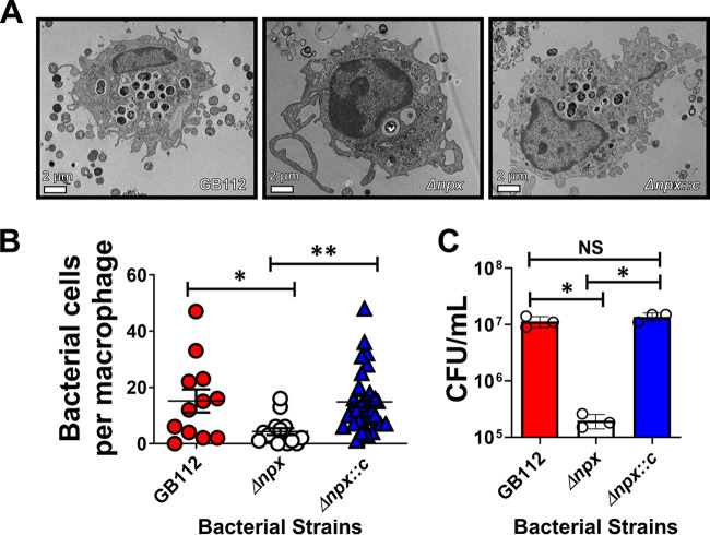
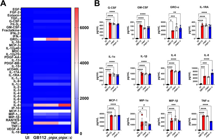
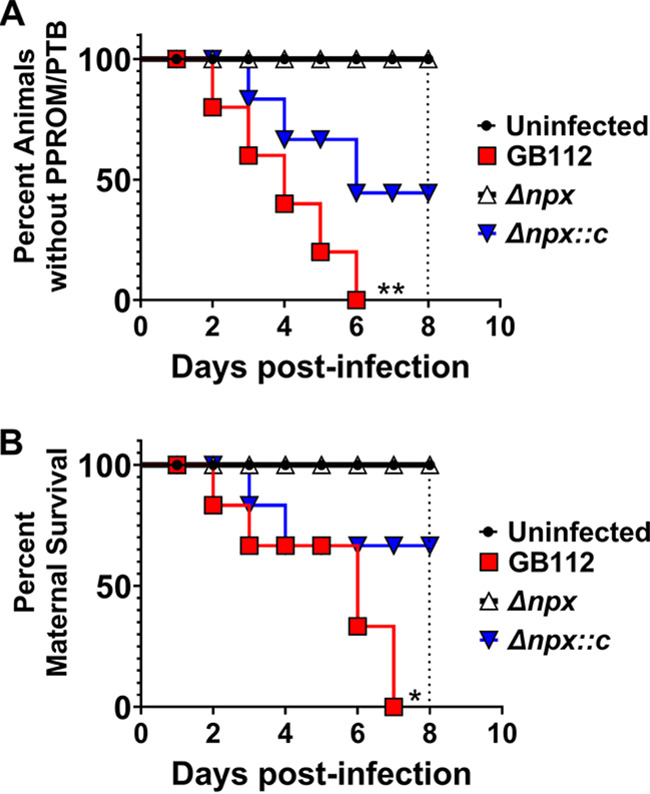
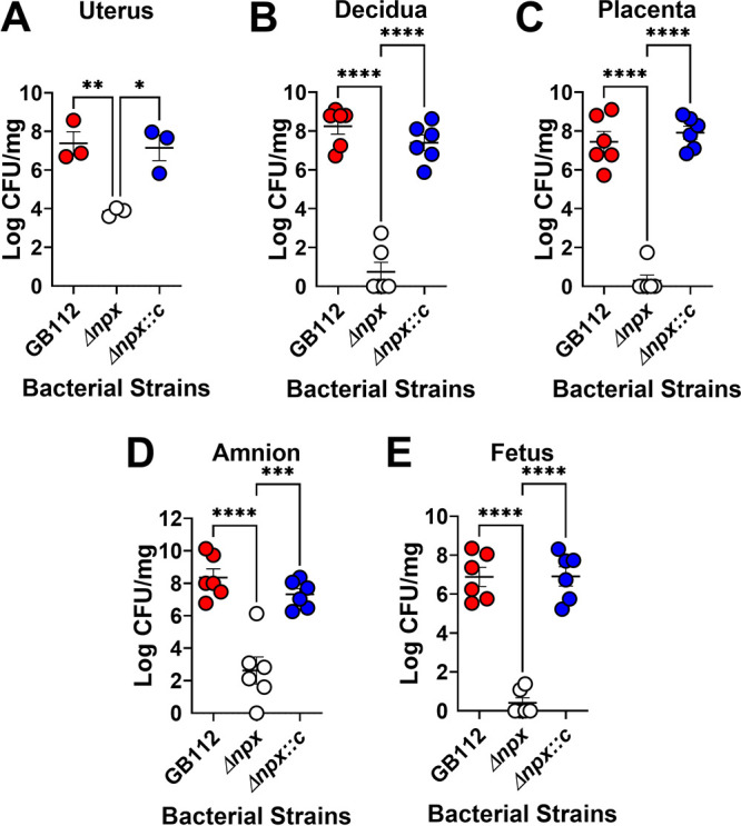
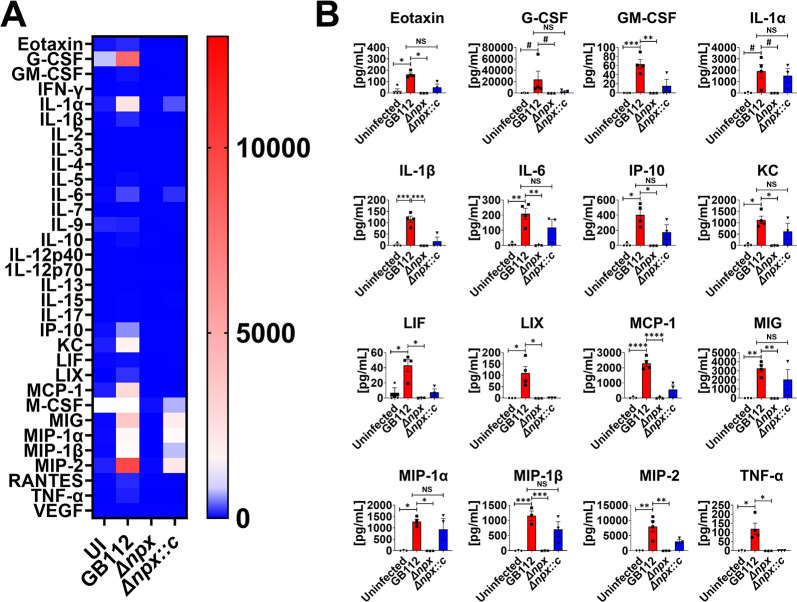
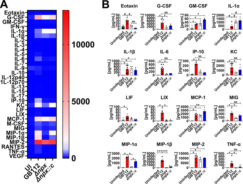
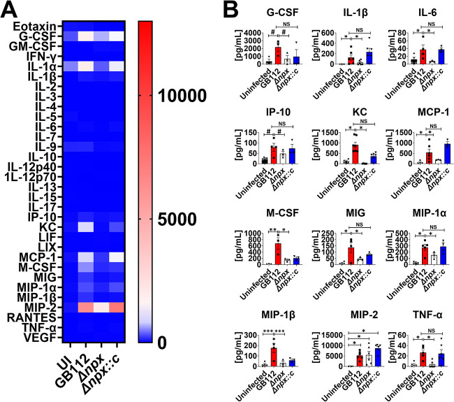
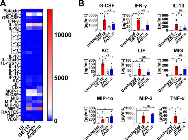
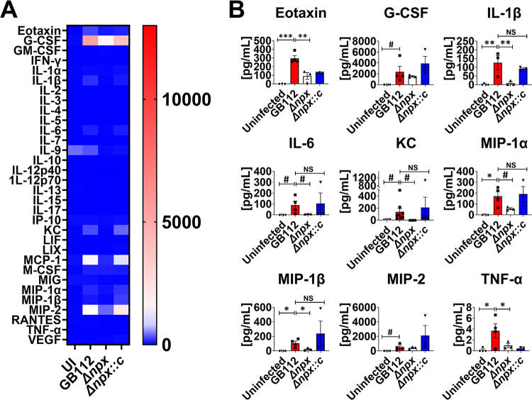
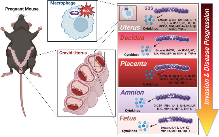
Streptococcus agalactiae, also known as group B Streptococcus (GBS), is a Gram-positive encapsulated bacterium that colonizes the gastrointestinal tract of 30 to 50% of humans. GBS causes invasive infection during pregnancy that can lead to chorioamnionitis, funisitis, preterm prelabor rupture of membranes (PPROM), preterm birth, neonatal sepsis, and maternal and fetal demise. Upon infecting the host, GBS encounters sentinel innate immune cells, such as macrophages, within reproductive tissues. Once phagocytosed by macrophages, GBS upregulates the expression of the gene npx, which encodes an NADH peroxidase. GBS mutants with an npx deletion (Δnpx) are exquisitely sensitive to reactive oxygen stress. Furthermore, we have shown that npx is required for GBS survival in both THP-1 and placental macrophages. In an in vivo murine model of ascending GBS vaginal infection during pregnancy, npx is required for invading reproductive tissues and is critical for inducing disease progression, including PPROM and preterm birth. Reproductive tissue cytokine production was also significantly diminished in Δnpx mutant-infected animals compared to that in animals infected with wild-type (WT) GBS. Complementation in trans reversed this phenotype, indicating that npx is critical for GBS survival and the initiation of proinflammatory signaling in the gravid host. IMPORTANCE This study sheds new light on the way that group B Streptococcus (GBS) defends itself against oxidative stress in the infected host. The enzyme encoded by the GBS gene npx is an NADH peroxidase that, our study reveals, provides defense against macrophage-derived reactive oxygen stress and facilitates infections of the uterus during pregnancy. This enzyme could represent a tractable target for future treatment strategies against invasive GBS infections.

Keywords: ROS; Streptococcus; infection; innate immunity; reactive oxygen species.


Conflict of interest statement

The authors declare no conflict of interest.

Figures

FIG 1
FIG 1
Analysis of GBS survival in primary human placental macrophage cells. (A) Transmission electron microscopy analyses reveal that npx is required for GBS survival in placental macrophages. (B) Enumeration of bacterial cells per placental macrophage via electron microscopy analyses indicates that WT GBS (GB112) (red) survives within primary human placental macrophages, but an isogenic Δnpx mutant (white) is attenuated in intracellular survival compared to both the parental strain (GB112) and the complemented derivative (Δnpx::c) (blue). (C) Quantitative culture analysis of bacterial survival by gentamicin protection assays reveals that npx is required for GBS survival in placental macrophages. *, P < 0.05; **, P < 0.01 (by one-way ANOVA with Tukey’s post hoc multiple-comparison test). NS, statistically indistinguishable (3 biological replicates).
FIG 2
FIG 2
GBS npx expression is dispensable for the initiation of proinflammatory cytokine production by human placental macrophages. (A) Heat map results (blue, low levels; red, high levels) of multiplex cytokine analyses of primary human placental macrophage-secreted fractions after coculture with wild-type GB112, the isogenic Δnpx mutant, or the isogenic complemented Δnpx derivative (Δnpx::c) reveal that GBS infection induces the production of multiple proinflammatory cytokines compared to the uninfected controls (UI). EGF, epidermal growth factor; FGF, fibroblast growth factor; TGFα, transforming growth factor α; IFNα2, interferon alpha 2; PDGF-AA, platelet-derived growth factor AA; sCD40L, soluble CD40 ligand; VEGF-A, vascular endothelial growth factor A. (B) Quantitation of cytokine levels reveals the significantly enhanced production of G-CSF, GM-CSF, GRO-α, IL-1RA, IL-1α, IL-1β, IL-6, IL-8, MCP-1, MIP-1α, MIP-1β, and TNF-α in macrophages cocultured with the GB112 (red), Δnpx (white), and Δnpx::c (blue) strains compared to the uninfected controls (black). *, P < 0.05; **, P < 0.01; ***, P < 0.001; ****, P < 0.0001 (by one-way ANOVA with Tukey’s post hoc multiple-comparison test). #, P < 0.05 (by Student’s t test with Welch’s correction) (3 biological replicates).
FIG 3
FIG 3
Analysis of the role of GBS npx in disease progression in a mouse model of ascending vaginal infection during pregnancy. (A) Evaluation of the incidence of (percentage of animals exhibiting) preterm premature rupture of membranes (PPROM) and preterm birth (PTB) over time (8 days postinfection and term for gestation). Wild-type GB112-infected animals (red) had higher incidences of PPROM and PTB than did uninfected (black) and Δnpx mutant-infected (white) animals, with 100% of the GB112-infected cohort experiencing PPROM or PTB by 6 days postinfection. Complementation of the npx locus in transnpx::c) (blue) resulted in 65% PTB and PPROM by 6 days postinfection. (B) Evaluation of maternal mortality over time (8 days postinfection and term for gestation). GB112-infected animals had a 100% maternal mortality rate by 7 days postinfection, a rate that was significantly higher than those for the uninfected control and Δnpx mutant-infected cohorts, which exhibited 0% maternal mortality by 8 days postinfection. Complementation in trans partially restored mortality (33% maternal death by 8 days postinfection) (n = 6 to 9 dams total from 3 separate experiments) (*, P < 0.05; **, P < 0.01 [by a Mantel-Cox log rank test and a Gehan-Breslow-Wilcoxon test]).
FIG 4
FIG 4
Analysis of bacterial burdens within gravid reproductive tissues. Shown are data from quantitative culture analyses of the bacterial burdens within the uterus (A), decidua (B), placenta (C), amnion (D), and fetus (E) of pregnant mice on embryonic day 15.5, 2 days after infection with either WT GBS (GB112) (red), an isogenic Δnpx mutant (white), or the isogenic complemented derivative (Δnpx::c) (blue). GBS npx is required for full bacterial burdens in reproductive tissues in a model of ascending infection during pregnancy. *, P < 0.05; **, P < 0.01; ***, P < 0.001; ****, P < 0.0001 (by one-way ANOVA with Tukey’s post hoc multiple-comparison test) (n = 3 dams total, with 1 to 2 fetal-placental units analyzed per dam).
FIG 5
FIG 5
Analysis of cytokine production in uterus tissue in response to GBS infection. (A) Heat map results (blue, low levels; red, high levels) of multiplex cytokine analyses of gestational tissues after ascending vaginal infection with wild-type GB112, the Δnpx isogenic mutant, or the isogenic complemented Δnpx derivative (Δnpx::c) as well as the uninfected controls (UI). Uterine tissues were collected from pregnant mice on embryonic day 15.5, 2 days after vaginal infection with GBS. (B) Quantification of eotaxin, G-CSF, GM-CSF, IL-1α, IL-1β, IL-6, IP-10, KC, LIF, LIX, MCP-1, MIG, MIP-1α, MIP-1β, MIP-2, and TNF-α levels revealed that wild-type GB112 infection (red bars) significantly enhances the production of these cytokines compared to the levels in the uninfected controls (black bars), but the isogenic Δnpx mutant (white bars) is significantly attenuated in its ability to induce these cytokines compared to the parental strain. Conversely, the complemented derivative (Δnpx::c) (blue bars) is often statistically indistinguishable from the parental strain (not statistically significant [NS]). Bars indicate mean values ± standard errors of the means, with individual data points representing results from uterus tissues from individual dams. #, P < 0.05 (by Student’s t test). *, P < 0.05; **, P < 0.01; ***, P < 0.001; ****, P < 0.0001 (by one-way ANOVA with Tukey’s post hoc multiple-comparison test). The results indicate that GBS npx is required for the full initiation of proinflammatory cytokine responses in uterine tissues in a model of ascending infection during pregnancy.
FIG 6
FIG 6
Analysis of cytokine production in decidua tissue in response to GBS infection. (A) Heat map results (blue, low levels; red, high levels) of multiplex cytokine analyses of decidua tissues after ascending vaginal infection with wild-type GB112, the isogenic Δnpx mutant (Δnpx), or the isogenic complemented Δnpx derivative (Δnpx::c) as well as the uninfected controls (UI). Decidua tissues were collected from pregnant mice on embryonic day 15.5, 2 days after vaginal infection with GBS. (B) Quantification of eotaxin, G-CSF, GM-CSF, IL-1α, IL-1β, IL-6, IP-10, KC, LIF, LIX, MCP-1, MIG, MIP-1α, MIP-1β, MIP-2, and TNF-α levels revealed that wild-type GB112 infection (red bars) significantly enhances the production of these cytokines compared to the levels in the uninfected controls (black bars), but the isogenic Δnpx mutant (white bars) is significantly attenuated in its ability to induce these cytokines compared to the parental strain. Conversely, the complemented derivative (Δnpx::c) (blue bars) is often statistically indistinguishable from the parental strain (not statistically significant [NS]). Bars indicate mean values ± standard errors of the means, with individual data points representing results from decidual tissues from individual fetal-placental units from separate dams. #, P < 0.05 (by Student’s t test). *, P < 0.05; **, P < 0.01; ***, P < 0.001; ****, P < 0.0001 (by one-way ANOVA with Tukey’s post hoc multiple-comparison test). The results indicate that GBS npx is required for the full initiation of proinflammatory cytokine responses in decidual tissues in a model of ascending infection during pregnancy.
FIG 7
FIG 7
Analysis of cytokine production in placenta tissue in response to GBS infection. (A) Heat map results (blue, low levels; red, high levels) of multiplex cytokine analyses of placenta tissues after ascending vaginal infection with wild-type GB112, the Δnpx isogenic mutant, or the complemented isogenic Δnpx derivative (Δnpx::c) as well as the uninfected controls (UI). Placenta tissues were collected from pregnant mice on embryonic day 15.5, 2 days after vaginal infection with GBS. (B) Quantification of G-CSF, IL-1β, IL-6, IP-10, KC, MCP-1, M-CSF, MIG, MIP-1α, MIP-1β, MIP-2, and TNF-α levels revealed that wild-type GB112 infection (red bars) significantly enhances the production of these cytokines compared to the levels in the uninfected controls (black bars), but the isogenic Δnpx mutant (white bars) is significantly attenuated in its ability to induce these cytokines compared to the parental strain, except for MIP-2. Conversely, the complemented derivative (Δnpx::c) (blue bars) is often statistically indistinguishable from the parental strain (not statistically significant [NS]). Bars indicate mean values ± standard errors of the means, with individual data points representing results from placental tissues from individual fetal-placental units from separate dams. #, P < 0.05 (by Student’s t test). *, P < 0.05; **, P < 0.01; ***, P < 0.001; ****, P < 0.0001 (by one-way ANOVA with Tukey’s post hoc multiple-comparison test). The results indicate that GBS npx is required for the initiation of numerous proinflammatory cytokine responses in placental tissues in a model of ascending infection during pregnancy.
FIG 8
FIG 8
Analysis of cytokine production in amnion tissue in response to GBS infection. (A) Heat map results (blue, low levels; red, high levels) of multiplex cytokine analyses of amnion tissues after ascending vaginal infection with wild-type GB112, the Δnpx isogenic mutant, or the isogenic complemented Δnpx derivative (Δnpx::c) as well as the uninfected controls (UI). Amnion tissues were collected from pregnant mice on embryonic day 15.5, 2 days after vaginal infection with GBS. (B) Quantification of G-CSF, IFN-γ, IL-1β, KC, LIF, MIG, MIP-1α, MIP-2, and TNF-α levels revealed that wild-type GB112 infection (red bars) significantly enhances the production of these cytokines compared to the levels in the uninfected controls (black bars), but the isogenic Δnpx mutant (white bars) is significantly attenuated in its ability to induce these cytokines compared to the parental strain, except for MIP-1α. Conversely, the complemented derivative (Δnpx::c) (blue bars) is often statistically indistinguishable from the parental strain (not statistically significant [NS]). Bars indicate mean values ± standard errors of the means, with individual data points representing results from amnion tissues from individual fetal-placental units from separate dams. #, P < 0.05 (by Student’s t test). *, P < 0.05; **, P < 0.01; ***, P < 0.001; ****, P < 0.0001 (by one-way ANOVA with Tukey’s post hoc multiple-comparison test). The results indicate that GBS npx is required for the initiation of numerous proinflammatory cytokine responses in amnion tissues in a model of ascending infection during pregnancy.
FIG 9
FIG 9
Analysis of cytokine production in fetus tissue in response to GBS infection. (A) Heat map results (blue, low levels; red, high levels) of multiplex cytokine analyses of fetal tissues after ascending vaginal infection with wild-type GB112, the Δnpx isogenic mutant, or the isogenic complemented Δnpx derivative (Δnpx::c) as well as the uninfected controls (UI). Fetus tissues were collected from pregnant mice on embryonic day 15.5, 2 days after vaginal infection with GBS. (B) Quantification of eotaxin, G-CSF, IL-1β, IL-6, KC, MIP-1α, MIP-1β, MIP-2, and TNF-α levels revealed that wild-type GB112 infection (red bars) significantly enhances the production of these cytokines compared to the levels in the uninfected controls (black bars), but the isogenic Δnpx mutant (white bars) is significantly attenuated in its ability to induce eotaxin, IL-1β, IL-6, KC, MIP-1α, MIP-1β, and TNF-α compared to the parental strain. Conversely, the complemented derivative (Δnpx::c) (blue bars) is statistically indistinguishable from the parental strain (not statistically significant [NS]) in its ability to induce IL-1β, IL-6, KC, MIP-1α, and MIP-1β. Bars indicate mean values ± standard errors of the means, with individual data points representing results from fetal tissues from individual fetuses from separate dams. #, P < 0.05 (by Student’s t test). *, P < 0.05; **, P < 0.01; ***, P < 0.001; ****, P < 0.0001 (by one-way ANOVA with Tukey’s post hoc multiple-comparison test). The results indicate that GBS npx is required for the initiation of numerous proinflammatory cytokine responses in fetal tissues in a model of ascending infection during pregnancy.
FIG 10
FIG 10
Conceptual model of npx-dependent invasion and proinflammatory signal initiation in ascending GBS infections during pregnancy. GBS ascends the reproductive tract by surviving within placental macrophages, in part by encoding an NADH peroxidase to aid in detoxifying oxidative stress (such as peroxides [H2O2]). GBS invasion and full burdens within the gravid reproductive tract (uterus, decidua, placenta, amnion, and fetus) require npx expression and trigger the production of tissue compartment-specific proinflammatory cytokines, resulting in inflammation and disease progression, including rupture of membranes, preterm birth, and maternal demise. (Image created with BioRender.com.)

Similar articles

Cited by

References

    1. Goldenberg RL, Culhane JF, Iams JD, Romero R. 2008. Epidemiology and causes of preterm birth. Lancet 371:75–84. doi:10.1016/S0140-6736(08)60074-4. - DOI - PMC - PubMed
    1. Martin JA, Hamilton BE, Sutton PD, Ventura SJ, Menacker F, Kirmeyer S, Munson ML, Centers for Disease Control and Prevention National Center for Health Statistics National Vital Statistics System . 2007. Births: final data for 2005. Natl Vital Stat Rep 56:1–103. - PubMed
    1. Purisch SE, Gyamfi-Bannerman C. 2017. Epidemiology of preterm birth. Semin Perinatol 41:387–391. doi:10.1053/j.semperi.2017.07.009. - DOI - PubMed
    1. McCormick MC. 1985. The contribution of low birth weight to infant mortality and childhood morbidity. N Engl J Med 312:82–90. doi:10.1056/NEJM198501103120204. - DOI - PubMed
    1. Saigal S, Doyle LW. 2008. An overview of mortality and sequelae of preterm birth from infancy to adulthood. Lancet 371:261–269. doi:10.1016/S0140-6736(08)60136-1. - DOI - PubMed

Supplementary concepts