. 2022 Dec 20;13(6):e0289122.
doi: 10.1128/mbio.02891-22. Epub 2022 Nov 21.

The RNA-Binding Protein ProQ Promotes Antibiotic Persistence in Salmonella

Affiliations

The RNA-Binding Protein ProQ Promotes Antibiotic Persistence in Salmonella

Alisa Rizvanovic et al. mBio. .

Abstract

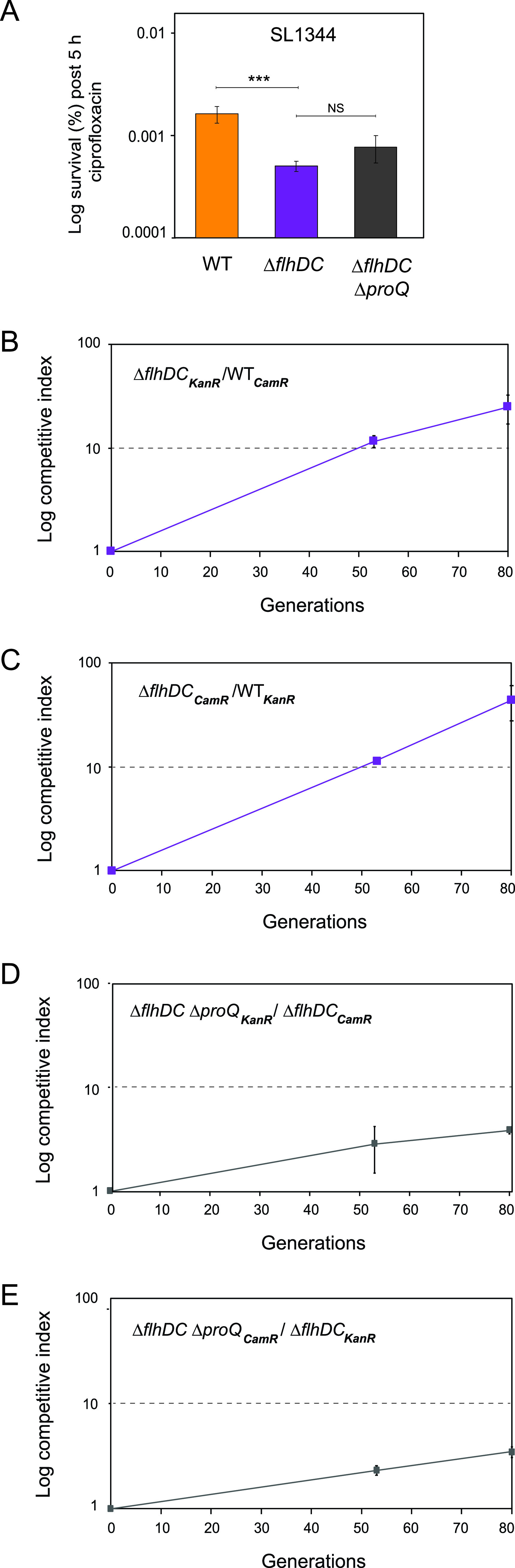
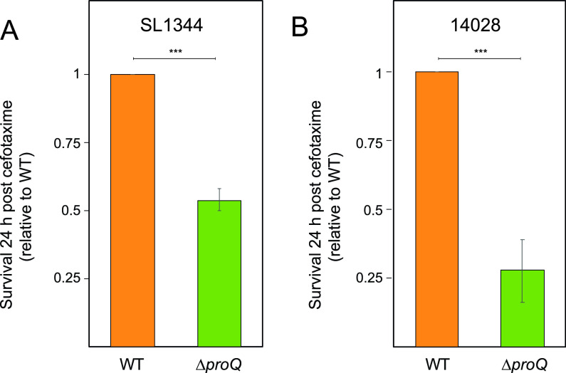
Bacterial populations can survive exposure to antibiotics through transient phenotypic and gene expression changes. These changes can be attributed to a small subpopulation of bacteria, giving rise to antibiotic persistence. Although this phenomenon has been known for decades, much remains to be learned about the mechanisms that drive persister formation. The RNA-binding protein ProQ has recently emerged as a global regulator of gene expression. Here, we show that ProQ impacts persister formation in Salmonella. In vitro, ProQ contributes to growth arrest in a subset of cells that are able to survive treatment at high concentrations of different antibiotics. The underlying mechanism for ProQ-dependent persister formation involves the activation of metabolically costly processes, including the flagellar pathway and the type III protein secretion system encoded on Salmonella pathogenicity island 2. Importantly, we show that the ProQ-dependent phenotype is relevant during macrophage infection and allows Salmonella to survive the combined action of host immune defenses and antibiotics. Together, our data highlight the importance of ProQ in Salmonella persistence and pathogenesis. IMPORTANCE Bacteria can avoid eradication by antibiotics through a phenomenon known as persistence. Persister cells arise through phenotypic heterogeneity and constitute a small fraction of dormant cells within a population of actively growing bacteria, which is susceptible to antibiotic killing. In this study, we show that ProQ, an RNA-binding protein and global regulator of gene expression, promotes persisters in the human pathogen Salmonella enterica serovar Typhimurium. Bacteria lacking the proQ gene outcompete wild-type bacteria under laboratory conditions, are less prone to enter growth dormancy, and form fewer persister cells. The basis for these phenotypes lies in ProQ's ability to activate energy-consuming cellular processes, including flagellar motility and protein secretion. Importantly, we show that ProQ contributes to the persister phenotype during Salmonella infection of macrophages, indicating an important role of this global regulator in Salmonella pathogenesis.

Keywords: ProQ; RNA-binding protein; Salmonella; antibiotic persistence; antibiotic persisters; flagella; flagellar gene regulation; persister formation.


Conflict of interest statement

The authors declare no conflict of interest.

Figures

FIG 1
FIG 1
Effects of ProQ on Salmonella growth. (A) Growth curves of Salmonella SL1344 wild-type or ΔproQ cells carrying an empty control vector (WT, ΔproQ) or an IPTG-inducible proQ overexpression construct (pProQ). The optical density was monitored at 600 nm during growth in LB medium supplemented with IPTG (500 μM) in 96-well plates. The average values of six replicates with standard deviations (SD) are shown. (B and C) Growth competition experiments between Salmonella SL1344 strains. Salmonella wild-type and ΔproQ strains carrying antibiotic resistance marker genes (CamR, gene conferring chloramphenicol resistance; KanR, gene conferring kanamycin resistance) were mixed at a ratio of 1:1 in LB medium and incubated at 37°C. At roughly 24-h intervals, mixtures were plated on selective agar plates to determine CFU counts, and the remaining mixtures were serially passaged by 10,000-fold dilution in fresh LB medium for regrowth. Competitive indexes were calculated as the ratio of mutant cells to wild-type cells at the indicated generation divided by the initial ratio. Average values of six (generations 0 to 40) and three (generations 50 to 80) replicates with standard errors of the means (SEM) are shown.
FIG 2
FIG 2
Single-cell analysis of Salmonella growth using fluorescence dilution in vitro. (A) Schematic of the fluorescence dilution method (2, 3, 12). Bacterial cells were labeled by inducing the expression of green fluorescence protein (GFP). After the accumulation of GFP, bacterial cells were transferred into fresh medium without an inducer, and changes in the GFP signal intensity were monitored by flow cytometry analysis. In growing cells, the GFP signal intensity decreases with each cell division. In nongrowing cells, the GFP signal intensity is retained. (B) Flow cytometry detection of green fluorescence in a Salmonella SL1344 wild-type population carrying plasmid pFCcGi (64). Bacterial cultures were grown to stationary phase in LB medium supplemented with arabinose (0.2%) to induce GFP expression. Upon regrowth in fresh LB medium without an inducer, dilution of the preformed pool of GFP was monitored during 0 h, 1 h, 2 h, and 3 h. Representative data are shown for one out of three replicates. Data for monitoring fluorescence dilution in SL1344 ΔproQ populations are shown in Fig. S1. a.u., arbitrary units. (C) Quantification of the percentage of nongrowing cells for Salmonella wild-type and ΔproQ strains (from panel B) 0.5 h, 1 h, 2 h, and 3 h after regrowth in fresh LB medium. The average values from three replicates with SD are shown. Statistical significance was determined using a two-tailed t test (*, P < 0.1; **, P < 0.01; NS, nonsignificant).
FIG 3
FIG 3
Effects of ProQ on Salmonella antibiotic persistence in vitro. Exponential-phase cultures of Salmonella SL1344 wild-type and ΔproQ strains were treated with ciprofloxacin (1 μg/mL; 60× MIC) (A) and ampicillin (50 μg/mL; 60× MIC) (B) for 5 h. CFU counts were determined before and after treatments to calculate the surviving fraction. Average values from 12 (A) and 6 (B) replicates with SEM are shown. Statistical significance was determined using a two-tailed t test (*, P < 0.1).
FIG 4
FIG 4
Effects of ProQ and FlhDC on antibiotic persistence in vitro and growth. (A) Exponential-phase cultures of Salmonella SL1344 wild-type, ΔflhDC, and ΔflhDC ΔproQ strains were treated with ciprofloxacin (1 μg/mL; 60× MIC) for 5 h. CFU counts were determined before and after treatment to calculate the surviving fraction. Average values from 5 (wild-type), 11 (ΔflhDC), and 8 (ΔflhDC ΔproQ) replicates with SEM are shown. Statistical significance was determined using a two-tailed t test (***, P < 0.001; NS, nonsignificant). (B to E) Growth competition experiments between Salmonella strains. Salmonella SL1344 wild-type and ΔflhDC strains (B and C) or ΔflhDC and ΔflhDC ΔproQ strains (D and E) carrying antibiotic resistance marker genes (CamR, gene conferring chloramphenicol resistance; KanR, gene conferring kanamycin resistance) were mixed at a ratio of 1:1 in LB medium and incubated at 37°C. At roughly 24-h intervals, mixtures were plated on selective agar plates to determine CFU counts, and the remaining mixtures were serially passaged by 10,000-fold dilution in fresh LB medium for regrowth. (B and C) Competitive indexes were calculated as the ratio of ΔflhDC cells to wild-type cells at the indicated generation divided by the initial ratio. (D and E) Competitive indexes were calculated as the ratio of ΔflhDC ΔproQ cells to ΔflhDC cells at the indicated generation divided by the initial ratio. The average values from three replicates with SD are shown.
FIG 5
FIG 5
Effects of ProQ and SlyA on antibiotic persistence in vitro. Exponential-phase cultures of Salmonella SL1344 wild-type, ΔproQ, ΔslyA, and ΔslyA ΔproQ strains in acidic SPI-2 medium were treated with cefotaxime (100 μg/mL) for 5 h. CFU counts were determined before and after treatment to calculate the surviving fraction. The average values from four replicates with SEM are shown. Statistical significance was determined using a two-tailed t test (***, P < 0.001; **, P < 0.01; NS, nonsignificant).
FIG 6
FIG 6
Effects of ProQ on Salmonella antibiotic persistence during macrophage infection. Mouse bone marrow-derived macrophages were infected with wild-type or ΔproQ strains of Salmonella SL1344 (A) and 14028 (B) and treated with cefotaxime (100 μg/mL; 100× MIC) for 24 h. For Salmonella SL1344, the infection medium was supplemented with 2 mM histidine. Intracellular Salmonella cells were released from macrophages, and the surviving fraction was determined by CFU counts and normalized to wild-type levels. Average values from six replicates (A) and four replicates (B) with SEM are shown. Statistical significance was determined using a two-tailed t test (***, P < 0.001).
FIG 7
FIG 7
Single-cell analysis of Salmonella growth using fluorescence dilution during macrophage infection. Salmonella SL1344 and 14028 wild-type and ΔproQ strains were grown to stationary phase in LB medium supplemented with arabinose (0.2%) to induce GFP expression. Mouse bone marrow-derived macrophages were infected with the preinduced Salmonella cells for 16 h. Intracellular Salmonella cells were released from macrophages and subjected to flow cytometry analysis. (A and C) Quantification of the fraction of nongrowing cells. (B and D) Quantification of the number of undergone generations. The average values from six replicates with SD are shown. Statistical significance was determined using a two-tailed t test (**, P < 0.01).
FIG 8
FIG 8
Schematic model of ProQ-dependent persister formation. During the growth of Salmonella monocultures under standard laboratory conditions, ProQ promotes the expression of energetically costly but dispensable processes such as flagella and the SPI-2 T3SS. This leads to decreased growth, an increased frequency of nongrowing cells, and higher persister levels. When Salmonella resides inside macrophages, ProQ promotes the expression of the SPI-2 T3SS, which is indispensable for the survival, growth, and maintenance of persisters under this condition.

Similar articles

Cited by

References

    1. Balaban NQ, Helaine S, Lewis K, Ackermann M, Aldridge B, Andersson DI, Brynildsen MP, Bumann D, Camilli A, Collins JJ, Dehio C, Fortune S, Ghigo JM, Hardt WD, Harms A, Heinemann M, Hung DT, Jenal U, Levin BR, Michiels J, Storz G, Tan MW, Tenson T, Van Melderen L, Zinkernagel A. 2019. Definitions and guidelines for research on antibiotic persistence. Nat Rev Microbiol 17:441–448. doi:10.1038/s41579-019-0196-3. - DOI - PMC - PubMed
    1. Helaine S, Cheverton AM, Watson KG, Faure LM, Matthews SA, Holden DW. 2014. Internalization of salmonella by macrophages induces formation of nonreplicating persisters. Science 343:204–208. doi:10.1126/science.1244705. - DOI - PMC - PubMed
    1. Helaine S, Thompson JA, Watson KG, Liu M, Boyle C, Holden DW. 2010. Dynamics of intracellular bacterial replication at the single cell level. Proc Natl Acad Sci USA 107:3746–3751. doi:10.1073/pnas.1000041107. - DOI - PMC - PubMed
    1. Roostalu J, Jõers A, Luidalepp H, Kaldalu N, Tenson T. 2008. Cell division in Escherichia coli cultures monitored at single cell resolution. BMC Microbiol 8:68. doi:10.1186/1471-2180-8-68. - DOI - PMC - PubMed
    1. Mulcahy LR, Burns JL, Lory S, Lewis K. 2010. Emergence of Pseudomonas aeruginosa strains producing high levels of persister cells in patients with cystic fibrosis. J Bacteriol 192:6191–6199. doi:10.1128/JB.01651-09. - DOI - PMC - PubMed

MeSH terms