

Otitis media and interna with or without polyps in cats: association between meningeal enhancement on postcontrast MRI, cerebrospinal fluid abnormalities, and clinician treatment choice and outcome
- PMID: 36409551
- PMCID: PMC10812352
- DOI: 10.1177/1098612X221125573
Otitis media and interna with or without polyps in cats: association between meningeal enhancement on postcontrast MRI, cerebrospinal fluid abnormalities, and clinician treatment choice and outcome
Abstract
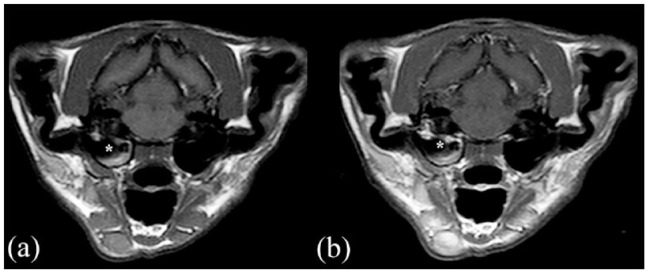
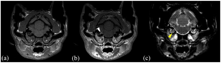
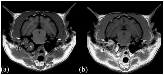
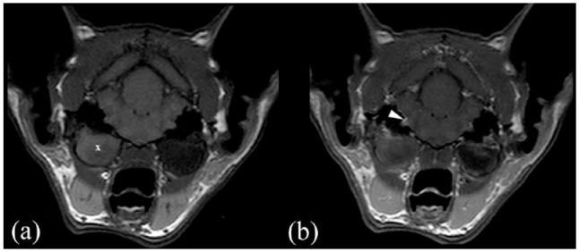
Objectives: The aim of this study was to evaluate the association between meningeal enhancement (MgE) and cerebrospinal fluid (CSF) analysis results, their individual association with bacteriology results from affected ear samples and whether these test results influenced clinicians' therapeutic choice in cats with otitis media and interna (OMI).
Methods: This was a multicentre retrospective study carried out over an 8-year period. Cats diagnosed with OMI, with or without a nasopharyngeal polyp, leading to peripheral vestibular signs were included. Only cats for which MRI with postcontrast T1-weighted sequences and CSF analyses available were included. Cats with intra-axial MRI lesions or empyema were excluded.
Results: Fifty-eight cats met the inclusion criteria. MgE was reported in 26/58 cases, of which nine had an abnormal CSF result (increased total nucleated cell count [TNCC] or total protein); 32/58 cases had no MgE, of which 10 showed abnormal CSF results. There was no association between bacteriology results (external ear canal or bulla) and MgE or abnormal CSF results. CSF abnormalities were statistically significantly more common in acute cases (n = 16/37) than in chronic cases (n = 3/21; Fischer's test P = 0.04). Prednisolone was prescribed in 10/16 cases with increased TNCC. Among the 42 cases with normal TNCC, 15 received prednisolone and 13 received non-steroidal anti-inflammatory drugs. Various antimicrobial drugs were prescribed in 53/58 cats. Duration of antimicrobial treatment was similar, regardless of positive bacterial culture (5.58 vs 4.22 weeks), abnormal CSF (5.83 vs 4.76 weeks) or MgE (5.33 vs 4.90 weeks).
Conclusions and relevance: No association was found between the CSF and MgE results. Furthermore, no association was found between MgE, CSF or bacteriology findings. In addition, abnormal CSF results might lead the clinician to treat with corticosteroids, but they did not have any impact on duration of antimicrobial treatment. CSF abnormalities were seen significantly less frequently in chronic cases. The outcome tended to be poorer when MgE was detected on MRI.
Keywords: MRI meningeal enhancement; Otitis media and interna; cerebrospinal fluid; peripheral vestibular signs.
Conflict of interest statement
The authors declared no potential conflicts of interest with respect to the research, authorship, and/or publication of this article.
Figures
Similar articles
-
A Pandora's box in feline medicine: presenting signs and surgical outcomes in 58 previously hoarded cats with chronic otitis media-interna.J Feline Med Surg. 2023 Sep;25(9):1098612X231197089. doi: 10.1177/1098612X231197089. J Feline Med Surg. 2023. PMID: 37728478 Free PMC article.
-
Suppression of inner ear signal intensity on fluid-attenuated inversion recovery magnetic resonance imaging in cats with vestibular disease.J Feline Med Surg. 2023 Apr;25(4):1098612X231168001. doi: 10.1177/1098612X231168001. J Feline Med Surg. 2023. PMID: 37102785 Free PMC article.
-
Retrospective study of the presentation, diagnosis and management of 16 cats with otitis media not due to nasopharyngeal polyp.J Feline Med Surg. 2018 Dec;20(12):1082-1086. doi: 10.1177/1098612X17746282. Epub 2017 Dec 13. J Feline Med Surg. 2018. PMID: 29235932 Free PMC article.
-
A review of techniques for the investigation of otitis externa and otitis media.Clin Tech Small Anim Pract. 2001 Nov;16(4):236-41. doi: 10.1053/svms.2001.27601. Clin Tech Small Anim Pract. 2001. PMID: 11793879 Review.
-
Otitis externa and otitis media: diagnostic and medical aspects.Semin Vet Med Surg Small Anim. 1993 Feb;8(1):3-9. Semin Vet Med Surg Small Anim. 1993. PMID: 8456201 Review. No abstract available.
Cited by
-
A Pandora's box in feline medicine: presenting signs and surgical outcomes in 58 previously hoarded cats with chronic otitis media-interna.J Feline Med Surg. 2023 Sep;25(9):1098612X231197089. doi: 10.1177/1098612X231197089. J Feline Med Surg. 2023. PMID: 37728478 Free PMC article.
-
Case report: Positioning head tilt observed in a dog and four cats with bilateral peripheral vestibular dysfunction.Front Vet Sci. 2024 Nov 18;11:1495807. doi: 10.3389/fvets.2024.1495807. eCollection 2024. Front Vet Sci. 2024. PMID: 39624061 Free PMC article.
References
-
- Harvey RG, ter Haar G. Ear, nose and throat diseases of the dog and cat. Boca Raton, FL: CRC Press, 2016.
-
- Ackermann AL, Lenz JA, May ER, et al.. Mycoplasma infection of the middle ear in three cats. Vet Dermatol 2017; 28: 417–e102. DOI: 10.1111/vde.12437. - PubMed
MeSH terms
LinkOut - more resources
Full Text Sources
Medical
Miscellaneous
