. 2023 Apr 12;227(7):888-900.
doi: 10.1093/infdis/jiac450.

Molecular Characterization of BK Polyomavirus Replication in Allogeneic Hematopoietic Cell Transplantation Patients

Affiliations

Molecular Characterization of BK Polyomavirus Replication in Allogeneic Hematopoietic Cell Transplantation Patients

Karoline Leuzinger et al. J Infect Dis. .

Abstract

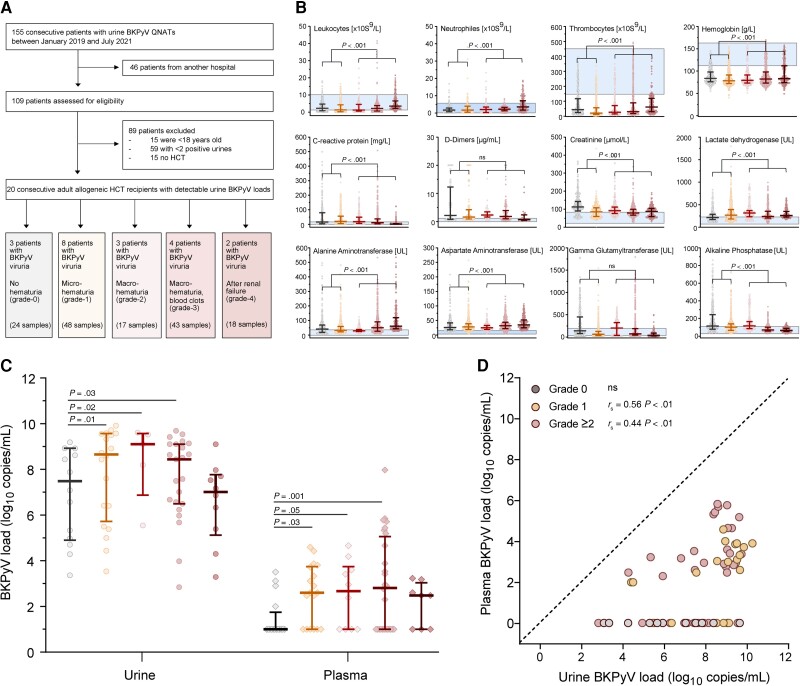
Background: High-level BK polyomavirus (BKPyV) replication in allogeneic hematopoietic cell transplantation (HCT) predicts failing immune control and BKPyV-associated hemorrhagic cystitis.

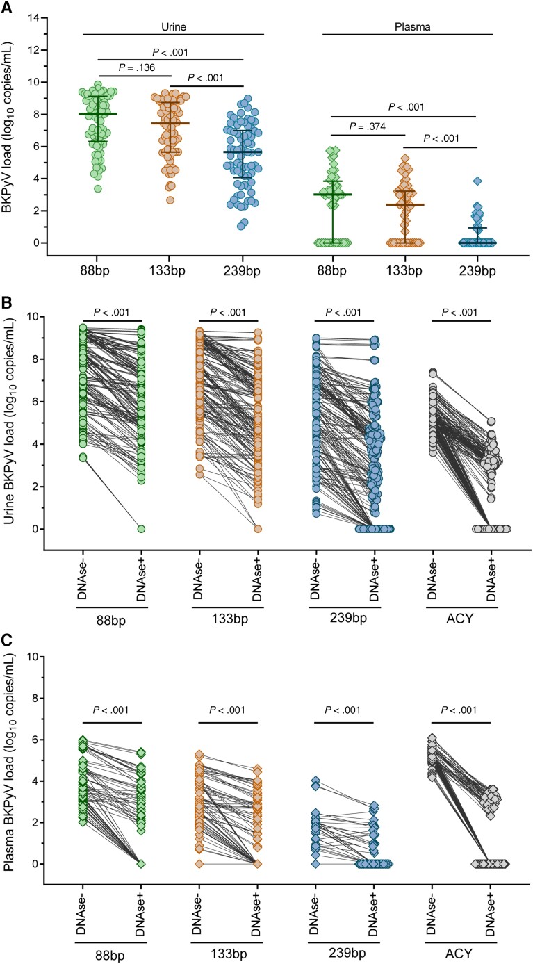
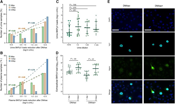
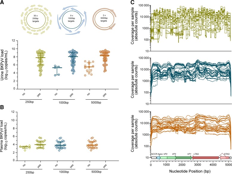
Methods: To identify molecular markers of BKPyV replication and disease, we scrutinized BKPyV DNA-loads in longitudinal urine and plasma pairs from 20 HCT patients using quantitative nucleic acid testing (QNAT), DNase-I treatment prior to QNAT, next-generation sequencing (NGS), and tested cell-mediated immunity.

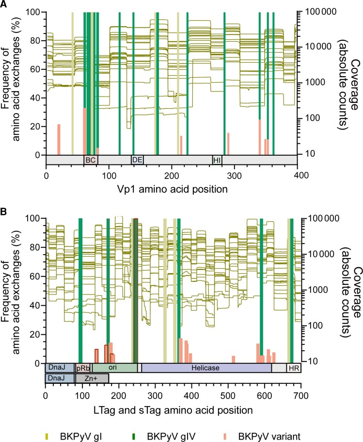
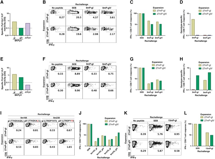
Results: We found that larger QNAT amplicons led to under-quantification and false-negatives results (P < .001). DNase-I reduced urine and plasma BKPyV-loads by >90% (P < .001), indicating non-encapsidated BKPyV genomes. DNase-resistant urine BKPyV-loads remained infectious in cell culture. BKPyV genome fragmentation of ≤250 bp impaired NGS coverage of genetic variation using 1000-bp and 5000-bp amplicons. Conversely, 250-bp amplicons captured viral minority variants. We identified genotype-specific and genotype-independent changes in capsid Vp1 or T-antigen predicted to escape from antibody neutralization or cytotoxic CD8 T-cells, respectively. Genotype-specific changes in immunodominant 9mers were associated with reduced or absent CD8 T-cell responses. Thus, failure to control BKPyV replication in HCT Patients may involve insufficient genotype-specific cytotoxic CD8 T-cell responses, potentially predictable by low neutralizing antibodies as well as genotype-independent immune escape.

Conclusions: Our results provide new insights for patient evaluation and for designing immune protection through neutralizing antibodies, adoptive T-cell therapy, or vaccines.

Keywords: BK polyomavirus; BKPyV; CD8; HCT; LTag; T cell; Vp1; epitope; hematopoietic cell transplantation; hemorrhagic cystitis; immune escape; neutralizing antibody.


Conflict of interest statement

Potential conflicts of interest. H. H. H. has received speaker honorarium from Gilead, Biotest, and Vera Therapeutics; and served as consultant for Roche, Molecular Partners, Vera Therapeutics, and AiCuris. All other authors report no potential conflicts. All authors have submitted the ICMJE Form for Disclosure of Potential Conflicts of Interest. Conflicts that the editors consider relevant to the content of the manuscript have been disclosed

Figures

Figure 1.
Figure 1.
Patients' characteristics. The epidemiology and patients' demographics are displayed in Supplementary Table 1. Patients provided longitudinal plasma (total n = 73) and urine samples (total n = 77) after HCT. Samples from patients without hematuria are displayed in grey, with microscopic (grade 1) hematuria in yellow, and hematuria grade ≥2 in shades of red according to severity. A, Flow diagram of the study. B, Cellular and blood chemistry parameters of the 20 HCT patients at the onset of BKPyV replication (± 7 d; median, 25th and 75th percentiles, minimum, maximum). Blue area indicates the reference values of the parameters in the general healthy population (P value by Mann-Whitney U test). C, Urine and plasma BKPyV loads of the 20 HCT patients by hematuria grade (median, 25th and 75th percentiles, minimum, maximum; P value by Mann-Whitney U test). D, Time-matched urine and plasma samples of the 20 HCT patients (n = 72; dashed line indicates 100% agreement level, P value by Spearman rank correlation). Abbreviations: BKPyV, BK polyomavirus; HCT, hematopoietic cell transplantation; ns, not significant; QNAT, quantitative nucleic acid testing.
Figure 2.
Figure 2.
Assessment of BKPyV genome load, DNase protection, and amplicon length. BKPyV loads were retrospectively analyzed in longitudinal urine (n = 77) and plasma (n = 73) samples from 20 HCT patients by 3 QNATs with different amplicon size (88 bp, 133 bp, and 239 bp). DNase I sensitivity of urine and plasma BKPyV genome loads as well as of cell-free human genomic DNA was assessed by DNAse I digestion prior to nucleic acid extraction, followed by BKPyV and ACY QNAT. Statistical comparison of nonparametric data was done using Mann-Whitney U test. A, Impact of BKPyV DNA fragment size on retrospective urine and plasma BKPyV load quantification (median, 25th and 75th percentiles). B, DNase I sensitivity of urine BKPyV loads and cell-free human genomic DNA. C, DNase I sensitivity of plasma BKPyV loads and cell-free human genomic DNA. Abbreviations: ACY, aspartoacylase; BKPyV, BK polyomavirus; HCT, hematopoietic cell transplantation; QNAT, quantitative nucleic acid testing.
Figure 3.
Figure 3.
Assessment of urine and plasma BKPyV load reduction after DNase I digestion. A, Reduction in urine BKPyV load after DNase I digestion. Linear regression analysis for 88-bp, 133-bp, and 239-bp QNATs. B, Reduction in plasma BKPyV load after DNase I digestion. Linear regression analysis for 88-bp, 133-bp, and 239-bp QNATs. C, Serial dilution and DNase I treatment of urines without significant reduction in urine BKPyV loads as determined by 88-bp QNAT in 15 urines with <0.5 log10 c/mL from (A); median, 25th and 75th percentiles; Mann-Whitney U test. D, BKPyV culture from untreated (DNase−) or DNase I treated (DNase+) urine samples in COS-7 cells. BKPyV loads were determined at 1 day and 6 days postinfection with the 88-bp QNAT (median, 25th and 75th percentiles; Mann-Whitney U test). E, Immunofluorescence staining of BKPyV culture from untreated (DNase−) or DNase I treated (DNase+) urine samples in COS-7 cells at 6 days postinfection. DAPI staining of the COS-7 cell nucleus (blue), BKPyV-specific viral Vp1 capsid (cyan), and agno protein (green). Scale bars represent 50 µm. Abbreviations: BKPyV, BK polyomavirus; DAPI, 4′,6-diamidino-2-phenylindole; QNAT, quantitative nucleic acid testing.
Figure 4.
Figure 4.
NGS coverage in urine and plasma samples. NGS was performed from longitudinal urine (n = 77) and plasma samples (n = 29) from the 20 hematopoietic cell transplantation patients using amplicons with different lengths (250 bp, 1000 bp, and 5000 bp). A, Targeted NGS from urine using different amplicon targets. B, Targeted NGS from plasma using different amplicon targets. C, NGS coverage of full BKPyV genome sequences from urine samples using 250-bp, 1000-bp, and 5000-bp targets (n = 63). Abbreviations: BKPyV, BK polyomavirus; NGS, next-generation sequencing; no, no target amplicon present; yes, target amplicon present.
Figure 5.
Figure 5.
Variation in immunogenic Vp1 and LTag epitopes determined by NGS. Proportion of sequences with BKPyV genotype-specific and variant (genotype-independent) amino acid exchanges in viral capsid Vp1 and the LTag protein compared to the archetype WW [32] reference sequence (BKPyV subtype Ib-1; accession No. AB211371.1) as determined by 250-bp NGS (left y-axis). Vp1 and LTag target region coverage of 250-bp NGS (right y-axis). A, Frequency of amino acid exchanges in the Vp1 protein. The external BC, DE, and HI loops in Vp1 are indicated below the diagram. B, Frequency of amino acid exchanges in the LTag and sTag proteins as determined by 250-bp NGS. The respective domains in LTag and sTag (according to DeCaprio et al [33]) are indicated below the diagram. Of note, the amino-terminal part including the DnaJ homology domain is identical for sTag and LTag, whereas the carboxyterminal domains indicated are specific for sTag or LTag. Changes in immunodominant 9mer T-cell epitopes with significant impact on HLA-A/HLA-B-binding scores are indicated by a red frame. Abbreviations: BKPyV, BK polyomavirus; DnaJ, DnaJ homology region; gI, genotype I; gIV, genotype IV; HR, host range domain; LTag, large tumor antigen; NGS, next-generation sequencing; ori, origin of DNA replication binding domain; pRb, retinoblastoma protein binding domain.
Figure 6.
Figure 6.
BKPyV gI- and gIV-specific CD8 T-cell response. PBMCs of 2 healthy donors were used to expand BKPyV genotype-specific T-cells in vitro. After expansion with gI- or gIV-specific LTag overlapping 27mer pools (27 mP-gI and 27 mP-gIV), cells were restimulated with a pool of immunodominant BKPyV gI or gIV 9mers (9 mP-gI and 9 mP-gIV), with gI 9mers (TTKEKAQIL and LTRDPYHTI) or gIV 9mers (TTKEKALIL and LTRDPYYII), or a genotype-independent 9mer (9m27). Stimulation by SEB was used to determine the proportion of IFN-γ–producing T-cells. IFN-γ production was measured by intracellular staining using FACS. BKPyV gI- and gIV-specific IgG antibodies and JCPyV IgG specific antibodies from donor 1 (A) and donor 2 (E). Assessment of functional T-cell responses by rechallenge with BKPyV gI- and gIV-specific LTag-9mers and measurement of IFN-γ production using FACS in donor 1 (B) and donor 2 (F). Normalization of CD8 T-cells responding to BKPyV gI- and gIV-specific LTag-9mers to the total number of T-cells that could be stimulated by SEB from donor 11 (C) and donor 2 (G). Exclusion of T-cells stimulated by the genotype-independent LTag-9mer (9m27; corresponding to 3.75% and 0.68% after 27mP-gI expansion and 2.2% and 0.27% after 27mP-gIV expansion for donor 1 and donor 2; respectively) leaving solely BKPyV gI- and gIV-specific CD8 T-cell responses from donor 1 (D) and donor 2 (H). Rechallenge of CD8 T-cells from donor 2 using single BKPyV gI- or gIV-specific LTag-9mers, 9m165 and 9m238 (I). Normalization of CD8 T-cells responding to BKPyV gI- and gIV-specific LTag-9mers to the total number of T-cells that could be stimulated by SEB (J). Rechallenge of CD4 T cells from donor 2 using a pool of BKPyV gI-specific LTag 15mers (K). Normalization of CD4 T cells responding to BKPyV gI-specific LTag 15mers to the total number of T cells that could be stimulated by SEB (L). Abbreviations: BKPyV, BK polyomavirus; FACS, fluorescence-activated cell sorting; gI, genotype I; gIV, genotype IV; IFN-γ, interferon-γ; IgG, immunoglobulin G; JCPyV, JC polyomavirus; LTag, large tumor antigen; OD, optical density; SEB, Staphylococcus enterotoxin B; VLP, virus-like particle.

Similar articles

Cited by

References

    1. Graf FE, Hirsch HH. BK polyomavirus after solid organ and hematopoietic cell transplantation: one virus–three diseases. In: Morris MI, Kotton CN, Wolfe C, eds. Emerging transplant infections. Cham, Switzerland: Springer Nature, 2020:1–26.
    1. Cesaro S, Dalianis T, Hanssen Rinaldo C, et al. . ECIL guidelines for the prevention, diagnosis and treatment of BK polyomavirus-associated haemorrhagic cystitis in haematopoietic stem cell transplant recipients. J Antimicrob Chemother 2018; 73:12–21. - PubMed
    1. Imlay H, Xie H, Leisenring WM, et al. . Presentation of BK polyomavirus-associated hemorrhagic cystitis after allogeneic hematopoietic cell transplantation. Blood Adv 2020; 4:617–28. - PMC - PubMed
    1. Egli A, Infanti L, Dumoulin A, et al. . Prevalence of polyomavirus BK and JC infection and replication in 400 healthy blood donors. J Infect Dis 2009; 199:837–46. - PubMed
    1. Arthur RR, Shah KV, Baust SJ, Santos GW, Saral R. Association of BK viruria with hemorrhagic cystitis in recipients of bone marrow transplants. N Engl J Med 1986; 315:230–4. - PubMed

Publication types

Substances