. 2023 Apr 11;7(7):1156-1167.
doi: 10.1182/bloodadvances.2021006485.

Anti-HK antibody inhibits the plasma contact system by blocking prekallikrein and factor XI activation in vivo

Affiliations

Anti-HK antibody inhibits the plasma contact system by blocking prekallikrein and factor XI activation in vivo

Zu-Lin Chen et al. Blood Adv. .

Abstract

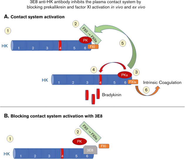
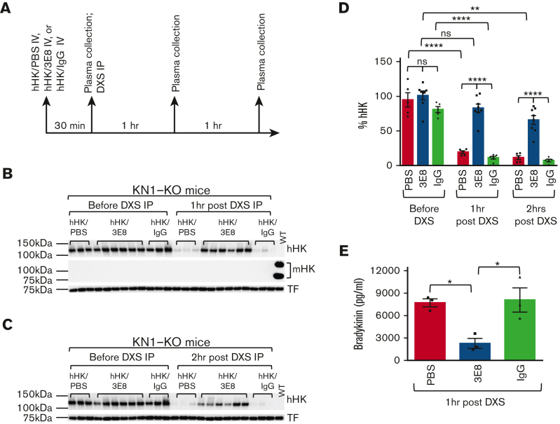
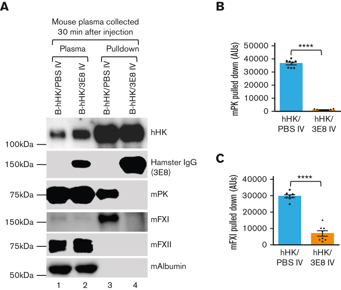
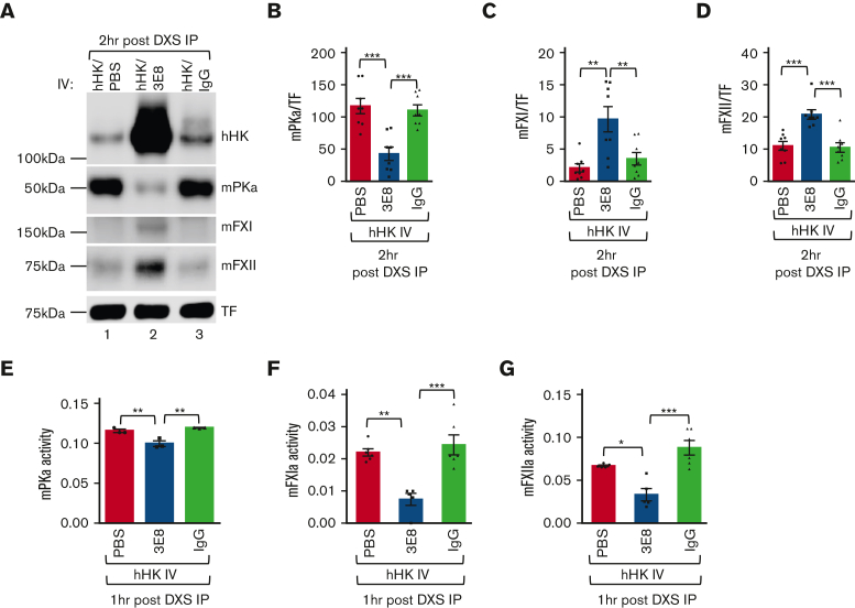
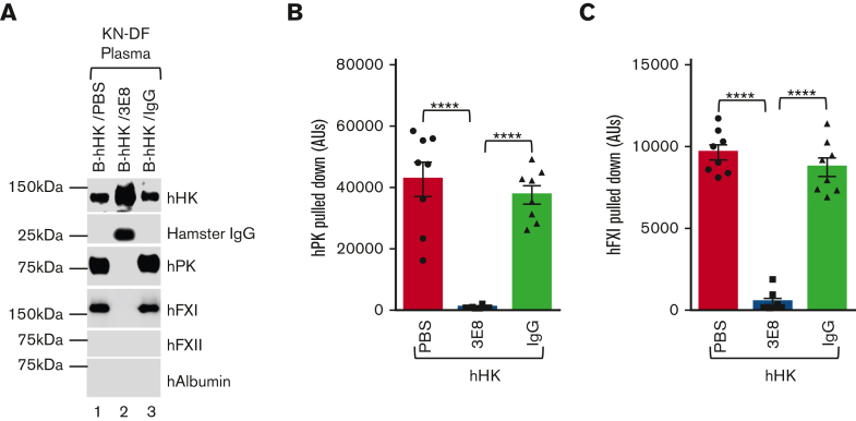
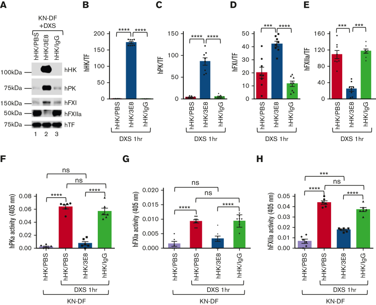
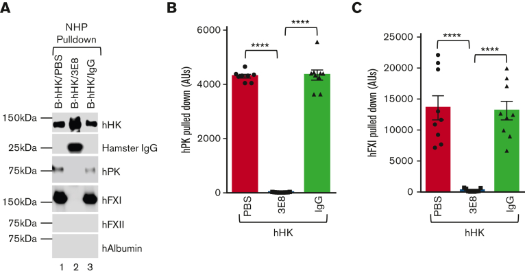
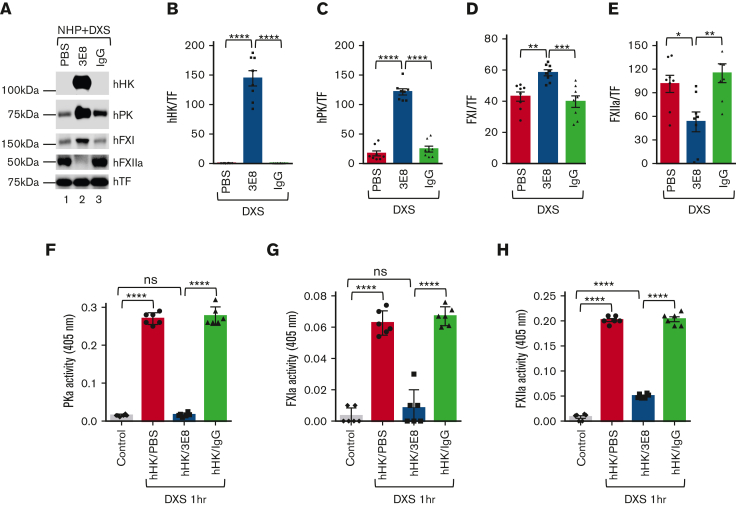
A dysregulated plasma contact system is involved in various pathological conditions, such as hereditary angioedema, Alzheimer disease, and sepsis. We previously showed that the 3E8 anti-high molecular weight kininogen (anti-HK) antibody blocks HK cleavage and bradykinin generation in human plasma ex vivo. Here, we show that 3E8 prevented not only HK cleavage but also factor XI (FXI) and prekallikrein (PK) activation by blocking their binding to HK in mouse plasma in vivo. 3E8 also inhibited contact system-induced bradykinin generation in vivo. Interestingly, FXII activation was also inhibited, likely because of the ability of 3E8 to block the positive feedback activation of FXII by kallikrein (PKa). In human plasma, 3E8 also blocked PK and FXI binding to HK and inhibited both thrombotic (FXI activation) and inflammatory pathways (PK activation and HK cleavage) of the plasma contact system activation ex vivo. Moreover, 3E8 blocked PKa binding to HK and dose-dependently inhibited PKa cleavage of HK. Our results reveal a novel strategy to inhibit contact system activation in vivo, which may provide an effective method to treat human diseases involving contact system dysregulation.


Conflict of interest statement

Conflict-of-interest disclosure: The 3E8 and 2B7 anti–HK antibodies have been licensed to MilliporeSigma.

Figures

None
Graphical abstract
Figure 1.
Figure 1.
3E8 antiHK antibodyinhibitsDXS-induced hHK cleavage and bradykinin release in KN1-KO mice in vivo. (A) KN1-KO mice were IV injected with hHK along with either PBS, 3E8, or hamster IgG as described in “Methods” (n = 5-8 mice per group). After 30 minutes, blood was collected and plasma was prepared. DXS was then intraperitoneally injected, and 1 or 2 hours after DXS injection, blood was collected and plasma was prepared and analyzed by western blotting. (B) Representative western blot shows that injected hHK levels were consistent between groups before DXS injection. One hour after DXS injection, hHK was cleaved and its levels were decreased in hHK/PBS and hHK/hamster IgG groups, but hHK was protected from cleavage in hHK/3E8-injected mice. Mouse HK was not detected in KN1-KO mouse plasma but was present in wild-type mouse plasma. The membrane was stripped and reprobed for transferrin (TF) for normalization. (C) Representative western blot shows hHK changes before and 2 hours after DXS injection into KN1-KO mice. Two hours after DXS injection, hHK was cleaved and dramatically decreased in hHK/PBS and hHK/hamster IgG–injected mice but was protected from cleavage in hHK/3E8-treated mice. (D) Statistical analyses show that the injected hHK levels in groups of mice were similar before DXS injection, but 1 and 2 hours after DXS injection, hHK in 3E8-injected mouse plasma was significantly protected from cleavage compared with hHK in PBS or hamster IgG–injected plasmas. hHK signal was normalized to TF. n = 5 to 8/group. (E) Plasma bradykinin levels in DXS-injected KN1-KO mice infused with hHK/PBS, hHK/3E8, or hHK/IgG were measured according to manufacturer’s instructions. 3E8 significantly inhibited DXS-induced bradykinin generation. n = 3 per group. Data are denoted as mean ± standard error of the mean (SEM). ∗P<.05, ∗∗P≤.01, ∗∗∗∗P≤.0001, P>.05 was not significant (ns).
Figure 2.
Figure 2.
3E8 anti–HK antibody blocks binding of mPK and mFXI to hHK in KN1-KO mice in vivo. B-hHK with or without 3E8 anti–HK antibody was IV injected in KN1-KO mice (n = 7/group), and 30 minutes later mouse plasma was collected and prepared. (A) Lanes 1 and 2: western blot was used to analyze mouse plasma after injections. Plasma contained B-hHK, mPK, mFXI, and mFXII. Plasma from mice also injected with 3E8 showed presence of hamster IgG (lane 2). Mouse albumin was used as a loading control. Lanes 3 and 4: streptavidin beads pulled down B-hHK and any bound proteins, and samples were examined by western blot. B-hHK treated plasma pulled down hHK, mPK, and mFXI (through binding to HK). However, plasma treated with both B-hHK and 3E8 only pulled down hHK and IgG, because 3E8 blocked the binding of mPK and mFXI to HK. (B, C) Statistical analysis (n = 8 mice per group) showed 3E8 antibody significantly blocked mPK and mFXI binding to B-hHK. Data are denoted as mean ± SEM. ∗∗∗∗P≤.0001.
Figure 3.
Figure 3.
3E8 blocks binding of mPK or mFXI to hHK and inhibits mouse contact system thrombotic and inflammatory pathways in vivo. KN1-KO mice were IV injected with hHK along with either PBS, 3E8, or hamster IgG, and then intraperitoneally injected with DXS as described in “Methods”. (A) Two hours after DXS injection, plasma was prepared and analyzed by western blotting. Cleavage of hHK, mFXI, and mFXII was protected in mice treated with 3E8 antibody, but not in PBS- or hamster IgG–treated mice. mPKa levels were lower in 3E8-treated mice, as 3E8 protected against activation of PK. TF was used for normalization. (B-D) Quantification of mPKa, mFXI, and mFXII levels from western blot analyses. mPKa levels (cleaved and activated prekallikrein) were significantly less in 3E8-injected mouse plasma than in PBS- or hamster IgG–injected mouse plasma, indicating less mPK cleavage and activation by 3E8. mFXI levels were significantly higher in 3E8-injected mouse plasma than those in PBS- or IgG-injected mouse plasma, indicating less mFXI cleavage and activation because of 3E8 protection. Levels of mFXII were significantly higher in 3E8 vs PBS or IgG-treated mouse plasma, indicating less mFXII cleavage and activation because of 3E8 treatment. (E-G) 3E8 antibody significantly inhibited mouse PKa activity (E), mouse FXIa activity (F), and mouse FXIIa activity (G) when compared with PBS or IgG-treated mouse plasma. Activity bar graphs were prepared from cumulative data at 15 minute measurements. n = 3 to 8 per group. Data are denoted as mean ± SEM. ∗P<.05, ∗∗P≤.01, ∗∗∗P≤.001.
Figure 4.
Figure 4.
Exogenous purified hHK binds to hPK and hFXI, and 3E8 anti–HK antibody blocked this binding in KN-DF human plasma ex vivo. B-hHK along with either PBS, 3E8, or hamster IgG were added to KN-DF human plasma and incubated at 37°C for 20 minutes. Streptavidin was used to pull down B-hHK–bound proteins complex. (A) Representative western blot shows that B-hHK was pulled down from all samples. B-hHK pulled down 3E8 anti–HK antibody (as denoted by hamster IgG band), but B-hHK did not pull down hamster IgG in the absence of 3E8 (indicating specificity of the antibody; lane 2 vs lanes 1 and 3). hPK and hFXI were pulled with B-hHK only in the absence of 3E8 in KN-DF plasma, suggesting that 3E8 blocked the binding of hPK and hFXI to B-hHK. hFXII and human albumin were not pulled down from any of the samples. (B,C) Quantification of amount of hPK and hFXI pulled down under all KN-DF plasma conditions. n = 8 per group, Data are denoted as mean ± SEM. ∗∗∗∗P ≤ .0001.
Figure 5.
Figure 5.
3E8 blocks binding of of hPK or hFXI to hHK and inhibits contact system thrombotic and inflammatory pathway activation in KN-DF human plasma ex vivo. hHK was added to KN-DF human plasma along with either PBS, 3E8, or hamster IgG. After incubation at 37°C for 20 minutes, DXS was added for an additional 60 minutes at 37°C to activate plasma contact system. (A) Representative western blot of samples. TF was used for normalization. (B) As expected, hHK cleavage was blocked by 3E8 antibody, but not by PBS or IgG in KN-DF human plasma. (C,D) Levels of hPK and hFXI were significantly higher in 3E8-treated plasma than in PBS- or IgG-treated KN-DF human plasma, indicating that 3E8 inhibited binding and activation of hPK and hFXI. (E) Levels of hFXIIa were significantly lower in 3E8-treated plasma than in PBS- or IgG-treated KN-DF human plasma, suggesting that the prevention of hPK and hFXI activation by 3E8 also hinders hFXII activation through the feedback mechanism. (F-H) The 3E8 antibody significantly inhibited PKa activity (F), FXIa activity (G), and FXIIa activity (H) when compared with PBS- or IgG-treated KN-DF plasma. Activity bar graphs were generated from cumulative data at 15 minute measurements. n = 7 to 8 per group, Data are denoted as mean ± SEM. ∗∗∗P≤.001, ∗∗∗∗P≤.0001.
Figure 6.
Figure 6.
3E8 antiHK antibody blocks binding of exogenously added hHK to hPK and hFXI and displaces hPK and hFXI binding to hHK in NHP ex vivo. B-hHK was added to NHP with PBS, 3E8, or hamster IgG and incubated at 37°C for 20 minutes. Streptavidin was used to pull down B-hHK-bound proteins complex. (A) Representative western blot results of the pulldown experiments. hHK was pulled down in all samples. The 3E8 anti–HK antibody was pulled down with B-hHK, but control hamster IgG was not. (B,C) hPK and hFXI were pulled from PBS- and hamster IgG–treated NHP but not from 3E8-incubated KN-DF plasma, indicating that binding of hPK and hFXI to B-hHK was blocked by 3E8 anti–HK antibody. hFXII and human albumin were not pulled down from any of the samples. (D-G) NHP was incubated with PBS, 3E8, or hamster IgG at 37°C for 20 minutes. B-2B7 anti–HK antibody was added and incubated for additional 20 minutes. Streptavidin was used to pull down B-2B7–bound proteins. (D) Representative western blot results of the pulldown experiments. Although hHK was pulled down in all samples, hFXI and hPK were only pulled down in PBS- or IgG-treated plasma (Lanes 1 and 3 vs Lane 2). (E) 3E8 anti–HK antibody protected hHK cleavage during pulldown processes. (F & G) hFXI and hPK were pulled down from PBS- and hamster IgG–treated, but not 3E8-treated NHP, indicating that binding of hPK and hFXI to hHK was displaced by the 3E8 anti–HK antibody. hFXII and human albumin were not pulled down in any of the samples. n = 8 to 9/group. Data are denoted as mean ± SEM. ∗∗P ≤ .01, ∗∗∗∗P ≤ .0001.
Figure 7.
Figure 7.
Blocking hPK or hFXI binding to hHK inhibits activation of both contact system thrombotic and inflammatory pathways in NHP ex vivo. hHK as well as either PBS, 3E8, or hamster IgG were added to NHP and incubated at 37°C for 20 minutes. DXS was then added and incubated at 37°C for additional 60 minutes to activate the plasma contact system. (A) Samples were analyzed via western blotting. TF was used for normalization. (B) As expected, hHK cleavage by DXS-induced contact system activation was blocked by 3E8 antibody but occurred in plasma samples containing PBS or IgG. (C,D) hPK and hFXI levels were significantly higher in NHP treated with 3E8 rather than in PBS or IgG, indicating that 3E8 prevented hPK and hFXI cleavage and activation by DXS. (E) Levels of hFXIIa were significantly lower in NHP treated with 3E8 than with PBS or hamster IgG, indicating that 3E8 indirectly allows for less hFXII cleavage and activation in NHP ex vivo. (F-H) The 3E8 antibody significantly inhibited PKa (F), FXIa (G), and FXIIa (H) activities when compared with PBS- or IgG-treated NHP. Activity bar graphs were prepared from cumulative data at 15 minute measurements. n = 8 per group. Data are denoted as mean ± SEM. ∗P≤.05, ∗∗P≤.01, ∗∗∗P≤.001, ∗∗∗∗P≤.0001.

Similar articles

Cited by

References

    1. Jukema BN, de Maat S, Maas C. Processing of factor XII during inflammatory reactions. Front Med (Lausanne) 2016;3 - PMC - PubMed
    1. Schmaier AH. The contact activation and kallikrein/kinin systems: pathophysiologic and physiologic activities. J Thromb Haemost. 2016;14(1):28–39. - PubMed
    1. Simao F, Ustunkaya T, Clermont AC, Feener EP. Plasma kallikrein mediates brain hemorrhage and edema caused by tissue plasminogen activator therapy in mice after stroke. Blood. 2017;129(16):2280–2290. - PMC - PubMed
    1. Wu Y. Contact pathway of coagulation and inflammation. Thromb J. 2015;13 - PMC - PubMed
    1. Maas C, Govers-Riemslag JW, Bouma B, et al. Misfolded proteins activate factor XII in humans, leading to kallikrein formation without initiating coagulation. J Clin Invest. 2008;118(9):3208–3218. - PMC - PubMed

Publication types