. 2023 Jan 1;103(1):855-918.
doi: 10.1152/physrev.00061.2021. Epub 2022 Sep 15.

A systematic review of the biological mediators of fat taste and smell

Affiliations

A systematic review of the biological mediators of fat taste and smell

Rosario B Jaime-Lara et al. Physiol Rev. .

Abstract

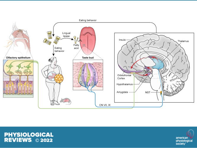
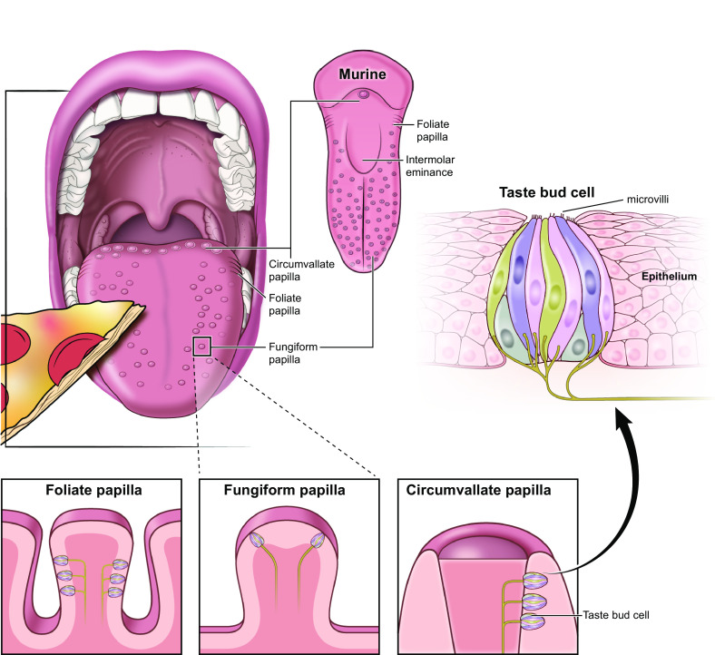
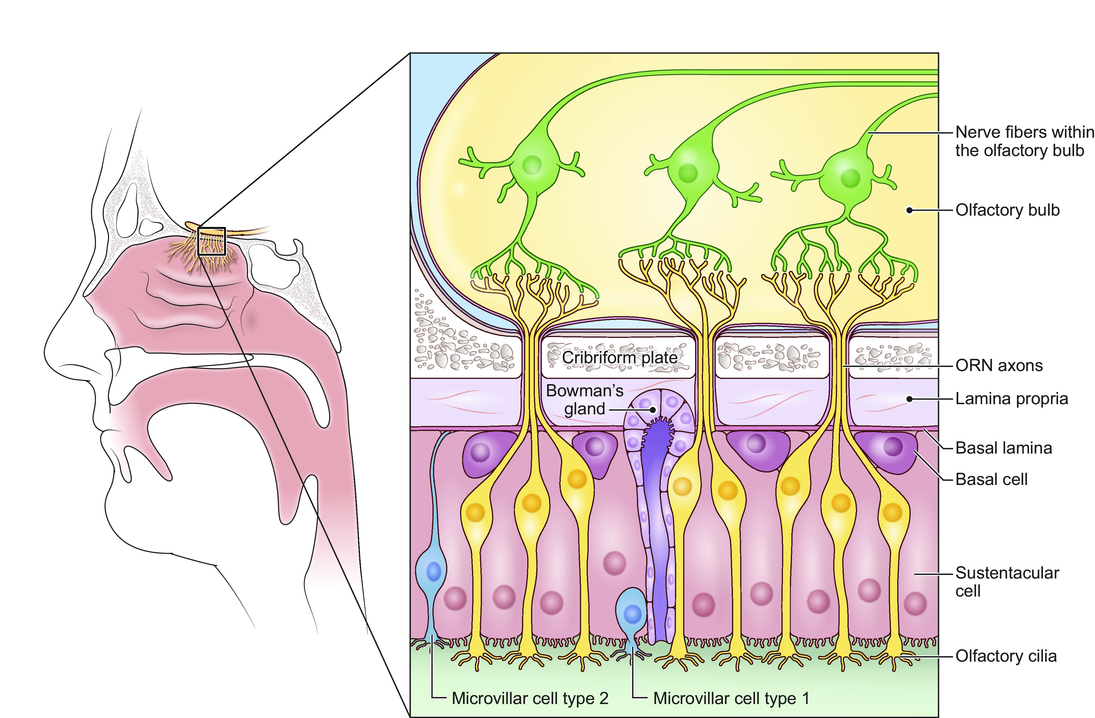
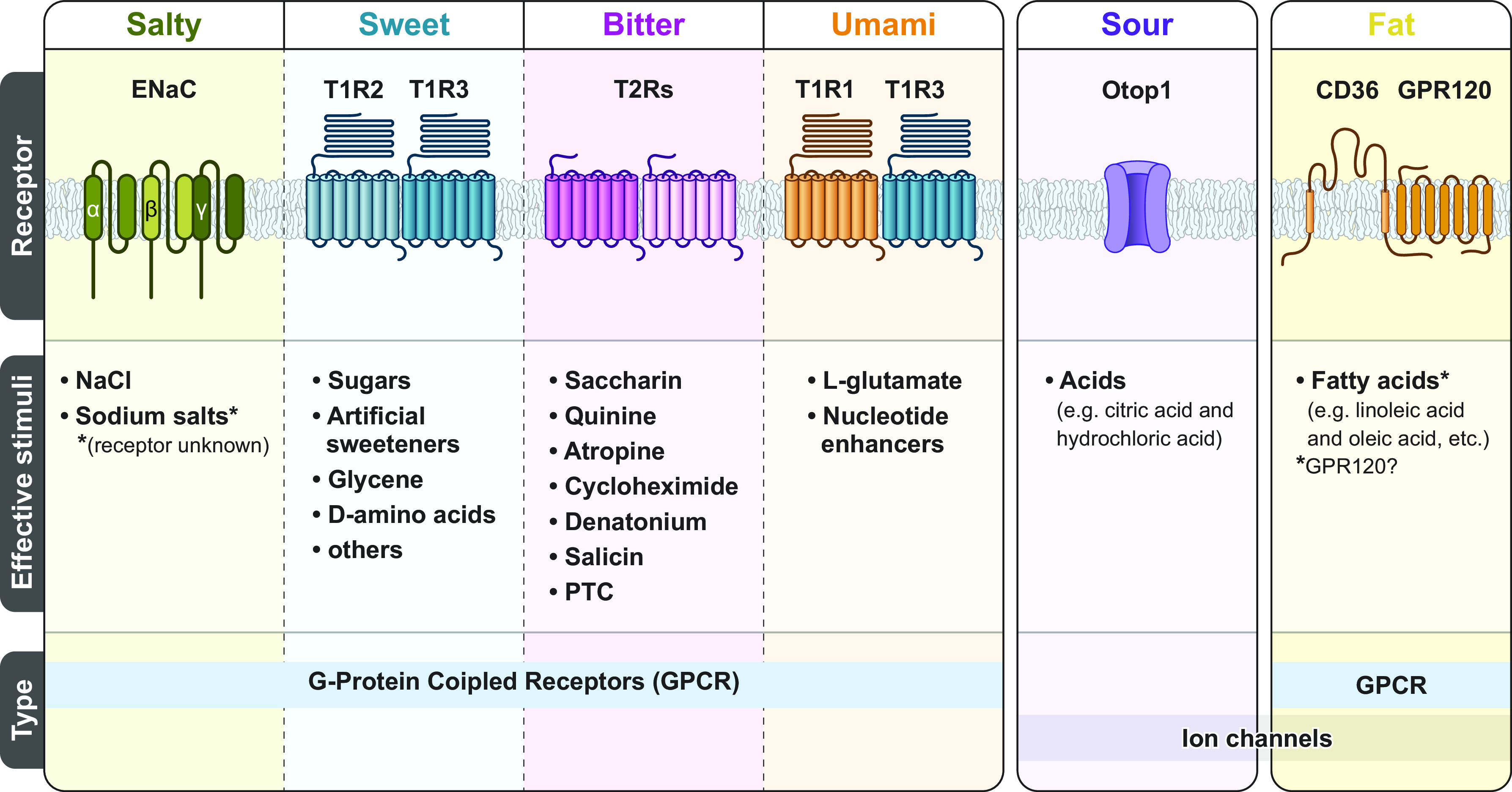
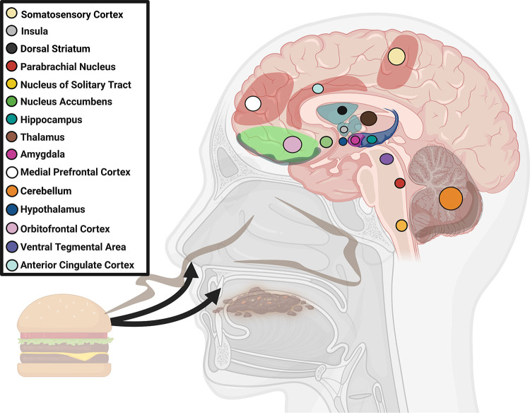
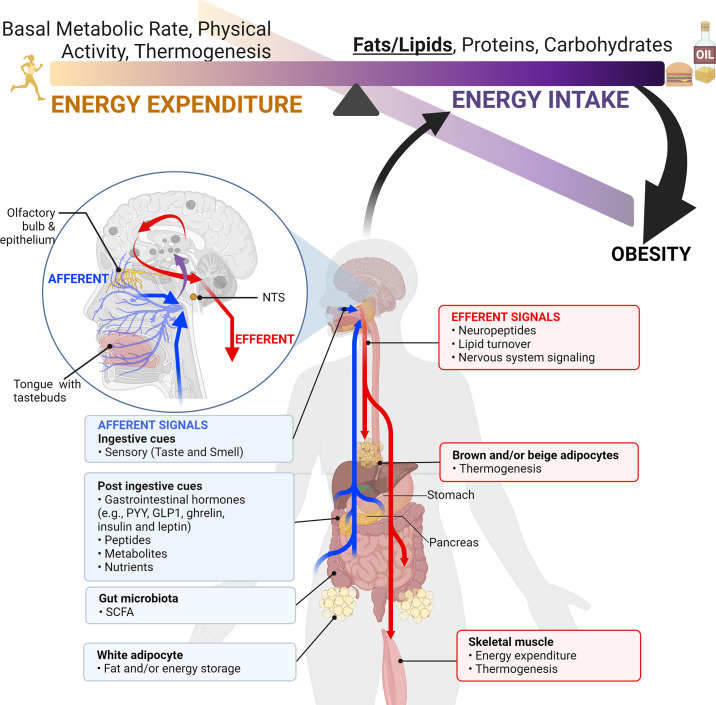
Taste and smell play a key role in our ability to perceive foods. Overconsumption of highly palatable energy-dense foods can lead to increased caloric intake and obesity. Thus there is growing interest in the study of the biological mediators of fat taste and associated olfaction as potential targets for pharmacologic and nutritional interventions in the context of obesity and health. The number of studies examining mechanisms underlying fat taste and smell has grown rapidly in the last 5 years. Therefore, the purpose of this systematic review is to summarize emerging evidence examining the biological mechanisms of fat taste and smell. A literature search was conducted of studies published in English between 2014 and 2021 in adult humans and animal models. Database searches were conducted using PubMed, EMBASE, Scopus, and Web of Science for key terms including fat/lipid, taste, and olfaction. Initially, 4,062 articles were identified through database searches, and a total of 84 relevant articles met inclusion and exclusion criteria and are included in this review. Existing literature suggests that there are several proteins integral to fat chemosensation, including cluster of differentiation 36 (CD36) and G protein-coupled receptor 120 (GPR120). This systematic review will discuss these proteins and the signal transduction pathways involved in fat detection. We also review neural circuits, key brain regions, ingestive cues, postingestive signals, and genetic polymorphism that play a role in fat perception and consumption. Finally, we discuss the role of fat taste and smell in the context of eating behavior and obesity.

Keywords: chemosensation; fat taste; obesity; oleogustus; olfaction.


Conflict of interest statement

No conflicts of interest, financial or otherwise, are declared by the authors.

Figures

None
Graphical abstract
FIGURE 1.
FIGURE 1.
Taste bud cell. Taste buds are onion-shaped clusters of taste receptor cells. They are found in the tongue, the soft palate, the pharynx, and the esophagus. There are three different kinds of papillae that contain taste bud cells: foliate, fungiform, and circumvallate papillae. Food particles that dissolve in liquids and/or saliva (tastants) bind to microvilli to stimulate taste receptors and initiate transduction cascades that give rise to tate.
FIGURE 2.
FIGURE 2.
Olfactory epithelium and olfactory bulb. The olfactory epithelium is located within the nasal cavity and is comprised of olfactory sensory neurons (ORNs), basal cells, and supporting cells (i.e., sustentacular and microvillar cells). ORN dendrites project to the mucus layer that lines the olfactory epithelium. Smell/odors stimulate the olfactory cilia initiating olfactory signal transduction.
FIGURE 3.
FIGURE 3.
Taste receptors by taste modality. There are five commonly recognized primary taste modalities: salty, sweet, bitter, sour, and umami. However, there is growing evidence suggesting fat may be an additional taste modality. Each primary taste modality is characterized by multiple elements including having dedicated receptors [i.e., G protein-coupled receptors (GPCRs) and ion channels] and a defined class of effective stimuli. CD36, cluster of differentiation 36; ENaC, epithelial sodium channel; PTC, phenylthiocarbamide. Figure adapted from Ref. , with permission from Springer Nature.
FIGURE 4.
FIGURE 4.
Triglyceride breakdown and fatty acid types. Triglycerides are a common type of dietary fat, composed of three fatty acids joined to glycerol. Lingual lipase can breakdown dietary triglycerides into fatty acids and glycerol. Rodents synthesize salivary lipase (LIPF). Humans also have salivary lipase forms (e.g., LIPK, LIPM, LIPN). Studies selected in this review often studied human and animal mode responses to common fatty acids, including oleic acid and linoleic acid. Image created with BioRender.com, with permission.
FIGURE 5.
FIGURE 5.
PRISMA flow diagram. The diagram shows the selection of reports included in this systematic review.
FIGURE 6.
FIGURE 6.
Taste transduction by taste modality. Taste buds contain three cell types specific to taste modalities: type I, type II, and type III cells. Type I cells are glial-like cells and are involved in salt detection. Type II cells are involved in sweet, bitter, and umami. Presynaptic type III cells detect sour stimuli and potentially salty stimuli. Each cell type and each taste modality are characterized by their own combination of receptors and transduction pathways. Taste information is then relayed to the brain via afferent nerves (cranial nerves VII, IX, and X). ENaC, epithelial sodium channel; IP3, inositol (1,4,5)-triphosphate; NTs, neurotransmitters; PIP2, phosphatidylinositol 4,5-bisphophate; VGSC, voltage-gated sodium channel. Figure adapted from Ref. , with permission from Springer Nature.
FIGURE 7.
FIGURE 7.
Fat Transduction signaling in taste bud cells. In taste bud cells: (1) free fatty acids (FFAs) binds to cluster of differentiation 36 (CD36), and CD36 interacts with GPR120. This triggers a signaling cascade involving α-gustducin which activates Ca2+, dependent phospholipase (PLC). (2) PLC cleaves phosphatidylinositol 4,5-bisphophate (PIP2; bound to the membrane) into diacylglycerol (DAG) (stays in membrane) and inositol (1,4,5)-triphosphate (IP3) (free in cytosol). DAG can phosphorylate protein kinase C (PKC), which helps to activate the extracellular signal-regulated kinases (ERK) pathway. (3) IP3 leaves the membrane and binds to IP3-gated calcium ion channels on the endoplasmic reticulum (ER) membrane. This opens the IP3 channels and releases Ca2+ stores from the ER into the cytosol, increasing intracellular calcium levels ([Ca2+]i). (4) The increased [Ca2+]i triggers several events that lead to membrane depolarization of the cell. (4a) TRPM5 channels (a Ca2+-gated sodium channel) open, allowing Na+ to come into the cell. Additionally, Delayed rectifying K+ (DRK) channels close, preventing potassium from leaving the cell. (4b) The calcium release from the ER requires that stromal interaction molecule 1 (STIM1) replenish the ER’s calcium stores. (4c) STIM1 activates Orai1 (calcium release-activated calcium channel) to enable the influx of calcium into the cytosol. Subsequent opening of calcium homeostasis modulator 1 (CALHM1) channels allows for additional calcium influx into the cell. (5) Increased [Ca2+]i and CD36-induced Fyn/Src kinase activation contribute to phosphorylation of the ERK1/2 pathway. This can activate cAMP signaling, which promotes transcription of cellular regulation factors. (6) Depolarization throughout the cell leads to the release of serotonin (5-HT) and ATP, which act as neurotransmitters. Serotonin binds to its receptors, and ATP binds to purinergic P2X2 and P2X3 receptors that cause neuronal excitability, further relaying fat chemosensory signals to the brain. CD36, cluster of differentiation 36; GPR120, G protein-coupled receptor 120. Image created with BioRender.com, with permission.
FIGURE 8.
FIGURE 8.
Brain regions involved in fat taste and smell. The perception of fat taste and smell activates of multiple brain regions involved in fat chemosensation. Image created with BioRender.com, with permission.
FIGURE 9.
FIGURE 9.
Postingestive and ingestive cues mediate energy intake and energy balance. The brain receives afferent signals including ingestive cues (e.g., taste and smell), and postingestive cues (e.g., hunger and satiety hormones, peptides, metabolites, and nutrient sensing). Together these afferent signals are processed by the brain to inform homeostatic status/energy balance. In response to these cues, efferent signals can stimulate physiological events, including lipid turnover and energy expenditure. Importantly, postingestive cues, can also trigger efferent signals that impact future ingestive cues. For example, an animal or human who may not initially prefer a fatty substance may develop a preference following rewarding postingestive cues. Altogether, these afferent and efferent signals can impact energy intake and excessive energy intake can lead to obesity. NTS, nucleus of the solitary tract; GLP-1, glucagon-like-peptide-1; SCFA, short-chain fatty acid. Image created with BioRender.com, with permission.

References

    1. Dudine L, Canaletti C, Giudici F, Lunardelli A, Abram G, Santini I, Baroni V, Paris M, Pesavento V, Manganotti P, Ronchese F, Gregoretti B, Negro C. Investigation on the loss of taste and smell and consequent psychological effects: a cross-sectional study on healthcare workers who contracted the COVID-19 infection. Front Public Health 9: 666442, 2021. doi:10.3389/fpubh.2021.666442. - DOI - PMC - PubMed
    1. Hur K, Choi JS, Zheng M, Shen J, Wrobel B. Association of alterations in smell and taste with depression in older adults. Laryngoscope Investig Otolaryngol 3: 94–99, 2018. doi:10.1002/lio2.142. - DOI - PMC - PubMed
    1. Parma V, Ohla K, Veldhuizen MG, Niv MY, Kelly CE, Bakke AJ, , et al. . More than smell—COVID-19 is associated with severe impairment of smell, taste, and chemesthesis. Chem Senses 45: 609–622, 2020. doi:10.1093/chemse/bjaa041. - DOI - PMC - PubMed
    1. Hannum ME, Koch RJ, Ramirez VA, Marks SS, Toskala AK, Herriman RD, Lin C, Joseph PV, Reed DR. Taste loss as a distinct symptom of COVID-19: a systematic review and meta-analysis. Chemical Senses 47: bjac001, 2022. doi:10.1093/chemse/bjac001. - DOI - PMC - PubMed
    1. Hannum ME, Ramirez VA, Lipson SJ, Herriman RD, Toskala AK, Lin C, Joseph PV, Reed DR. Objective sensory testing methods reveal a higher prevalence of olfactory loss in COVID-19-positive patients compared to subjective methods: a systematic review and meta-analysis. Chem Senses 45: 865–874, 2020. doi:10.1093/chemse/bjaa064. - DOI - PMC - PubMed

Publication types

LinkOut - more resources