. 2022 Nov 29;119(48):e2209441119.
doi: 10.1073/pnas.2209441119. Epub 2022 Nov 21.

Titin force in muscle cells alters lattice order, thick and thin filament protein formation

Affiliations

Titin force in muscle cells alters lattice order, thick and thin filament protein formation

Anthony L Hessel et al. Proc Natl Acad Sci U S A. .

Abstract

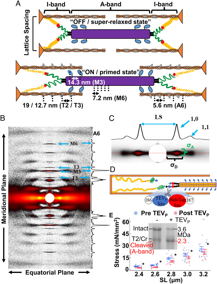
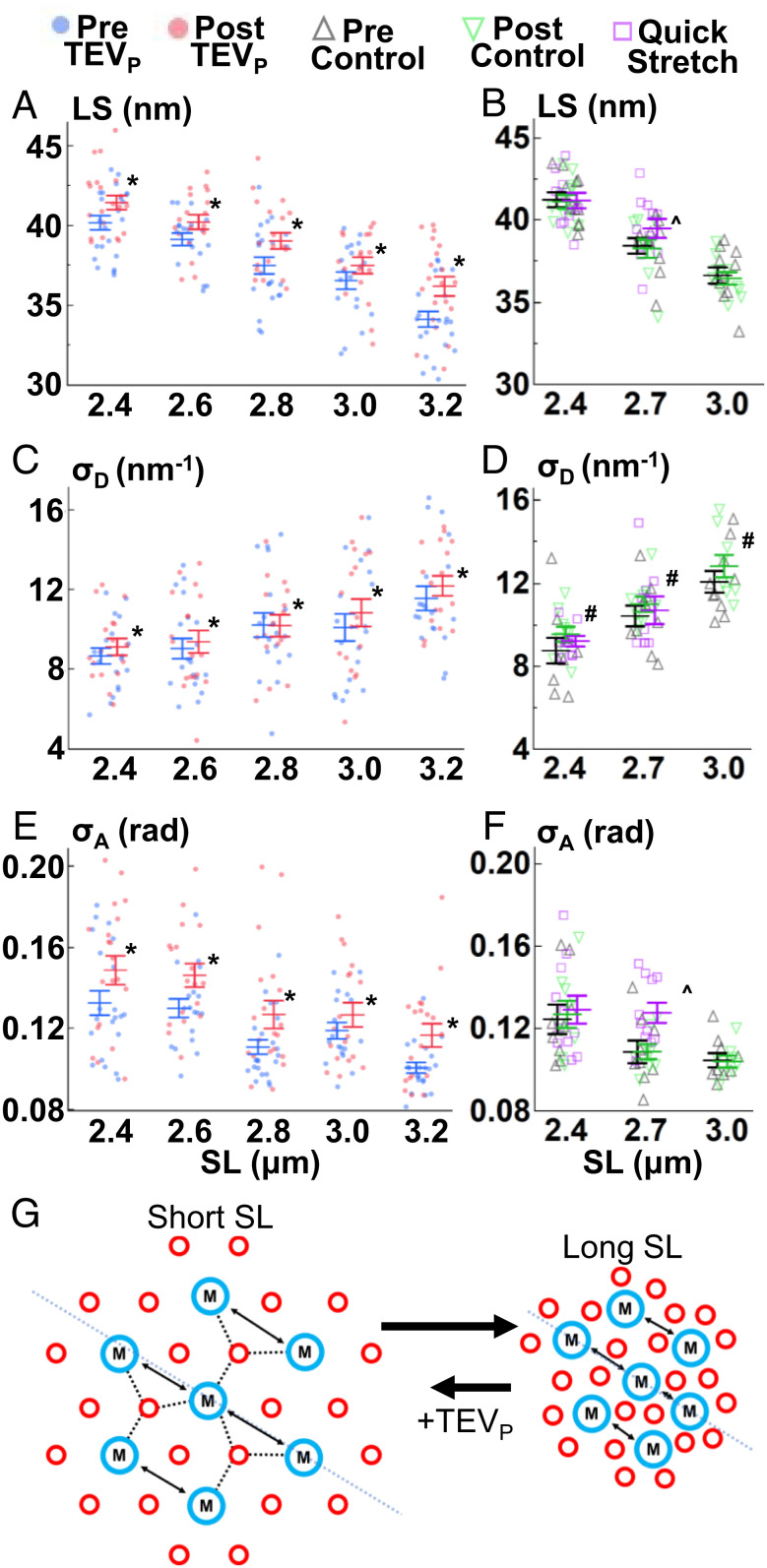
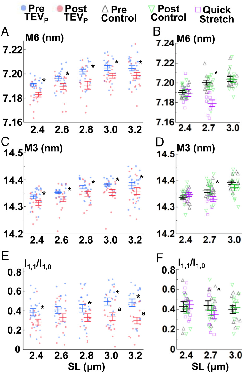
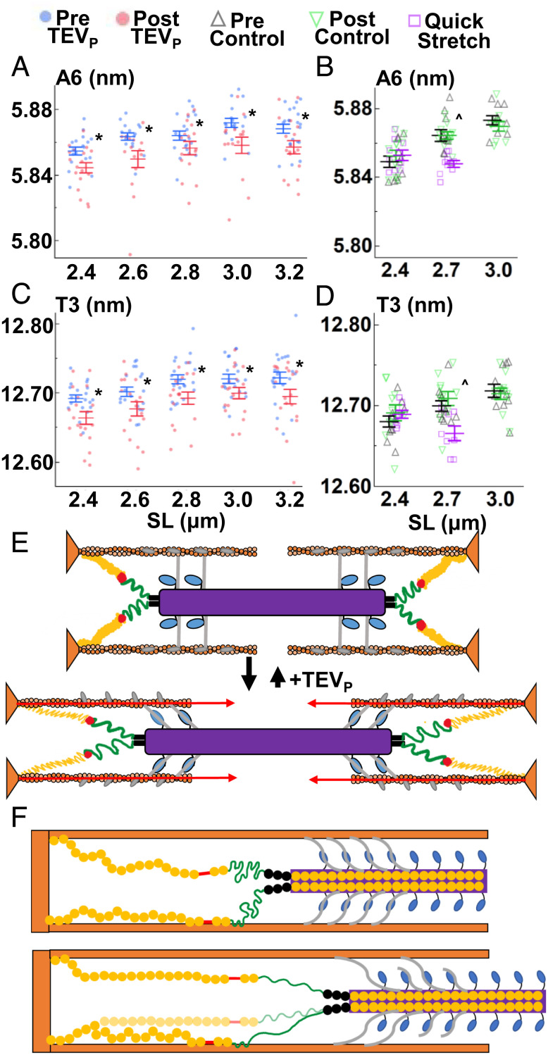
Skeletal muscle force production is increased at longer compared to shorter muscle lengths because of length-dependent priming of thick filament proteins in the contractile unit before contraction. Using small-angle X-ray diffraction in combination with a mouse model that specifically cleaves the stretch-sensitive titin protein, we found that titin cleavage diminished the length-dependent priming of the thick filament. Strikingly, a titin-sensitive, length-dependent priming was also present in thin filaments, which seems only possible via bridge proteins between thick and thin filaments in resting muscle, potentially myosin-binding protein C. We further show that these bridges can be forcibly ruptured via high-speed stretches. Our results advance a paradigm shift to the fundamental regulation of length-dependent priming, with titin as the key driver.

Keywords: X-ray diffraction; elasticity; length-dependent activation; mouse; ultrastructure.


Conflict of interest statement

The authors declare no competing interest.

Figures

Fig. 1.
Fig. 1.
X-ray diffraction of skeletal fibers. (A) The passive sarcomeres at short (Top) and long (Bottom) lengths are made up of organized repeating structures (approximate spacing and IDs indicated) including the thick filament backbone (purple), myosin heads (light blue), thin filament backbone (orange), and troponin complex (gray). Thick and thin filaments are attached in the I-band by titin, which is made up of the proximal Ig (yellow), N2A (red), PEVK (green), and distal Ig regions (black). From short to long lengths, proteins reorient from “OFF / superrelaxed state” to “ON / primed state”. Note that spacing values are indicative of the respective spacing parameter assessed. (B) Representative X-ray image of fibers (heatmap gradient) with reflections labeled with the ID shown in (A). (C) The main equatorial-plane reflections. 2D intensity projections are shown and critical features marked. (D) TC muscle has a TEV protease (TEVP) recognition site built into I-band titin, allowing for quick and targeted titin cleavage. HaloTag site can, e.g., be used for imaging purposes. (E) Genetically heterozygous TC psoas fibers before (blue) and after (red) TEVP treatment, where ~50% titin cleavage decreased force across SL. Inset: Coomassie-stained titin gel of Het TC fibers before and after TEVP treatment. Cr = Cronos. *P < 0.05 after titin cleavage. Data throughout reported as mean ± SEM.
Fig. 2.
Fig. 2.
Lattice changes with SL, titin cleavage, and velocity. LS (A and B), σD (C and D), and σA (E and F) were recorded before (blue) and after (red) titin cleavage (A, C, and E), before (black) and after (green) a control solution, and after a relatively quicker stretch (magenta) (B, D, and F). Details in main text. (G) Cartoon summary of the thick (blue) and thin (red) filament lattice (cross-section of sarcomeres) after stretch, and after TEVP treatment. *P < 0.05 from pre to post values. ^P < 0.05 between the control (pre) and quick stretch experiments. #P < 0.05 between pre and post control values. Also see SI Appendix, Tables S1, S2, S7, and S8.
Fig. 3.
Fig. 3.
Thick filament changes with SL, titin cleavage, and velocity. M6 spacings (A and B), M3 spacings (C and D), and I1,1/I1,0 (E and F) were recorded as explained in Fig. 2. Details in main text. *P < 0.05 from pre to post values. ^P < 0.05 between quick stretch and control (pre) data. ªP < 0.05 similar effect magnitude at those SLs. Also see SI Appendix, Tables S1, S2, S4, S5, S7, and S8.
Fig. 4.
Fig. 4.
Thin filament changes with SL, titin cleavage, and velocity. A6 (A and B), spacings and T3 spacings (C and D) were recorded as explained in Fig. 2. (E) Cartoon summary of the sarcomere at short and long lengths, with proposed A-band bridges added. The ON state is reduced at long lengths after titin cleavage. (F) Sarcomere stretch from short (Top) to long (Bottom) extends the entire I-band titin producing relatively low titin-based forces, but evidence indicates that titin extension can be modified by titin–thin filament interactions around the N2A/PEVK region, functionally shortening the free titin to the stiffer PEVK region, leading to relatively larger titin-based forces after stretch. Higher velocities can be expected to detach the interaction, reducing titin-based forces. *P < 0.05 from pre to post values. ^P < 0.05 between quick stretch and control (pre) data. Also see SI Appendix, Tables S1, S2, S4, S5, S7, and S8.

Similar articles

Cited by

References

    1. Huxley A. F., Simmons R. M., Proposed mechanism of force generation in striated muscle. Nature 233, 533–538 (1971). - PubMed
    1. Gordon A. M., Huxley A. F., Julian F. J., The variation in isometric tension with sarcomere length in vertebrate muscle fibres. J. Physiol. 184, 170–192 (1966). - PMC - PubMed
    1. Holt N. C., Williams C. D., Can strain dependent inhibition of cross-bridge binding explain shifts in optimum muscle length? Integr. Comp. Biol. 58, 174–185 (2018). - PMC - PubMed
    1. Martyn D. A., Gordon A. M., Length and myofilament spacing-dependent changes in calcium sensitivity of skeletal fibres: Effects of pH and ionic strength. J. Muscle. Res. Cell Motil. 9, 428–445 (1988). - PubMed
    1. de Tombe P. P., et al. , Myofilament length dependent activation. J. Mol. Cell Cardiol. 48, 851–858 (2010). - PMC - PubMed

Publication types

LinkOut - more resources