. 2022 Nov 29;119(48):e2203567119.
doi: 10.1073/pnas.2203567119. Epub 2022 Nov 21.

Regulation of the DEAH/RHA helicase Prp43 by the G-patch factor Pfa1

Affiliations

Regulation of the DEAH/RHA helicase Prp43 by the G-patch factor Pfa1

Marieke Enders et al. Proc Natl Acad Sci U S A. .

Abstract

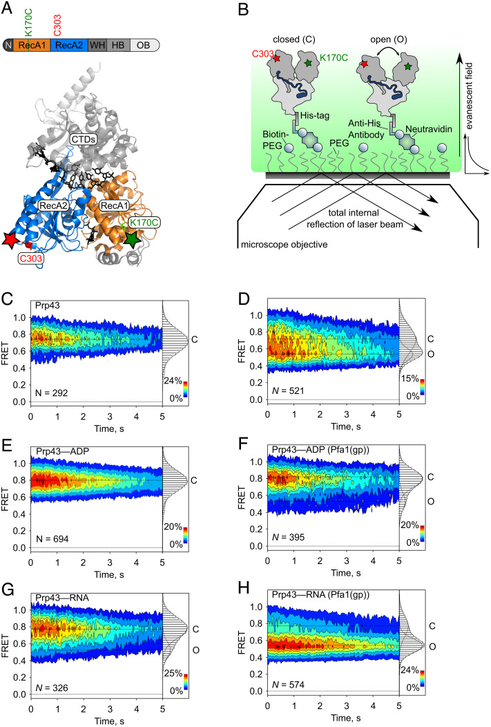
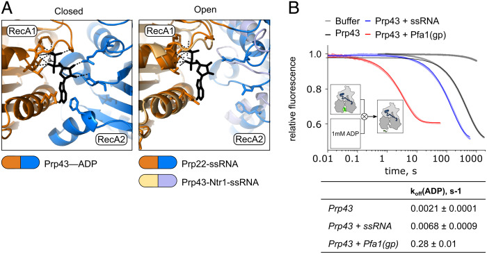
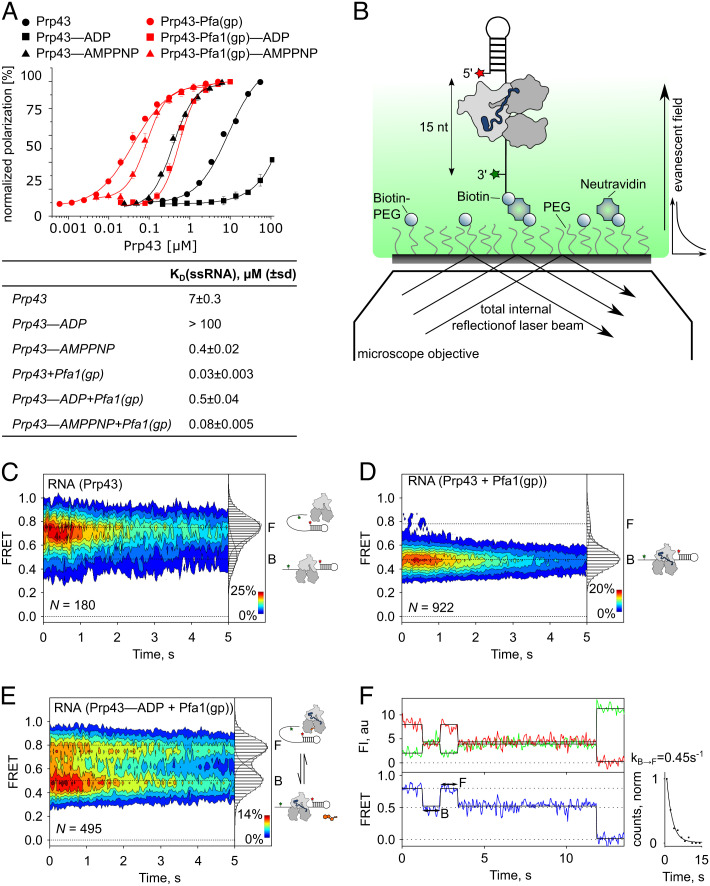
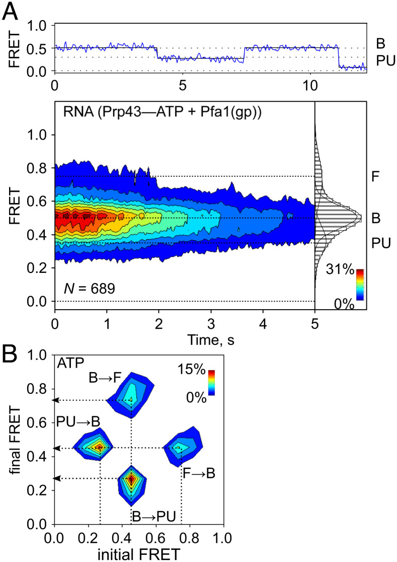
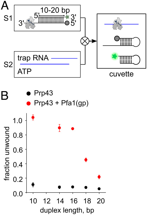
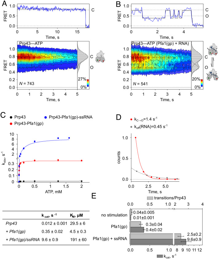
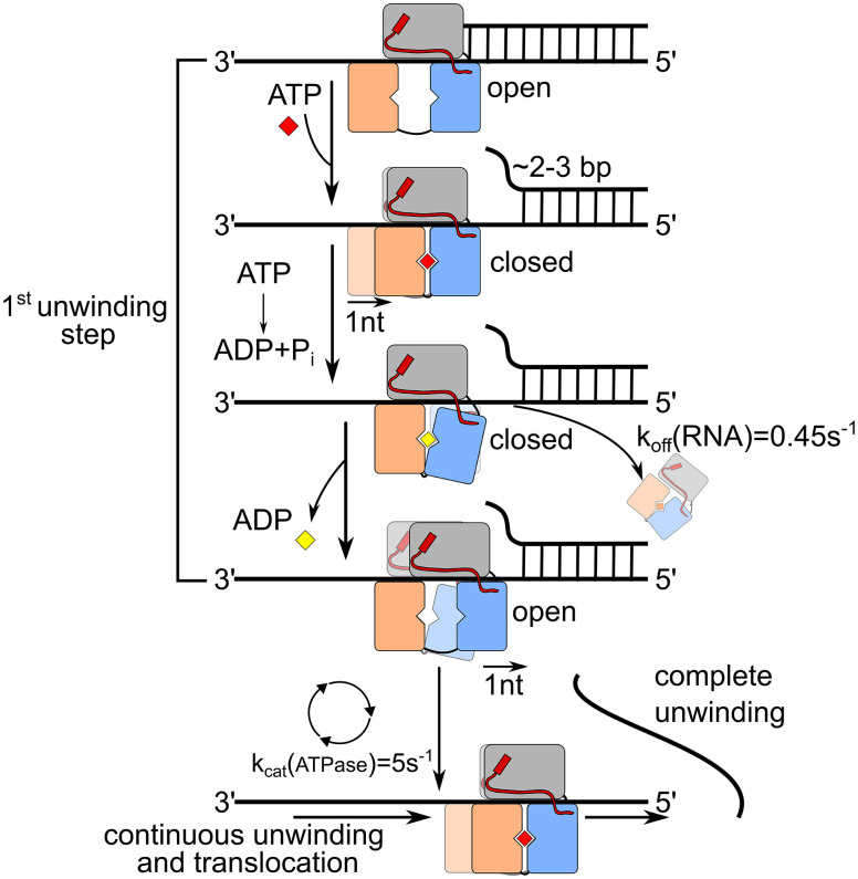
The DEAH/RHA helicase Prp43 remodels protein-RNA complexes during pre-messenger RNA (mRNA) splicing and ribosome biogenesis. The helicase activity and ATP turnover are intrinsically low and become activated by G-patch (gp) factors in the specific cellular context. The gp motif connects the helicase core to the flexible C-terminal domains, but it is unclear how this affects RecA domain movement during catalysis and the unwinding of RNA substrates. We developed single-molecule Förster Resonance Energy Transfer (smFRET) reporters to study RecA domain movements within Prp43 in real time. Without Pfa1(gp), the domains approach each other adopting predominantly a closed conformation. The addition of Pfa1(gp) induces an open state, which becomes even more prevalent during interaction with RNA. In the open state, Prp43 has reduced contacts with bound nucleotide and shows rapid adenosine diphosphate (ADP) release accelerating the transition from the weak (ADP) to the strong (apo) RNA binding state. Using smFRET labels on the RNA to probe substrate binding and unwinding, we demonstrate that Pfa1(gp) enables Prp43(ADP) to switch between RNA-bound and RNA-unbound states instead of dissociating from the RNA. ATP binding to the apo-enzyme induces the translocation along the RNA, generating the unwinding force required to melt proximal RNA structures. During ATP turnover, Pfa1(gp) stimulates alternating of the RecA domains between open and closed states. Consequently, the translocation becomes faster than dissociation from the substrate in the ADP state, allowing processive movement along the RNA. We provide a mechanistic model of DEAH/RHA helicase motility and reveal the principles of Prp43 regulation by G-patch proteins.

Keywords: DEAH/RHA helicases; G-patch proteins; molecular machines; ribosome biogenesis; splicing.


Conflict of interest statement

The authors declare no competing interest.

Figures

Fig. 1.
Fig. 1.
Pfa1(gp) promotes opening of RecA domains in Prp43. (A) Domain organization and structure of ctPrp43 (residues 61–764) with U7-RNA and ADP-BeF3 (Protein Data Bank (PDB) ID code 5lta). Remainders of the N-terminal domain (residues 61–96) and the C-terminal domains (winged-helix: residues 459–526, helix-bundle: residues 527–640, and OB-fold: residues 641–764) are shown in grey shades; RecA1 (residues 97–273) and RecA2 (residues 274–458) domains are shown in orange and blue, respectively. RNA and ADP-BeF3 are shown in black. Cy3- and Cy5-label positions at K170C and C303 are indicated as red and green stars. (B) TIRF microscopy experiment scheme monitoring RecA domain movement in Prp43. The C-terminal His-tag of Prp43 interacts with a biotinylated antibody for attachment on neutravidin-functionalized coated coverslips. Distance changes between Cy3 and Cy5 report on the conformation (closed or open) of the helicase core. (CH) Contour plots and 2D histograms showing the distribution of FRET values (mean ± SD) of (C) Prp43 in the apo state (0.77 ± 0.03), (D) Prp43–Pfa1(gp) complex (0.75 ± 0.05 and 0.56 ± 0.04), (E) Prp43–ADP (0.81 ± 0.02), (F) Prp43–ADP–Pfa1(gp) complex (0.82 ± 0.03 and 0.54 ± 0.03), (G) Prp43–RNA (0.79 ± 0.02 and 0.53 ± 0.04), and (H) Prp43–RNA–Pfa1(gp) complex (0.77 ± 0.02 and 0.54 ± 0.01) corresponding to closed (C) or open (O) conformations. Contour plots show that traces are stable over time and last for several seconds. Normalization was performed here and in all further FRET distributions by the number of FRET counts. N is here and all further plots the number of individual traces. Data are from N = 3 independent experiments.
Fig. 2.
Fig. 2.
The opening of RecA domains facilitates ADP release. (A) Modeling of ADP binding by Prp43 with closed and open RecA domains. Left panel: Nucleotide binding site of ctPrp43–ADP in the closed state ((43); PDB 5d0u), where ADP is sandwiched between RecA1 (orange) and RecA2 (blue). The adenine moiety (black) is bound by π–π electron stacking with F360 in the RecA2 and R162 in the RecA1 domain. The ribose is bound by hydrogen bonding with D391 and R435 in Rec2 and phosphates hydrogen bond with G122, G124, K125, T126, and T127 in RecA1. Right panel: Overlay of the nucleotide binding site of scPrp43 as part of the intron-lariat spliceosome ((16); PDB: 5y88, pale orange and light blue) and the closely related ctPrp22 bound to U12-RNA ((10); PDB: 6i3p, orange and blue) with open RecA domains. The position of ADP was derived by alignment of RecA1 domains in open and closed state structures, assuming that contacts with RecA1 are maintained during domain opening. RecA2 is too far distant to maintain the interactions with ADP. Due to limited resolution, the scPrp43 structure does not contain side chains. Therefore, the residues that interact with ADP in the closed state structure are only shown for ctPrp22. (B) Release of mant-ADP from Prp43 monitored by stopped-flow apparatus. Experiment scheme: Syringe 1 contains Prp43 preincubated with mant-ADP, where mant fluorescence (bright green) is induced by tryptophane residues acting as FRET donor. Syringe 2 contains 1 mM unlabeled ADP. Upon rapid mixing the release of mant-ADP becomes apparent by loss of mant fluorescence. Averages of fluorescence time traces, normalized by fluorescence intensity, are shown for Prp43, Prp43–ssRNA, and Prp43-Pfa1(gp). Table indicates mant-ADP dissociation rates (koff(ADP) ± SD) derived from at least N = 3 independent measurements.
Fig. 3.
Fig. 3.
Pfa1(gp) enhances RNA binding of Prp43. (A) scPrp43 binding affinity to ssRNA determined by fluorescence polarization spectroscopy in the absence (black) or presence (red) of Pfa1(gp) in the nt-free (circles), ADP- (squares), or AMPPNP- (triangles) state. Shown are mean values; error bars correspond to the SD derived from N = 3 independent measurements. Normalization was performed by polarization values. The table indicates affinity constants (KD(ssRNA) ± SD). For Prp43–ADP less than 50% polarization was reached, indicated KD is an estimate of the lower limit. (B) TIRF microscopy experiment scheme to monitor single-strand RNA binding and duplex unwinding by Prp43. The 3′-end of the RNA probe is biotinylated for attachment on neutravidin-functionalized coated coverslips. Distance changes between Cy3 (position 76, red star) and Cy5 (position 1, green star) report on conformational changes of the probe related to the binding of Prp43 and the unwinding of the stem loop. (CE) Contour plots and 2D histograms showing the distribution of FRET values (mean ± SD, derived from N = 3 independent data sets) of the RNA probe in the presence of (C) Prp43(apo) (0.79 ± 0.01 and 0.51 ± 0.02), (D) Prp43-Pfa1(gp) (0.78 ± 0.03 and 0.48 ± 0.02), or (E) Prp43–ADP-Pfa1(gp) (0.80 ± 0.02 and 0.51 ± 0.02). Cartoons indicate the state of the RNA (free (F) or bound (B) by Prp43). (F) Representative time traces of Cy3- (green) and Cy5- (red) fluorescence intensity (FI) and FRET (blue, Bottom plot) corresponding to transitions between the B and the F state. Solid lines represent the Hidden–Markov fit of the traces. The distribution of dwell times of the B state, normalized by the number of FRET counts, was fitted by an exponential function to determine the dissociation rate of Prp43 from the RNA (kB→F = 0.45 s−1). n = 76 transitions were included in the analysis.
Fig. 4.
Fig. 4.
Duplex RNA unwinding by Prp43–Pfa1(gp) complex upon ATP binding. (A) Representative FRET trace (Above), contour plot, and 2D histogram (Below) showing the distribution of FRET values (mean ± SD, derived from N = 3 independent data sets) of the RNA probe in the presence of the Prp43–Pfa1(gp) complex at 2 µM ATP (0.76 ± 0.02 (F), 0.51 ± 0.02 (B) and 0.35 ± 0.05 (partially unwound, PU)). (B) Transition density plot visualizing the frequency of transitions between B and F (B→F, F→B) or B and PU (B→PU, PU→B) states.
Fig. 5.
Fig. 5.
Separation of long RNA duplexes by Prp43–Pfa1(gp) complex. (A) Stopped-flow experiment scheme monitoring unwinding of duplex RNA. Syringe 1 (S1) contains duplex RNA constructs labeled by ATTO488 (green star) and Eclipse quencher (grey circle) preincubated with Prp43 or Prp43–Pfa1(gp) complex. Syringe 2 (S2) contains Poly U RNA (100 µg/ml) and 4 mM ATP. Upon rapid mixing, the separation of duplex strands becomes apparent by dequenching of the ATTO488 fluorophore (bright green star). Single strands fold into stem loops to prevent reannealing. (B) Fraction of duplex RNA unwound by Prp43 in the presence (red) and absence (black) of Pfa1(gp), depending on duplex length. The maximum amplitude of the normalized fluorescence time traces was used to indicate the fraction of unwound RNA. Shown are mean values; error bars correspond to the SD derived from N = 3 independent measurements.
Fig. 6.
Fig. 6.
Pfa1(gp) and ssRNA accelerate conformational cycling of RecA domains during ATP turnover. (A and B) Representative FRET trace (Above), contour plots and 2D histograms (Below) showing the distribution of FRET values (mean ± SD, derived from N = 3 independent data sets) of (A) Cy3-/Cy5-labeld Prp43–ATP (0.80 ± 0.02 and 0.53 ± 0.02) or (B) Cy3-/Cy5-labeld Prp43–ATP in complex with Pfa1(gp) and in the presence of ssRNA (0.80 ± 0.01 and 0.52 ± 0.01). (C) Michaelis–Menten titration and table with ATP turnover numbers (kcat) and dissociation constants (KM) showing the stimulation of Prp43 ATPase activity by Pfa1(gp) and ssRNA. (D) Rates of RecA domain opening (kC→O, red) and the RNA dissociation rate (koff(RNA), grey) of the Prp43–Pfa1(gp) complex on ssRNA. (E) Comparison between transitions per molecule Prp43 as observed in the smFRET experiment (light grey) and the ATPase turnover numbers (kcat) determined in C (dark grey). The transitions per molecule Prp43 were determined by dividing the number of transitions between C and O state by the number of individual traces (N). In the case of stimulation by Pfa1(gp) and ssRNA only detectably activated molecules, i.e., traces that showed at least one transition, were included.
Fig. 7.
Fig. 7.
Model of the motility cycle of the Prp43–Pfa1(gp) complex. Pfa1(gp) is depicted in red, the C-terminal domains of Prp43 are colored in grey, and RecA1 and RecA2 are shown in orange and blue, respectively. The model describes how the ATP-dependent conformational cycling of the Prp43–Pfa1(gp) complex induces weak and strong RNA binding states leading to processive translocation and unwinding of RNA. Prp43 affinity constants for ssRNA are derived from fluorescence polarization experiments (Fig. 3A), the Prp43 drop-off rate corresponds to kB→F of the RNA probe (Fig. 3 E and F) and the ATP turnover rate was determined by Michaelis–Menten titration (SI Appendix, Fig. S9).

Similar articles

Cited by

References

    1. Pyle A. M., Translocation and unwinding mechanisms of RNA and DNA helicases. Annu. Rev. Biophys. 37, 317–336 (2008). - PubMed
    1. Fairman-Williams M. E., Guenther U. P., Jankowsky E., SF1 and SF2 helicases: Family matters. Curr. Opin. Struct. Biol. 20, 313–324 (2010). - PMC - PubMed
    1. Caruthers J. M., McKay D. B., Helicase structure and mechanism. Curr. Opin. Struct. Biol. 12, 123–133 (2002). - PubMed
    1. Cordin O., Banroques J., Tanner N. K., Linder P., The DEAD-box protein family of RNA helicases. Gene 367, 17–37 (2006). - PubMed
    1. He Y., Andersen G. R., Nielsen K. H., Structural basis for the function of DEAH helicases. EMBO Rep. 11, 180–186 (2010). - PMC - PubMed

Publication types

MeSH terms

LinkOut - more resources