. 2022 Nov 29;119(48):e2212436119.
doi: 10.1073/pnas.2212436119. Epub 2022 Nov 21.

Escherichia coli O157:H7 senses microbiota-produced riboflavin to increase its virulence in the gut

Affiliations

Escherichia coli O157:H7 senses microbiota-produced riboflavin to increase its virulence in the gut

Bin Liu et al. Proc Natl Acad Sci U S A. .

Abstract

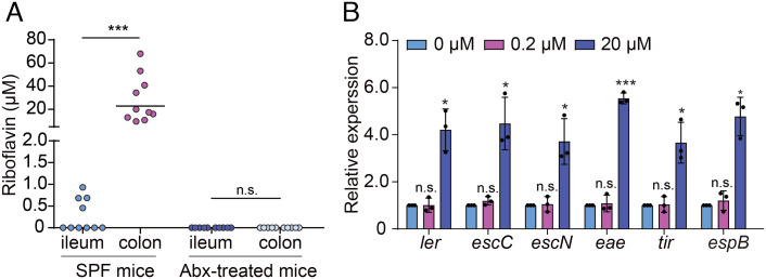
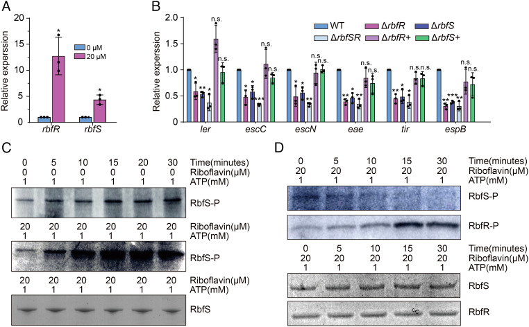
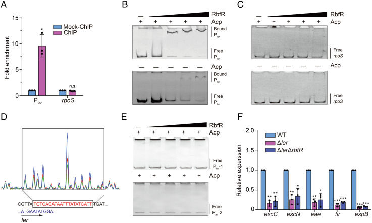
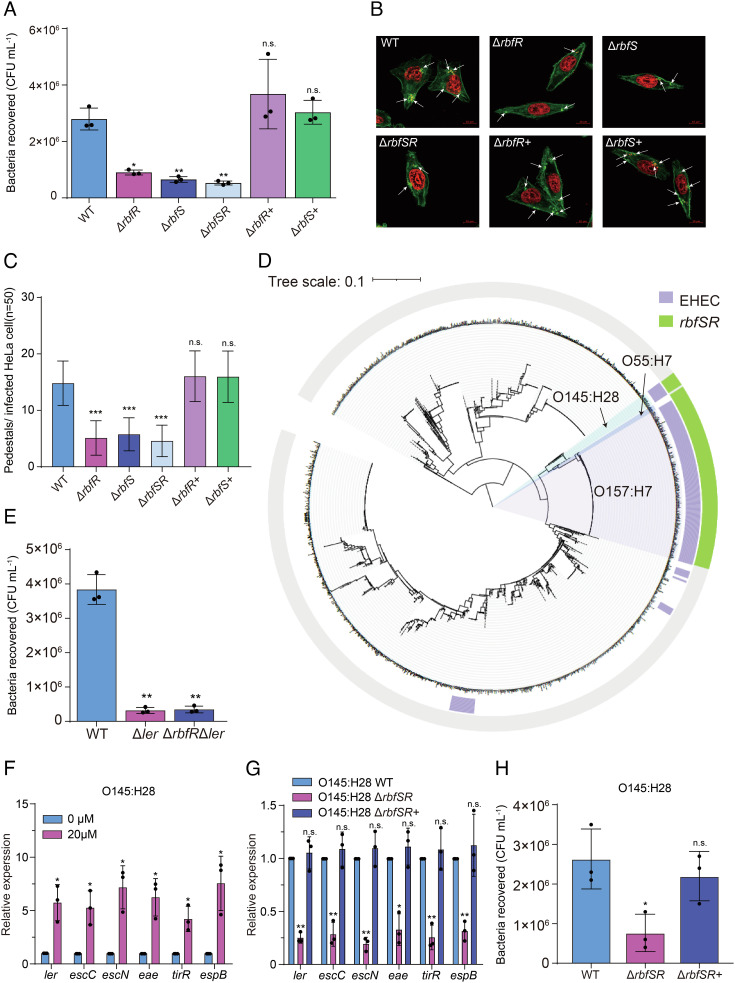
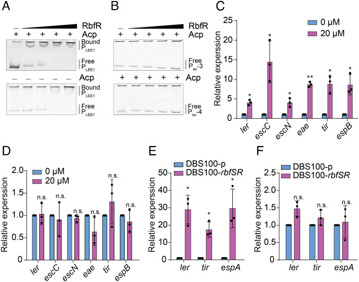
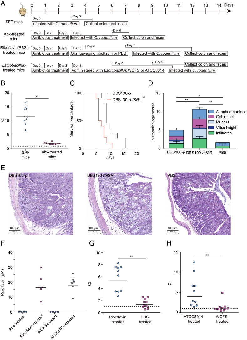
Riboflavin is produced by most commensal bacteria in the human colon, where enterohemorrhagic Escherichia coli (EHEC) colonizes and causes diseases. Sensing environmental signals to site-specifically express the type-III secretion system (T3SS), which injects effectors into host cells leading to intestinal colonization and disease, is key to the pathogenesis of EHEC. Here, we reveal that EHEC O157:H7, a dominant EHEC serotype frequently associated with severe diseases, acquired a previously uncharacterized two-component regulatory system rbfSR, which senses microbiota-produced riboflavin to directly activate the expression of LEE genes encoding the T3SS in the colon. rbfSR is present in O157:H7 and O145:H28 but absent from other EHEC serotypes. The binding site of RbfR through which it regulates LEE gene expression was identified and is conserved in all EHEC serotypes and Citrobacter rodentium, a surrogate for EHEC in mice. Introducing rbfSR into C. rodentium enabled bacteria to sense microbiota-produced riboflavin in the mouse colon to increase the expression of LEE genes, causing increased disease severity in mice. Phylogenic analysis showed that the O55:H7 ancestor of O157:H7 obtained rbfSR which has been kept in O157:H7 since then. Thus, acquiring rbfSR represents an essential step in the evolution of the highly pathogenic O157:H7. The expression of LEE genes and cell attachment ability of other EHEC serotypes in the presence of riboflavin significantly increased when rbfSR was introduced into them, indicating that those serotypes are ready to use RbfSR to increase their pathogenicity. This may present a potential public health issue as horizontal gene transfer is frequent in enteric bacteria.

Keywords: enterohaemorrhagic Escherichia coli; evolution; gut microbiota; two-component regulatory system; virulence regulation.


Conflict of interest statement

The authors declare no competing interest.

Figures

Fig. 1.
Fig. 1.
Riboflavin increases LEE gene expression in vitro. (A) HPLC measurement of riboflavin concentration in the ileum and colon contents of SPF or abx-treated mice (n = 10 mice per group). (B) qRT-PCR analysis of LEE gene expression in EDL933 grown in SCEM medium containing 0.2 or 20 μM riboflavin. Data are represented the mean ± SD (n = 3). Significant differences were assessed by an unpaired t test. Error bars represent SD. *P < 0.05, **P < 0.01, ***P < 0.001; n.s. no significant difference.
Fig. 2.
Fig. 2.
Riboflavin increases LEE gene expression through RbfSR(Z5692/Z5684). (A) qRT-PCR analysis of rbfR(z5684) and rbfS(z5692) expression in WT grown in SCEM medium containing 0 or 20 μM riboflavin. Data are presented as the mean ± SD (n = 3). (B) qRT-PCR analysis of LEE gene expression in WT, ΔrbfRz5684rbfSz5692rbfSRz5684Δz5692rbfR+z5684+), and ΔrbfS+z5692+) grown in SCEM medium containing 20 μM riboflavin. Data are presented as the mean ± SD (n = 3). Significant differences were assessed by an unpaired t test. Error bars represent SD. *P < 0.05, **P < 0.01, ***P < 0.001; n.s. no significant difference. (C) Autophosphorylation of RbfS(Z5692). Coomassie gel of RbfS(Z5692) served as loading control. (D) Phosphotransfer from RbfS(Z5692) to RbfR(Z5684) (HK: RR=1:10). Coomassie gel of RbfS(Z5692) and RbfR(Z5684) served as loading control.
Fig. 3.
Fig. 3.
RbfR specifically binds to the promoter of ler. (A) Fold enrichment of the promoter region of ler (Pler) and coding region of rpoS in RbfR-ChIP samples, as measured by qRT-PCR. rpoS served as negative control. Data are presented as the mean ± SD (n = 3). (B and C) EMSA of the binding of purified RbfR to Pler(b) and rpoS(c) with or without acetyl phosphate (Acp). (D) RbfR binds to a motif in Pler. The protected region shows a significantly reduced peak intensities (green) pattern compared with those of the control (blue and red). The identified RbfR-binding motif is shown in a box at the Bottom of the figure. (E) EMSA of the binding of purified RbfR to Pler-1 (the binding motif was deleted) and Pler-2(the binding motif was mutated to 5- CTCTGTGCGGCCCGCGCTGCC-3) of EDL933 with or without Acp. (F) qRT-PCR analysis of LEE gene expression in WT, Δler, and ΔlerΔrbfR grown in SCEM medium containing 20 μM riboflavin. Data are presented as the mean ± SD (n = 3). Significant differences were assessed by an unpaired t test. Error bars represent SD. *P < 0.05, **P < 0.01, ***P < 0.001; n.s. no significant difference.
Fig. 4.
Fig. 4.
Expression of rbfSR increases the cell attachment ability of EHEC. (A) Adherence of WT, ΔrbfR, ΔrbfS, ΔrbfRΔrbfS, ΔrbfR+, and ΔrbfS+ to HeLa cells. Data are presented as the mean ± SD (n = 3). (B) Detection of AE lesion formation by WT, ΔrbfR, ΔrbfS, ΔrbfRΔrbfS, ΔrbfR+, and ΔrbfS+ by FAS in HeLa cells at 3 h post infection. The HeLa cell actin cytoskeleton (green) and nuclei of bacterial and HeLa cells (red) are shown. (C) FAS assay quantification of the number of pedestals per infected cell (n = 50). (D) Phylogenetic analysis of 2,134 publicly available E. coli complete genomes. Purple semicircle on the inner ring indicates the EHEC strains. The presence of rbfSR is labeled with a green semicircle on the outer ring. (E) Adherence of WT, Δler, and ΔrbfRΔler to HeLa cells. Data are presented as the mean ± SD (n = 3). (F) qRT-PCR analysis of LEE gene expression in EHEC O145:H28 grown in SCEM medium containing 0–20 μM riboflavin. Data are presented as the mean ± SD (n = 3). (G) qRT-PCR analysis of LEE gene expression in O145:H28 WT, O145:H28 ΔrbfSR, and O145:H28 ΔrbfSR+ grown in an SCEM medium containing 20 μM riboflavin. Data are presented as the mean ± SD (n = 3). (H) Adherence of O145:H28 WT, O145:H28 ΔrbfSR, and O145:H28 ΔrbfSR+ to HeLa cells. Data are presented as the mean ± SD (n = 3). Significant differences were assessed by an unpaired t test. Error bars represent SD. *P < 0.05, **P < 0.01, ***P < 0.001; n.s. no significant difference.
Fig. 5.
Fig. 5.
Expression of rbfSR in DBS100 increases LEE gene expression in vitro and in vivo. (A and B) EMSA of the binding of purified RbfR to Pler (a), Pler-3 (b, the binding motif was deleted), and Pler-4(b, the binding motif was mutated to 5-CTCCGTGCGGCCCGCGGTGCC-3) of DBS100 with or without Acp. (C and D) qRT-PCR analysis of LEE gene expression in DBS100-rbfSR(c) and DBS100-p(d) grown in SCEM medium containing 20 μM riboflavin. Data are presented as the mean ± SD (n = 3). (E and F) qRT-PCR analysis of LEE gene expression in DBS100-p and DBS100-rbfSR in SPF mice(E) and abx-treated mice(F). Data are presented as the mean ± SD (n = 3). Significant differences were assessed by an unpaired t test. Error bars represent SD. *P < 0.05, **P < 0.01, ***P < 0.001; n.s. no significant difference.
Fig. 6.
Fig. 6.
Riboflavin produced by microbiota increases C. rodentium intestinal colonization through RbfSR. (A) Schematic of C. rodentium colonization in different infection models. (B) Competition assay comparing the intestinal colonization ability of DBS100-p and DBS100-rbfSR in SPF mice and abx-treated mice (n = 10 mice per group). (C) Survival curve of mice infected with DBS100-p or DBS100-rbfSR (n = 10 mice per group). (D and E) Histological score (D) and representation (E) of colon 3 d after DBS100-p and DBS100-rbfSR infection (n = 3). (F) HPLC measurement of riboflavin concentrations in the colon contents of abx-treated miceabx-treated mice fed with 20 ug riboflavin (Riboflavin-treated), and abx-treated mice administered with WCFS (WCFS-treated) or ATCC8014 (ATCC8014-treated) (n = 6 mice per group). (G) Competition assay comparing the intestinal colonization ability of DBS100-p and DBS100-rbfSR in abx-treated mice fed with 20 ug riboflavin (Riboflavin-treated) or PBS(PBS-treated) (n = 10 mice per group). (H) Competition assay comparing the intestinal colonization ability of DBS100-p and DBS100-rbfSR in abx-treated mice administered with WCFS (WCFS-treated) or ATCC8014 (ATCC8014-treated mice) (n = 10 mice per group). Significant differences were assessed by log-rank (Mantel-Cox) test (C), unpaired t test (D), and Mann–Whitney U test (B, G, and H). Each symbol represents an individual mouse. Error bars represent SD. *P < 0.05, **P < 0.01, ***P < 0.001; n.s. no significant difference.

Similar articles

Cited by

References

    1. Kaper J. B., Nataro J. P., Mobley H. L., Pathogenic Escherichia coli. Nat. Rev. Microbiol. 2, 123–140 (2004). - PubMed
    1. Vallance B. A., Chan C., Robertson M. L., Finlay B. B., Enteropathogenic and enterohemorrhagic Escherichia coli infections: Emerging themes in pathogenesis and prevention. Can. J. Gastroenterol. 16, 771–778 (2002). - PubMed
    1. Jubelin G., et al. , Modulation of Enterohaemorrhagic Escherichia coli Survival and Virulence in the Human Gastrointestinal Tract. Microorganisms 6, 115 (2018). - PMC - PubMed
    1. Cameron E. A., Sperandio V., Frenemies: Signaling and nutritional integration in pathogen-microbiota-host interactions. Cell Host Microbe. 18, 275–284 (2015). - PMC - PubMed
    1. Pacheco A. R., et al. , Fucose sensing regulates bacterial intestinal colonization. Nature 492, 113–117 (2012). - PMC - PubMed

Publication types

LinkOut - more resources