

Wasteosomes (corpora amylacea) as a hallmark of chronic glymphatic insufficiency
- PMID: 36409907
- PMCID: PMC9860256
- DOI: 10.1073/pnas.2211326119
Wasteosomes (corpora amylacea) as a hallmark of chronic glymphatic insufficiency
Abstract
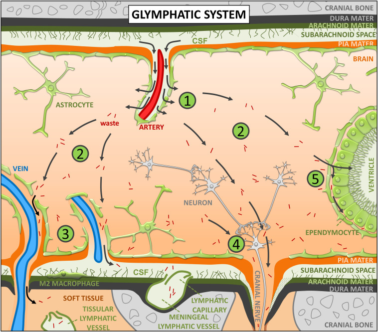
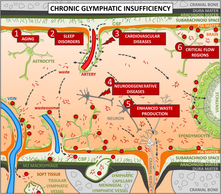
In different organs and tissues, the lymphatic system serves as a drainage system for interstitial fluid and is useful for removing substances that would otherwise accumulate in the interstitium. In the brain, which lacks lymphatic circulation, the drainage and cleaning function is performed by the glymphatic system, called so for its dependence on glial cells and its similar function to that of the lymphatic system. In the present article, we define glymphatic insufficiency as the inability of the glymphatic system to properly perform the brain cleaning function. Furthermore, we propose that corpora amylacea or wasteosomes, which are protective structures that act as waste containers and accumulate waste products, are, in fact, a manifestation of chronic glymphatic insufficiency. Assuming this premise, we provide an explanation that coherently links the formation, distribution, structure, and function of these bodies in the human brain. Moreover, we open up new perspectives in the study of the glymphatic system since wasteosomes can provide information about which variables have the greatest impact on the glymphatic system and which diseases occur with chronic glymphatic insufficiency. For example, based on the presence of wasteosomes, it seems that aging, sleep disorders, and cerebrovascular pathologies have the highest impact on the glymphatic system, whereas neurodegenerative diseases have a more limited impact. Furthermore, as glymphatic insufficiency is a risk factor for neurodegenerative diseases, information provided by wasteosomes could help to define the strategies and actions that can prevent glymphatic disruptions, thus limiting the risk of developing neurodegenerative diseases.
Keywords: brain; corpora amylacea; glymphatic system; wasteosomes.
Conflict of interest statement
The authors declare no competing interest.
Figures
Similar articles
-
Analyzing the Virchow pioneering report on brain corpora amylacea: shedding light on recurrent controversies.Brain Struct Funct. 2023 Jul;228(6):1371-1378. doi: 10.1007/s00429-023-02664-5. Epub 2023 Jun 26. Brain Struct Funct. 2023. PMID: 37358661 Free PMC article. Review.
-
Uncovering tau in wasteosomes (corpora amylacea) of Alzheimer's disease patients.Front Aging Neurosci. 2023 Mar 30;15:1110425. doi: 10.3389/fnagi.2023.1110425. eCollection 2023. Front Aging Neurosci. 2023. PMID: 37065464 Free PMC article.
-
Corpora amylacea act as containers that remove waste products from the brain.Proc Natl Acad Sci U S A. 2019 Dec 17;116(51):26038-26048. doi: 10.1073/pnas.1913741116. Epub 2019 Dec 3. Proc Natl Acad Sci U S A. 2019. PMID: 31796594 Free PMC article.
-
Wasteosomes (corpora amylacea) of human brain can be phagocytosed and digested by macrophages.Cell Biosci. 2022 Oct 28;12(1):177. doi: 10.1186/s13578-022-00915-2. Cell Biosci. 2022. PMID: 36307854 Free PMC article.
-
From corpora amylacea to wasteosomes: History and perspectives.Ageing Res Rev. 2021 Dec;72:101484. doi: 10.1016/j.arr.2021.101484. Epub 2021 Oct 9. Ageing Res Rev. 2021. PMID: 34634491 Review.
Cited by
-
Hallmarks of aging: middle-aging hypovascularity, tissue perfusion and nitric oxide perspective on healthspan.Front Aging. 2025 Jan 7;5:1526230. doi: 10.3389/fragi.2024.1526230. eCollection 2024. Front Aging. 2025. PMID: 39839443 Free PMC article.
-
Temporopolar blurring signifies abnormalities of white matter in mesial temporal lobe epilepsy.Ann Clin Transl Neurol. 2024 Nov;11(11):2932-2945. doi: 10.1002/acn3.52204. Epub 2024 Sep 28. Ann Clin Transl Neurol. 2024. PMID: 39342438 Free PMC article.
-
Distribution of corpora amylacea in the human midbrain: using synchrotron radiation phase-contrast microtomography, high-field magnetic resonance imaging, and histology.Front Neurosci. 2023 Oct 4;17:1236876. doi: 10.3389/fnins.2023.1236876. eCollection 2023. Front Neurosci. 2023. PMID: 37869518 Free PMC article.
-
New Pathways Identify Novel Drug Targets for the Prevention and Treatment of Alzheimer's Disease.Int J Mol Sci. 2023 Mar 11;24(6):5383. doi: 10.3390/ijms24065383. Int J Mol Sci. 2023. PMID: 36982456 Free PMC article. Review.
-
Corpora amylacea negatively correlate with hippocampal tau pathology in Alzheimer's disease.Front Neurosci. 2024 Feb 29;18:1286924. doi: 10.3389/fnins.2024.1286924. eCollection 2024. Front Neurosci. 2024. PMID: 38486969 Free PMC article.
References
Publication types
MeSH terms
LinkOut - more resources
Full Text Sources
Medical
