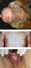
Approach to Investigation of Hyperandrogenism in a Postmenopausal Woman
- PMID: 36409990
- PMCID: PMC10099172
- DOI: 10.1210/clinem/dgac673
Approach to Investigation of Hyperandrogenism in a Postmenopausal Woman
Abstract
Postmenopausal hyperandrogenism is a condition caused by relative or absolute androgen excess originating from the ovaries and/or the adrenal glands. Hirsutism, in other words, increased terminal hair growth in androgen-dependent areas of the body, is considered the most effective measure of hyperandrogenism in women. Other symptoms can be acne and androgenic alopecia or the development of virilization, including clitoromegaly. Postmenopausal hyperandrogenism may also be associated with metabolic disorders such as abdominal obesity, insulin resistance, and type 2 diabetes. Mild hyperandrogenic symptoms can be due to relative androgen excess associated with menopausal transition or polycystic ovary syndrome, which is likely the most common cause of postmenopausal hyperandrogenism. Virilizing symptoms, on the other hand, can be caused by ovarian hyperthecosis or an androgen-producing ovarian or adrenal tumor that could be malignant. Determination of serum testosterone, preferably by tandem mass spectrometry, is the first step in the endocrine evaluation, providing important information on the degree of androgen excess. Testosterone >5 nmol/L is associated with virilization and requires prompt investigation to rule out an androgen-producing tumor in the first instance. To localize the source of androgen excess, imaging techniques are used, such as transvaginal ultrasound or magnetic resonance imaging (MRI) for the ovaries and computed tomography and MRI for the adrenals. Bilateral oophorectomy or surgical removal of an adrenal tumor is the main curative treatment and will ultimately lead to a histopathological diagnosis. Mild to moderate symptoms of androgen excess are treated with antiandrogen therapy or specific endocrine therapy depending on diagnosis. This review summarizes the most relevant causes of hyperandrogenism in postmenopausal women and suggests principles for clinical investigation and treatment.
Keywords: androgen-producing tumor; hirsutism; hyperandrogenism; ovarian hyperthecosis; postmenopausal women; virilization.
© The Author(s) 2022. Published by Oxford University Press on behalf of the Endocrine Society.
Figures


References
-
- Ludwig E. Classification of the types of androgenetic alopecia (common baldness) occurring in the female sex. Br J Dermatol. 1977;97(3):247‐254. - PubMed
-
- Davison SL, Bell R, Donath S, Montalto JG, Davis SR. Androgen levels in adult females: changes with age, menopause, and oophorectomy. J Clin Endocrinol Metab. 2005;90(7):3847‐3853. - PubMed
-
- Longcope C. Adrenal and gonadal androgen secretion in normal females. Clin Endocrinol Metab. 1986;15(2):213‐228. - PubMed
-
- Labrie F, Martel C, Belanger A, Pelletier G. Androgens in women are essentially made from DHEA in each peripheral tissue according to intracrinology. J Steroid Biochem Mol Biol. 2017;168(April):9‐18. - PubMed
Publication types
MeSH terms
Substances
LinkOut - more resources
Full Text Sources
Medical

