. 2023 Jan 1;324(1):L64-L75.
doi: 10.1152/ajplung.00116.2022. Epub 2022 Nov 21.

Activation of TREK-1 (K2P2.1) potassium channels protects against influenza A-induced lung injury

Affiliations

Activation of TREK-1 (K2P2.1) potassium channels protects against influenza A-induced lung injury

Tatiana Zyrianova et al. Am J Physiol Lung Cell Mol Physiol. .

Abstract

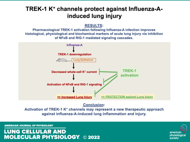
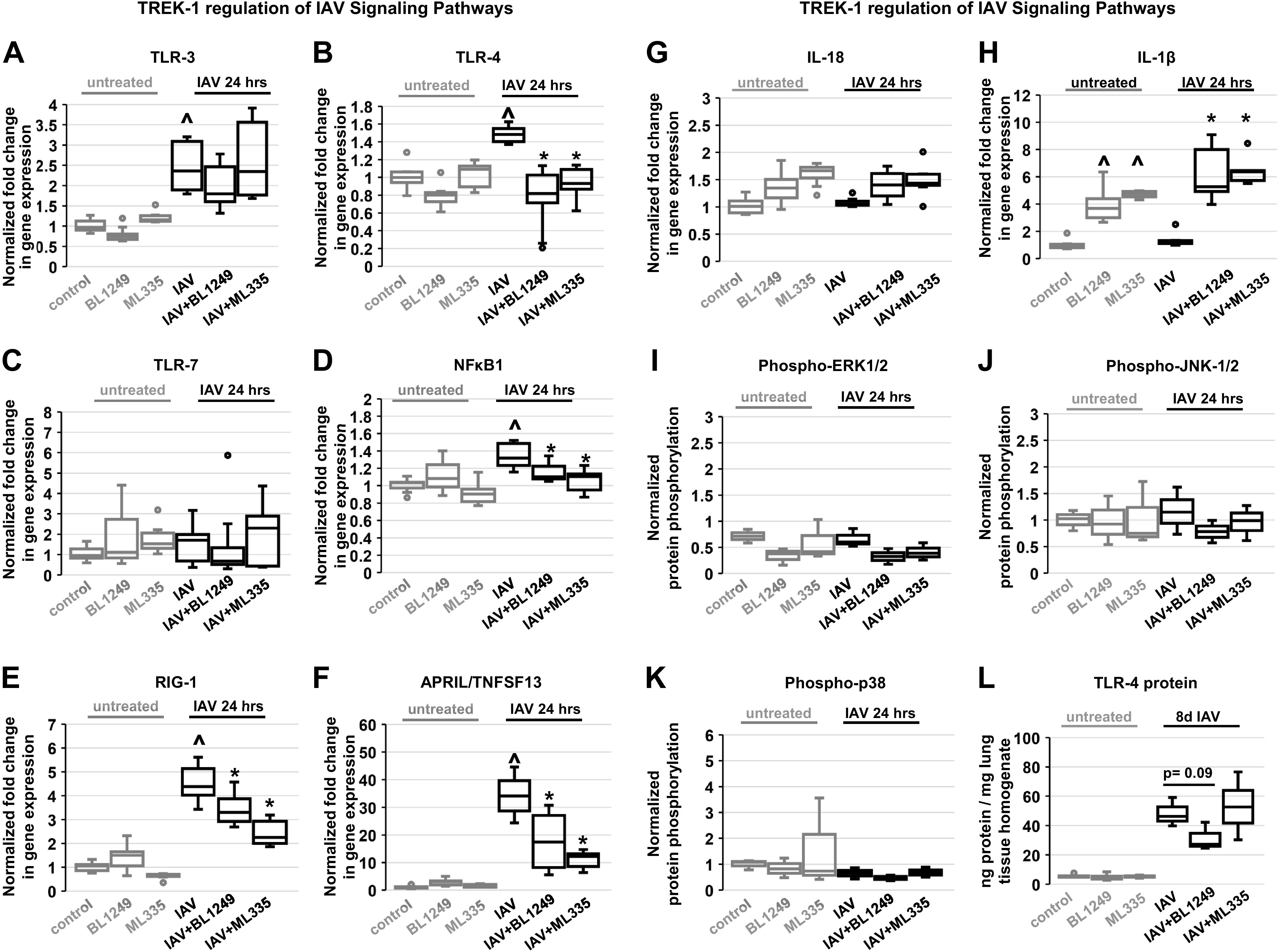
Influenza-A virus (IAV) infects yearly an estimated one billion people worldwide, resulting in 300,000-650,000 deaths. Preventive vaccination programs and antiviral medications represent the mainstay of therapy, but with unacceptably high morbidity and mortality rates, new targeted therapeutic approaches are urgently needed. Since inflammatory processes are commonly associated with measurable changes in the cell membrane potential (Em), we investigated whether Em hyperpolarization via TREK-1 (K2P2.1) K+ channel activation can protect against influenza-A virus (IAV)-induced pneumonia. We infected mice with IAV, which after 5 days caused 10-15% weight loss and a decrease in spontaneous activity, representing a clinically relevant infection. We then started a 3-day intratracheal treatment course with the novel TREK-1 activating compounds BL1249 or ML335. We confirmed TREK-1 activation with both compounds in untreated and IAV-infected primary human alveolar epithelial cells (HAECs) using high-throughput fluorescent imaging plate reader (FLIPR) assays. In mice, TREK-1 activation with BL1249 and ML335 counteracted IAV-induced histological lung injury and decrease in lung compliance and improved BAL fluid total protein levels, cell counts, and inflammatory IL-6, IP-10/CXCL-10, MIP-1α, and TNF-α levels. To determine whether these anti-inflammatory effects were mediated by activation of alveolar epithelial TREK-1 channels, we studied the effects of BL1249 and ML335 in IAV-infected HAEC, and found that TREK-1 activation decreased IAV-induced inflammatory IL-6, IP-10/CXCL10, and CCL-2 secretion. Dissection of TREK-1 downstream signaling pathways and construction of protein-protein interaction (PPI) networks revealed NF-κB1 and retinoic acid-inducible gene-1 (RIG-1) cascades as the most likely targets for TREK-1 protection. Therefore, TREK-1 activation may represent a novel therapeutic approach against IAV-induced lung injury.

Keywords: TREK-1 (KCNK2) ion channels; acute lung injury; cytokines; inflammation; influenza virus.


Conflict of interest statement

No conflicts of interest, financial or otherwise, are declared by the authors.

Figures

None
Graphical abstract
Figure 1.
Figure 1.
TREK-1 activation protects mice against influenza-A virus (IAV)-induced acute lung injury: representative hematoxylin-eosin (H&E) pictures of mouse lung sections (×10 magnification) are shown in (A), and composite histological lung injury scores (LIS) are depicted in (B). LIS were determined by an investigator blinded to the experimental conditions on H&E-stained lung sections using a 3-criteria LIS (22): 1) interstitial edema, 2) inflammatory cell infiltrate, and 3) parenchymal, peribronchial, and perivascular hemorrhage. Each of the three criteria was assigned a score between 0 and 3, with “0” representing no injury, “1” representing mild injury, “2” representing moderate injury, and “3” representing severe injury. Four random high-power fields per slide were scored under ×10 and ×20 magnifications and averaged for each criterion. Similar protective effects of TREK-1 activation were observed on lung compliance (C), bronchoalveolar lavage (BAL) total cell counts (D), and BAL total protein concentrations (E). BAL albumin (F) and IgM levels (G) were increased after IAV infection but not affected by TREK-1 activation. Intracellular reactive oxygen species (ROS) production by BAL cells was not affected by IAV infection or TREK-1 activation (H; representative 3-h time point is shown). After 5 days, acute IAV infection caused a 10–15% body weight loss (I), representative of a clinically relevant acute IAV infection. Additional weight loss occurred by day 8, which improved with BL1249 treatment. TREK-1 activation did not affect IAV viral copy numbers in mouse lung tissue after 8 days of infection as quantified by qPCR (J). All data are represented as Box-Whisker plots (medians, 1st and 3rd quartiles, maximum and minimum values; outliers > 1.5 times the interquartile range are shown as circles) and compared with unpaired Student t test, or ANOVA/Tukey–Kramer tests; n (number of mice) = 5–12, lowest n = 5 in BL1249 and ML335 alone groups; ^compared with noninfected mice on day 8, *compared with IAV-infected mice on day 8; #compared with untreated controls on day 0, $compared with IAV-infected mice on day 5; P ≤ 0.05.
Figure 2.
Figure 2.
TREK-1 activation regulates bronchoalveolar lavage (BAL) fluid cytokine concentrations in influenza-A virus (IAV)-infected mice: Once-daily intratracheal treatment of mice with BL1249 or ML335 6 h following IAV infection inhibits the IAV-induced increases in IL-6 (A), IP-10/CXCL-10 (B), MIP-1α (C), and TNF-α (D), but not CCL-2 (E), IL-10 (F), or IL-1β (G). Data are represented as Box-Whisker plots (medians, 1st and 3rd quartiles, maximum and minimum values; outliers > 1.5 times the interquartile range are shown as circles) and compared with ANOVA/Tukey–Kramer tests; n = 4–10, lowest n = 4 in BL1249 and ML335 alone groups; ^compared with noninfected mice on day 8, *compared with IAV-infected mice on day 8; P ≤ 0.05.
Figure 3.
Figure 3.
TREK-1 activation regulates cytokine secretion from influenza-A virus (IAV)-infected primary human alveolar epithelial cells (HAECs): acute IAV infection increases IL-6 (A), IP-10/CXCL-10 (B), and CCL-2 (C) secretion from HAEC after 24 h, but not MIP-1α (D), TNF-α (E), and IL-10 (F). Treatment of HAEC with BL1249 and ML335 6 h after IAV infection counteracts the IAV-induced effects on IL-6, IP-10/CXCL-10, and CLL-2, but has no effect on MIP-1α, TNF-α, and IL-10 levels. Data are represented as Box-Whisker plots (medians, 1st and 3rd quartiles, maximum and minimum values, and outliers > 1.5 times the interquartile range are shown as circles) and compared with ANOVA/Tukey–Kramer tests; n = 4–6, lowest n = 4 in BL1249 and ML335 alone groups; ^compared with noninfected cells after 24 h, *compared with IAV-infected cells after 24 h; P ≤ 0.05.
Figure 4.
Figure 4.
TREK-1 currents in primary human alveolar epithelial cells (HAECs): BL1249 activates TREK-1 currents in untreated HAEC, and counteracts the IAV-induced decrease in overall whole cell K+ currents. Representative curves are shown in (AC) (also see Inset C1), and a summary of n = 4–5 independent fluorescent imaging plate reader (FLIPR) curves is depicted in (D) (analyzed at the 60 min time point). E: TREK-1 gene expression in influenza-A virus (IAV)-infected mouse lungs (8 days) and primary mouse AT2 cells (24 h) by real-time qPCR. F: TREK-1 activation with BL1249 or ML335 does not affect viral IAV copy numbers in HAEC as measured by qPCR. DF: depicted as Box-Whisker plots (medians, 1st and 3rd quartiles, maximum and minimum values, and outliers > 1.5 times the interquartile range are shown as circles); n = 4–6, lowest n = 4 in BL1249 and ML335 alone groups; ^compared with noninfected mouse lung tissue or noninfected cells, P ≤ 0.05; unpaired Student t test was used for A, B, and E; ANOVA/Tukey–Kramer for C and D. *IAV-infected HAEC vs. IAV+ML335 treated HAEC.
Figure 5.
Figure 5.
Regulation of influenza-A virus (IAV)-induced signaling cascades by TREK-1 activation: data show gene expression levels of well-established IAV signaling pathways [Toll-like receptor (TLR)-3, TLR-4, TLR-7, NF-κB1, retinoic acid-inducible gene-1 (RIG-1), APRIL/TNFSF13, IL-18, and IL-1β explored by real-time qPCR after normalization to GAPDH (AH), MAPK protein phosphorylation by Mesoscale technology (IK), and TLR-4 protein levels in lung tissue by ELISA (L). Graphs depict Box-Whisker plots (medians, 1st and 3rd quartiles, maximum and minimum values; outliers > 1.5 times the interquartile range are shown as circles) and compared with ANOVA/Tukey–Kramer tests, n = 9 (number of independent replicates); ^compared with noninfected cells after 24 h, *compared with IAV-infected cells after 24 h; P ≤ 0.05.
Figure 6.
Figure 6.
Protein-protein interaction (PPI) network analysis (Cytoscape StringApp) reveals that the TREK-1 mediated decrease in IL-6 and IP-10/CXCL-10 secretion occurs primarily via inhibition of retinoic acid-inducible gene-1 (RIG-1) and NF-κB1 pathways, with a lower likelihood of TNFSF13 and Toll-like receptor (TLR)-4 involvement: a putative graphic model was created by inputting three experimentally validated data sets as query proteins into the model (purple boxes): 1) TREK-1 (KCNK2), 2) the TREK-1-regulated cytokines: IL-6 and IP-10/CXCL-10, which are altered in both bronchoalveolar lavage (BAL) fluid and human alveolar epithelial cells (HAECs) (Figs. 2 and 3), and 3) the proposed TREK-1-regulated signaling molecules: NF-κB1, RIG-1, TLR-4, and APRIL/TNFSF13 (Fig. 5). Model analysis revealed five additional predicted downstream interaction partners (blue boxes) with a “high” confidence level (0.7, scale: 0–1) and specific to the lung (tissue filter set to “Lung”). Proteins are arranged according to their respective cellular location: extracellular space, plasma membrane, cytoplasm, and nucleus. Red dashed arrows were added to indicate the experimentally validated primary TREK-1 targets.

Similar articles

Cited by

References

    1. Mertz D, Kim TH, Johnstone J, Lam P-P, Science M, Kuster SP, Fadel SA, Tran D, Fernandez E, Bhatnagar N, Loeb M. Populations at risk for severe or complicated influenza illness: systematic review and meta-analysis. BMJ 347: f5061, 2013. doi:10.1136/bmj.f5061. - DOI - PMC - PubMed
    1. Kalil AC, Thomas PG. Influenza virus-related critical illness: pathophysiology and epidemiology. Crit Care 23: 258, 2019. doi:10.1186/s13054-019-2539-x. - DOI - PMC - PubMed
    1. Lobo SM, Watanabe ASA, Salomão MLM, Queiroz F, Gandolfi JV, de Oliveira NE, Covello LHS, Sacillotto GH, de Godoy LG, Simões ES, Frini ICM, Da Silva Teixeira RER, Furlan NP, Dutra KR, Nogueira ML. Excess mortality is associated with influenza A (H1N1) in patients with severe acute respiratory illness. J Clin Virol 116: 62–68, 2019. doi:10.1016/j.jcv.2019.05.003. - DOI - PubMed
    1. Michael P, Brabant D, Bleiblo F, Ramana CV, Rutherford M, Khurana S, Tai TC, Kumar A, Kumar A. Influenza A induced cellular signal transduction pathways. J Thorac Dis 5, Suppl 2: S132–S141, 2013. doi:10.3978/j.issn.2072-1439.2013.07.42. - DOI - PMC - PubMed
    1. Jefferson T, Jones M, Doshi P, Spencer EA, Onakpoya I, Heneghan CJ. Oseltamivir for influenza in adults and children: systematic review of clinical study reports and summary of regulatory comments. BMJ 348: g2545, 2014. doi:10.1136/bmj.g2545. - DOI - PMC - PubMed

Publication types

MeSH terms

Substances