Review
. 2022 Dec:291:121903.
doi: 10.1016/j.biomaterials.2022.121903. Epub 2022 Nov 9.

Topography-mediated immunomodulation in osseointegration; Ally or Enemy

Affiliations
Review

Topography-mediated immunomodulation in osseointegration; Ally or Enemy

Sajjad Shirazi et al. Biomaterials. 2022 Dec.

Abstract

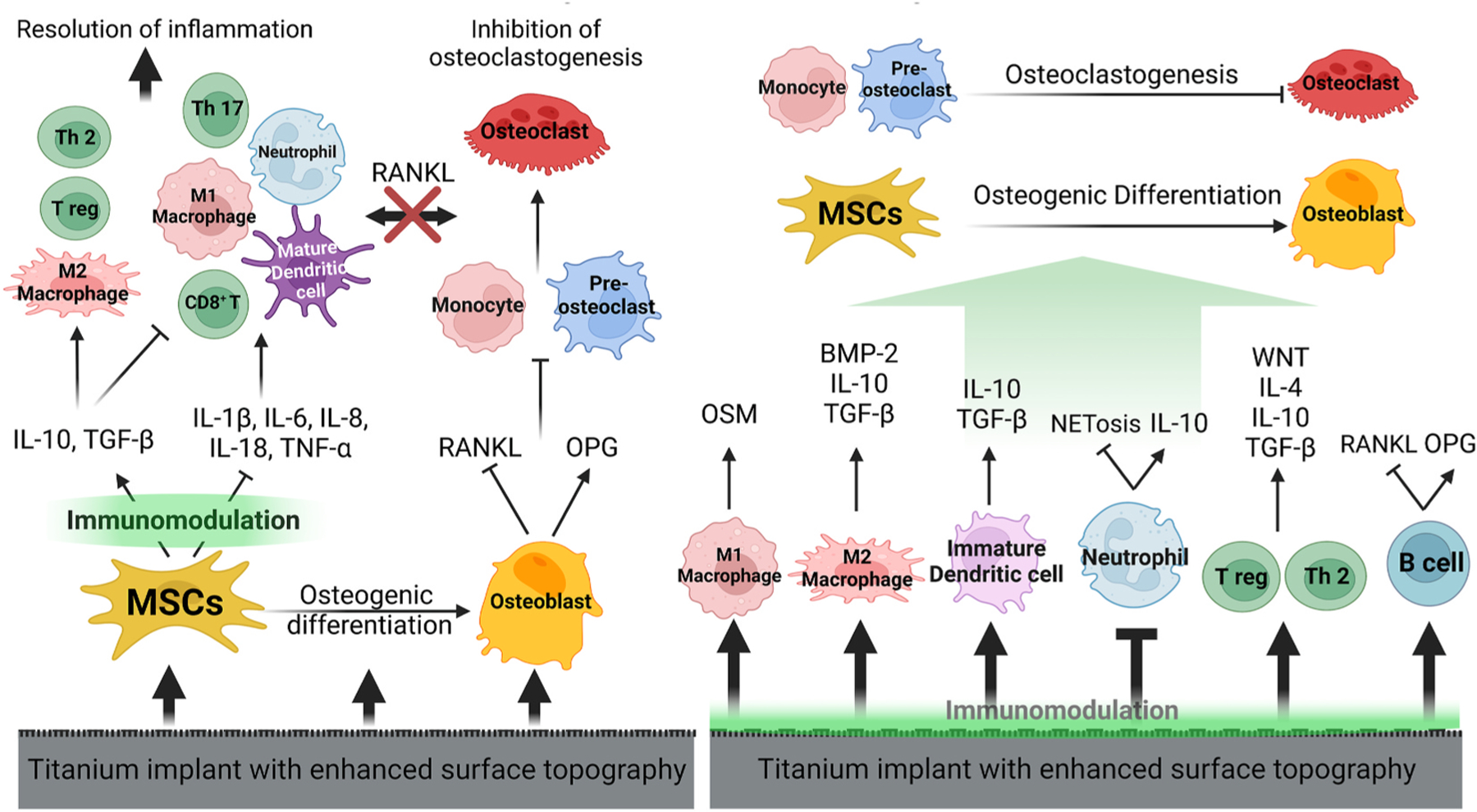
Osteoimmunology is at full display during endosseous implant osseointegration. Bone formation, maintenance and resorption at the implant surface is a result of bidirectional and dynamic reciprocal communication between the bone and immune cells that extends beyond the well-defined osteoblast-osteoclast signaling. Implant surface topography informs adherent progenitor and immune cell function and their cross-talk to modulate the process of bone accrual. Integrating titanium surface engineering with the principles of immunology is utilized to harness the power of immune system to improve osseointegration in healthy and diseased microenvironments. This review summarizes current information regarding immune cell-titanium implant surface interactions and places these events in the context of surface-mediated immunomodulation and bone regeneration. A mechanistic approach is directed in demonstrating the central role of osteoimmunology in the process of osseointegration and exploring how regulation of immune cell function at the implant-bone interface may be used in future control of clinical therapies. The process of peri-implant bone loss is also informed by immunomodulation at the implant surface. How surface topography is exploited to prevent osteoclastogenesis is considered herein with respect to peri-implant inflammation, osteoclastic precursor-surface interactions, and the upstream/downstream effects of surface topography on immune and progenitor cell function.

Keywords: BMP-2; Immunoengineering; Implant surface topography; Macrophage; Nano; Oncostatin M; Roughness.


Conflict of interest statement

Declaration of competing interest The authors declare that they have no known competing financial interests or personal relationships that could have appeared to influence the work reported in this paper.

Figures

Fig. 1.
Fig. 1.
Conceptual evolution of osseointegration. Historically, titanium was considered bioinert and osseointegration was viewed as an unimpeded bone formation. The principles advanced for the clinical success of osseointegration were based on the concept that proper bone surgery enabled subsequent formation of bone from the surgical margins toward the implant surface. The attribution of active interactions of the implant surface with adherent cells began with the concept of contact osteogenesis where enhanced surface topography guided the osteoprogenitor cell and their osteogenic functions to the implant surface. Bone formation occurred both from the surface (contact osteogenesis) as well as toward the surface (distance osteogenesis) both accelerating formation of and expanding the bone to implant contact. Continued studies of osteoprogenitor/osteoblast interactions with the implant surface demonstrated that surface topographic modifications were able to increase the rate and extents of osteoblastic differentiation and osteogenesis in vitro and were able to increase the bone-to-implant contact at the implant surface in animal and human studies. The osteogenesis-promoting nature of the implant surface was firmly established by studies demonstrating surface-mediated increases in osteoinductive protein and genes expression. This paradigm has most recently shifted to include the concept that the implant surface (principally, but not exclusively, Titanium) is immunomodulatory and bioactive. Investigations clearly demonstrated the presence of other cell types adherent to the implant surface and promoted more recent investigations of implant surface – immune cell interactions. The understanding of osseointegration has evolved to include the role of immune cells in modulating osteogenesis and osteoclastogenesis to affect bone accrual at the endosseous implant interface.
Fig. 2.
Fig. 2.. Topography is a critical variable in titanium surface biomimicry and immunomodulation.
Biologically inspired endosseous implant surfaces mimic the topographical features of an osteoclast resorption pit. Demonstrated are scanning electron micrographs of an osteoclast-resorbed bone surface (right panel) and titanium surfaces that have been acid etched and sandblasted with large-grit corundum. The action of osteoclasts on the bone surface leaves osteoclast resorption pits with microscale, hybrid, and nanoscale textures as well as biochemical cues for MSCs and osteoprogenitor cell recruitment, attachment and differentiation. Current studies suggest that surface topography influences adherent osteoprogenitor cell, immune cell and osteoclastic cell function and their interactions. The physical properties associated with osteoclast resorption pits can be generated on titanium using a variety of techniques, enhancing osseointegration through adsorption of various proteins, recruitment and attachment of pro-healing macrophages, MSCs and osteoprogenitor cells; and osteoblast differentiation, subsequent bone formation and downstream remodeling. The figure for bone resorption pit is adopted by kind permission of Tim Arnett (t.arnett@ucl.ac.uk) & Javier Manzano, from UCL https://boneresearchsociety.org/resources/image/40/#top.
Fig. 3.
Fig. 3.. Signaling pathways activated by titanium surface topography to induce osteoblastic differentiation and decrease the release of inflammatory factors in MSCs.
Surface roughness induces the osteoblastic differentiation of MSCs through multiple pathways. Runx2 and Osterix are major hubs where all osteogenic pathways converge. Surface roughness reduces the release of inflammatory cytokines from MSCs/osteoblasts possibly by blocking NF-κB and MAPK pathways. The detailed description of these pathways are studied in Refs. [,,–108,125].
Fig. 4.
Fig. 4.. Immunomodulation in osseointegration; bidirectional regulation of adherent cell function.
The modulation of adherent/adjacent osteoprogenitor cells may represent indirect influence of surface topography that is mediated by adherent immune cells. The cross-talk between immune cells, bone cells and surface topography is discussed in detail in the following sections. The effect of surface topography is shown with arrows arising from the surface.
Fig. 5.
Fig. 5.
Immunomodulation of osteoblast differentiation. A multitude of immune-derived factors promote or hinder osteoblast differentiation and activity.
Fig. 6.
Fig. 6.. The possible role of neutrophils in immunomodulation of osseointegration.
Neutrophils may modulate the process of osseointegration by secreting various factors that affect macrophages and T cells activation. An enhanced implant surface topography (rough hydrophilic) decreases NETosis and the release of inflammatory factors from neutrophils and improves osseointegration via downstream signals (dark green arrows). Bone stromal cells secrete factors that regulate neutrophils. Factors secreted from MSCs are depicted in orange arrows and factors secreted from neutrophils are shown in light green arrows. (For interpretation of the references to colour in this figure legend, the reader is referred to the Web version of this article.)
Fig. 7.
Fig. 7.
The possible role of macrophages in immunomodulation of osseointegration. Macrophages secret various factors that affect osteoprogenitor and other implant adherent cells leading to the resolution of inflammation and improved osteoblastogenesis. The initial M1 macrophage response is independent of surface topography. An enhanced implant surface topography eliminates M1 macrophages and increases M2 macrophages to improve osseointegration via downstream signals (dark green arrows). Both M1 (via OSM) and M2 macrophages (via BMP-2, TGF-β etc.) promote osteoinduction and osteoblast function in bone repair. Bone stromal cells secrete factors that regulate macrophages. Factors secreted from MSCs and osteoblasts are depicted in orange arrows and factors secreted from neutrophils are shown in light green arrows. (For interpretation of the references to colour in this figure legend, the reader is referred to the Web version of this article.)
Fig. 8.
Fig. 8.. The possible role of T cells in immunomodulation of osseointegration.
T cells may play roles in the process of osseointegration by secreting various factors that affect osteoblastogenesis and osteoclastogenesis. An enhanced implant surface topography (rough hydrophilic) decreases Th1 cells and increases Th2 and Treg cells leading to the resolution of inflammation and osteoblastic differentiation of MSCs via downstream signals (dark green arrows). Bone stromal cells secrete factors that regulate T cells. Factors secreted from MSCs and osteoblasts are depicted in orange arrows and factors secreted from T cells are shown in light green arrows. (For interpretation of the references to colour in this figure legend, the reader is referred to the Web version of this article.)
Fig. 9.
Fig. 9.. The possible role of B cells in immunomodulation of osseointegration.
B cells release various factors that affect other immune and bone cells. B cells may participate in the process of osseointegration by secreting OPG and increasing osteoblastogenesis, and increasing M2 macrophage leading to the resolution of inflammation. B cells secrete RANKL during inflammation. The effect of surface topography on B cells is not clear. Factors released from MSCs are depicted in orange arrows and factors secreted from B cells are shown in light green arrows. (For interpretation of the references to colour in this figure legend, the reader is referred to the Web version of this article.)
Fig. 10.
Fig. 10.
Immunomodulation of osteoclastogenesis. Osteoclastogenesis is a complex process as illustrated by the many of immune-derived factors that promote or hinder osteoclast differentiation and activity.
Fig. 11.
Fig. 11.. Immunomodulation of osteoclastogenesis by implant surface topography.
The effect of implant surface topography (thick green arrows) leading to the inhibition of osteoclastogenesis via downstream signals is illustrated. Surface-mediated factors activate inflammasome in various bone and immune cells (osteoblasts, osteoclasts, monocytes, macrophages, neutrophils, and adaptive immune cells, such as T helper 17 cells) leading to the release of IL-1β and IL-18. Elevated IL-1β expression promotes osteoclastogenesis, decreases osteoblast activity, and enhances inflammation by creating a pro-inflammatory milieu in a context- and cell type-dependent manner. IL-1β modulates osteoclast differentiation and activity by direct effects on osteoclasts or by indirectly modulating the expression of RANKL by other cell types. Surface topography can directly activate inflammasome pathway in adherent cells. Topographic cues directly increase OPG and reduce RANKL in bone cells. Increasing OPG/RANKL ratio is central to direct and/or indirect surface mediated anti-osteoclastogenic effects. An enhanced surface topography decreases the activity of pro-inflammatory immune cells and the subsequent release of osteoclastogenic factors, while inducing the activity of pro-healing immune cells and the release of anti-osteoclastogenic factors. The direct effect of topography to reduce osteoclast differentiation has been demonstrated. (For interpretation of the references to colour in this figure legend, the reader is referred to the Web version of this article.)
Fig. 12.
Fig. 12.
Harnessing osteoimmunology and surface topography for modulation of osseointegration. An enhanced titanium surface topography modulates the series of events after implant placement towards the resolution of inflammation and increased osteogenesis. The arrows arising from the surface denote to topographical cues.

Similar articles

Cited by

References

    1. Brånemark PI, Osseointegration and its experimental background, J. Prosthet. Dent 50 (1983) 399–410, 10.1016/s0022-3913(83)80101-2. - DOI - PubMed
    1. Albrektsson T, Albrektsson B, Osseointegration of bone implants. A review of an alternative mode of fixation, Acta Orthop. Scand 58 (1987) 567–577, 10.3109/17453678709146401. - DOI - PubMed
    1. Cooper LF, Shirazi S, Osseointegration—the biological reality of successful dental implant therapy: a narrative review, Front. Oral Maxillof. Med (2022), 10.21037/fomm-21-77. - DOI
    1. Arron JR, Choi Y, Bone versus immune system, Nature 408 (2000) 535–536, 10.1038/35046196. - DOI - PubMed
    1. Fischer V, Haffner-Luntzer M, Interaction between bone and immune cells: implications for postmenopausal osteoporosis, Semin. Cell Dev. Biol (2021) S1084–S9521, 10.1016/j.semcdb.2021.05.014 (21)00123–3. - DOI - PubMed