. 2022 Nov 21;41(1):328.
doi: 10.1186/s13046-022-02533-9.

OTUD4-mediated GSDME deubiquitination enhances radiosensitivity in nasopharyngeal carcinoma by inducing pyroptosis

Affiliations

OTUD4-mediated GSDME deubiquitination enhances radiosensitivity in nasopharyngeal carcinoma by inducing pyroptosis

Muping Di et al. J Exp Clin Cancer Res. .

Abstract

Background: Radioresistance is the primary cause of nasopharyngeal carcinoma (NPC) treatment failure. Previous studies have focused on the deficits in cellular apoptosis as a mechanism for radioresistance; however, additional potential death modes involved in modulating radiosensitivity of NPC have not been explored.

Methods: Pyroptosis was assessed by phase-contrast imaging, LDH release assays, live cell imaging, and Western blotting. In vitro and in vivo assays were used to investigate the function of gasdermin E (GSDME) and ovarian tumor family deubiquitinase 4 (OTUD4). NPC tissues were analyzed using Western blotting, immunohistochemistry, and real-time PCR. The molecular mechanism was determined using immunoprecipitation assays and mass spectrometry.

Results: Live cell imaging revealed that 40-75% of irradiation-induced dead NPC cells were pyroptotic cells. Furthermore, irradiation-induced pyroptosis is triggered by GSDME, which are cleaved by activated caspase-3 in the intrinsic mitochondrial pathway. Additionally, GSDME was significantly downregulated in radioresistant NPC specimens. Low GSDME expression was a predictor of worse prognosis and conferred NPC radioresistance both in vitro and in vivo. Mechanistically, OTUD4 deubiquitinated and stabilized GSDME, enhancing radiosensitivity of NPC cells by promoting pyroptosis. Clinically, OTUD4 was significantly correlated with GSDME in NPC biopsies, and patients with low expression of both OTUD4 and GSDME suffered the worst radiotherapy response and survival.

Conclusions: GSDME-dependent pyroptosis is a critical determinant of radiosensitivity in NPC, and is modulated by OTUD4 via deubiquitinating and stabilizing GSDME. These findings reveal a promising novel direction to investigate radioresistance and suggest potential therapeutic targets for sensitizing NPC to radiotherapy.

Keywords: GSDME; Nasopharyngeal carcinoma; OTUD4; Pyroptosis; Radioresistance.


Conflict of interest statement

Authors declare that they have no competing interests.

Figures
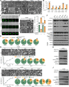
Fig. 1
Fig. 1
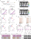
Radiotherapy induces GSDME-dependent pyroptosis in NPC cells through the intrinsic mitochondrial apoptotic pathway. A-B Six NPC cell lines were treated with or without 6 Gy irradiation. 72 h later, (A) cell morphological changes and (B) LDH release were measured. C Typical processes of cell pyroptosis, apoptosis and survival in HNE1 cells are shown. Brightfield and fluorescent images of stable EGFP-overexpressing NPC cells were taken at specific time-points post-6 Gy irradiation. D Percentage of pyroptotic, apoptotic, and viable cells in six NPC cells lines within 72 h after 6 Gy irradiation is indicated. E GSDME silencing inhibited pyroptosis (left), LDH release (upper right), and cell death (bottom right) in HONE1 cells at 72 h after 6 Gy irradiation. F Western blotting revealed the levels of indicated proteins in six NPC cell lines at 72 h after 6 Gy irradiation. Cyto C: cytochrome c. G Cell morphological changes (top), LDH release assay (bottom left), live cell imaging (bottom right), and (H) the levels of indicated proteins were assessed in HONE1 cell lines at the indicated time points after irradiation (6 Gy). (I) Cell morphological changes (top), LDH release assay (bottom left), live cell imaging (bottom right), and (J) the levels of indicated proteins were assessed in HONE1 cell lines at 72 h after the indicated irradiation dose. GAPDH was used to normalize the amount of protein loaded. All data are presented as the mean ± SD of three independent experiments. ns, No significant difference. ***P < 0.001, ****P < 0.0001
Fig. 2
Fig. 2
Upregulation of GSDME enhances pyroptosis and radiosensitivity in NPC cells in vitro. A The ratio of pyroptotic cells to dead cells in six NPC cell lines (HNE1, 5-8F, 6-10B, SUNE-1, HONE1, and HK1) within 72 h after 6 Gy irradiation, as calculated using live cell imaging data (left). Correlation analysis between the ratio of pyroptotic cells to dead cells and surviving fraction was performed (right). B Correlation analysis between relative GSDME-N levels and the surviving fraction in NPC cell lines within 72 h after 6 Gy irradiation was performed. C Western blotting showed GSDME expression in seven NPC cells and eight other cancer cells. MM: melanoma, EC: esophageal cancer, PCa: prostate cancer, BC: breast cancer. A375 cells were included as a positive control. D-M Stable GSDME-overexpressing SUNE1 cells (D, left upper) and stable GSDME-knockdown HK1 cells (E, left upper) were established. These cells were exposed to the indicated irradiation dose, and then phase-contrast cell imaging (D, E, right), LDH release assay (D, E, left lower), Western blotting analysis of GSDME-N (F, H), live cell imaging (G, I), Annexin/PI assay (J, K), and colony formation assay (L, M, left) were performed at designated time points. The survival curves of stable cell lines are indicated (L, M, right). GAPDH was used to determine the amount of loading proteins. All data are presented as the mean ± SD of three independent experiments. *P < 0.05, **P < 0.01, ***P < 0.001, ****P < 0.0001
Fig. 3
Fig. 3
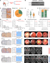
GSDME-dependent pyroptosis sensitizes NPC to radiotherapy in vivo. A Schematic view of the radiotherapy xenograft model. Cells stably expressing luciferase, SUNE1-Vector, SUNE1-GSDME, HONE1-Scramble, or HONE1-shGSDME, were subcutaneously injected into the nude mice (n = 28 mice per group). When tumor volumes reached 50–100 mm.3, mice bearing each type of tumor were randomized into a control group and a radiotherapy group (n = 14 in each subgroup). The mice in the radiotherapy group were irradiated with 12 Gy (2 Gy/day for 6 days). B Tumor size was monitored every 4 days (n = 5). C Representative bioluminescence images of tumors were taken on day 42. D Statistical analysis of photon flux (n = 5). E Images of resected tumors. F Tumor weight on day 42 (n = 5). G Serum LDH concentrations were determined pre-radiotherapy and following the third and sixth radiotherapy treatments (n = 3). H Representative images of HE (top) and IHC (bottom) staining of the tumor sections are shown. The dashed lines circumscribe the areas of tumor necrosis. I Western blotting showed the level of GSDME-N in resected tumor tissues. GAPDH was used to normalize the amount of protein loaded. Each bar represents the mean ± SD of three independent experiments. ns, No significant difference. *P < 0.05, **P < 0.01, ***P < 0.001, ****P < 0.0001
Fig. 4
Fig. 4
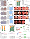
Low GSDME expression correlates with radioresistance and poor prognosis in NPC. A Schematic of specimens obtained from NPC via nasopharyngoscopy. B GSDME expression was analyzed by Western blotting in radiosensitive and radioresistant NPC tissues collected from 10 patients. GAPDH was used to normalize the amount of protein loaded. C Representative immunohistochemical images of GSDME low- and high- expression in NPC tissues (left and right, respectively). The magnified inset area is shown at the bottom. Scale bars represent 50 μm. D Percentage of GSDME low expression and high expression in NPC tissues (n = 150). Low GSDME expression, GSDME_L; High GSDME expression, GSDME_H. E The IHC score of GSDME in the radiosensitive and radioresistant tissues. F The correlation between GSDME expression and radiation therapy response. RS, radiosensitive; RR, radioresistant. G Association of GSDME expression with 5-year progression-free survival and 5-year locoregional recurrence-free survival was analyzed by Kaplan–Meier and Log Rank test. H Cox regression analysis of potential prognostic factors, including GSDME levels and other clinical characteristics. I Representative NPC cases received radiotherapy alone, showing the relationship between GSDME expression, serum LDH, and cancer regression. The yellow dashed lines indicate tumor in nasopharyngoscopy images. Scale bars represent 50 μm. RT, radiotherapy. J Representative NPC cases received chemoradiotherapy showing the relationship between GSDME expression, serum LDH, and cancer regression. The red lines indicate tumor tissue in magnetic resonance images. **P < 0.01, ***P < 0.001
Fig. 5
Fig. 5
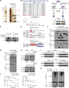
OTUD4 deubiquitinates and stabilizes GSDME. A Representative images of silver-stained gels of immunoprecipitation proteins using anti-IgG and anti-Flag affinity beads. B Upper panel: list of candidate proteins from tandem affinity purification-coupled mass spectrometry. Lower panel: MS profiles of OTUD4. C Upper panel: schematic of protein extraction from parental or radioresistant SUNE1 and HK1 cells. Lower panel: Western blotting showed OTUD4 protein expression in parental or radioresistant SUNE1 and HK1 cells. D Interaction of GSDME with OTUD4. Co-IP was performed using cell lysates obtained from HEK293T cells co-transfected with Flag-GSDME and Myc-OTUD4. E Upper panel: schematic of OTUD4 truncations. Lower panel: schematic of GSDME truncations. F HEK293T cells were transfected with Flag-GSDME and indicated Myc-OTUD4 truncations. Lysates were then IP with Myc-beads for Western blotting. G HEK293T cells were transfected with Myc-OTUD4 and indicated Flag-GSDME truncations. Lysates were then IP with Flag-beads for Western blotting. H Indicated cells were transfected with OTUD4-expressing plasmid, or infected with lentiviral mediated shRNA against OTUD4 (#1 and #2). Whole-cell extracts were collected for Western blotting analysis. I Cells were harvested for Western blotting analysis at indicated time points after cycloheximide (CHX) treatment. J Quantification of GSDME protein levels normalized to GAPDH. K SUNE1 and HK1 cells were transfected with HA-Ub and Flag-GSDME with or without Myc-OTUD4. After treatment with MG132 (10 µM, 6 h) GSDME ubiquitination was measured. Results are presented as mean ± SD from three independent experiments
Fig. 6
Fig. 6
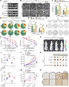
OTUD4 enhances radiosensitivity in NPC cells by promoting GSDME-dependent pyroptosis. A Western blotting showed the protein levels of OTUD4 and GSDME in HONE1 and 5-8F cells transfected with or without OTUD4 and shGSDME. B-G The above cells were exposed to the indicated irradiation dose, and then (B) phase-contrast cell imaging, (C) LDH release assay, (D) live cell imaging, (E) Annexin/PI assay, and (F) colony formation assay was performed at designated time points. G The dose-survival curves of the above cells are indicated. All data are presented as the mean ± SD of three independent experiments. H Representative bioluminescence images of tumors were taken on day 42. I Statistical analysis of photon flux (n = 5). J Tumor size was monitored every 4 days (n = 5). K Images of resected tumors. L Tumor weight on day 42 (n = 5). M Serum LDH concentrations were determined pre-radiotherapy and following the third and sixth radiotherapy treatments (n = 3). H Representative images of IHC staining of the tumor sections are shown. Scale bars represent 50 μm. ns, No significant difference. *P < 0.05, **P < 0.01, ***P < 0.001, ****P < 0.0001
Fig. 7
Fig. 7
The clinical relevance of the OTUD4/GSDME axis and radiotherapy response in NPC. A Representative immunohistochemical images of OTUD4 low- and high- expression in NPC tissues (left and right, respectively). The magnified inset area is shown at the bottom. Scale bars represent 50 μm. B The IHC score of OTUD4 in the radiosensitive and radioresistant tissues. C The association between OTUD4 expression and radiotherapy response, χ2 test. RS, radiosensitive; RR, radioresistant. D 5-year PFS for NPC was calculated by Kaplan–Meier analysis, compared using the Log Rank test, and stratified by low and high OTUD4 levels. Progression-free survival, PFS. E Representative NPC cases received radiotherapy alone, showing the relationship between OTUD4 expression, GSDME expression, serum LDH, and cancer regression. The yellow dashed lines indicate tumor in nasopharyngoscopy images. Scale bars represent 50 μm. RT, radiotherapy. F The correlation between OTUD4 expression and GSDME expression. G The correlation between OTUD4/GSDME expression and radiosensitivity. H Comparison of the progression-free survival and locoregional recurrence-free survival between 39 OTUD4low/GSDMElow, 56 OTUD4high/GSDME.high and 55 others. Actuarial probabilities were analyzed by Kaplan–Meier (Log Rank test). (I) Proposed model showing that GSDME-dependent pyroptosis is identified as a critical determinant of radiosensitivity in NPC. In addition, OTUD4/GSDME interaction inhibits OTUD4-mediated GSDME ubiquitination and stability, thereby promoting GSDME-dependent pyroptosis and enhancing radiosensitivity in NPC. *P < 0.05, **P < 0.01, ****P < 0.0001

References

    1. Chen YP, Chan ATC, Le QT, et al. Nasopharyngeal carcinoma. Lancet. 2019;394:64–80. doi: 10.1016/S0140-6736(19)30956-0. - DOI - PubMed
    1. Blanchard P, Lee A, Marguet S, et al. Chemotherapy and radiotherapy in nasopharyngeal carcinoma: an update of the MAC-NPC meta-analysis. Lancet Oncol. 2015;16:645–655. doi: 10.1016/S1470-2045(15)70126-9. - DOI - PubMed
    1. Miao J, Di M, Chen B, et al. A prospective 10-year observational study of reduction of radiation therapy clinical target volume and dose in early-stage nasopharyngeal carcinoma. Int J Radiat Oncol Biol Phys. 2020;107:672–682. doi: 10.1016/j.ijrobp.2020.03.029. - DOI - PubMed
    1. Zhang Y, Chen L, Hu GQ, et al. Gemcitabine and cisplatin induction chemotherapy in nasopharyngeal carcinoma. N Engl J Med. 2019;381:1124–1135. doi: 10.1056/NEJMoa1905287. - DOI - PubMed
    1. Zhan Y, Fan S. Multiple mechanisms involving in radioresistance of nasopharyngeal carcinoma. J Cancer. 2020;11:4193–4204. doi: 10.7150/jca.39354. - DOI - PMC - PubMed

Substances