Review
. 2022 Nov 21;20(1):180.
doi: 10.1186/s12964-022-00993-2.

How vascular smooth muscle cell phenotype switching contributes to vascular disease

Affiliations
Review

How vascular smooth muscle cell phenotype switching contributes to vascular disease

Genmao Cao et al. Cell Commun Signal. .

Abstract

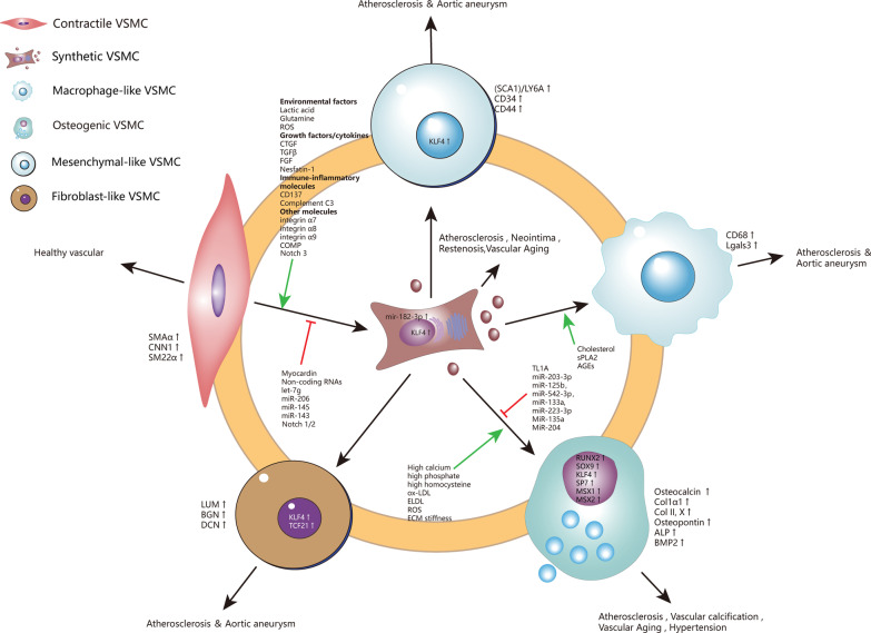
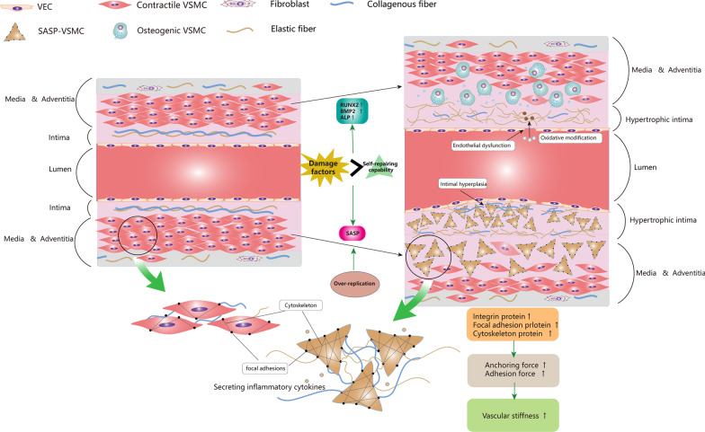
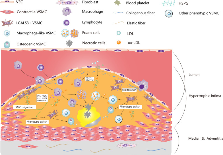
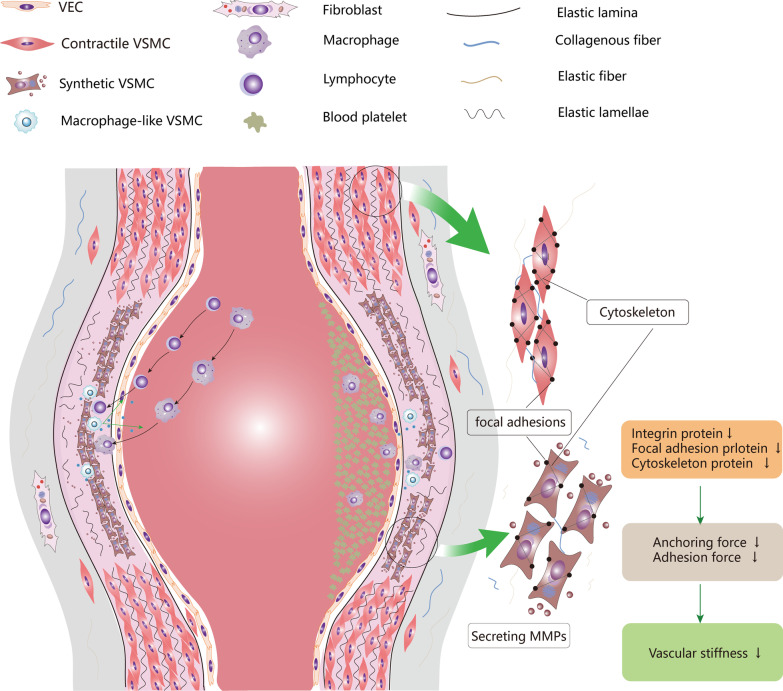
Vascular smooth muscle cells (VSMCs) are the most abundant cell in vessels. Earlier experiments have found that VSMCs possess high plasticity. Vascular injury stimulates VSMCs to switch into a dedifferentiated type, also known as synthetic VSMCs, with a high migration and proliferation capacity for repairing vascular injury. In recent years, largely owing to rapid technological advances in single-cell sequencing and cell-lineage tracing techniques, multiple VSMCs phenotypes have been uncovered in vascular aging, atherosclerosis (AS), aortic aneurysm (AA), etc. These VSMCs all down-regulate contractile proteins such as α-SMA and calponin1, and obtain specific markers and similar cellular functions of osteoblast, fibroblast, macrophage, and mesenchymal cells. This highly plastic phenotype transformation is regulated by a complex network consisting of circulating plasma substances, transcription factors, growth factors, inflammatory factors, non-coding RNAs, integrin family, and Notch pathway. This review focuses on phenotypic characteristics, molecular profile and the functional role of VSMCs phenotype landscape; the molecular mechanism regulating VSMCs phenotype switching; and the contribution of VSMCs phenotype switching to vascular aging, AS, and AA. Video Abstract.

Keywords: Aortic aneurysm; Atherosclerosis; Phenotype switching; Vascular aging; Vascular smooth muscle cells.


Conflict of interest statement

I declare that there is no competing interest.

Figures

Fig. 1
Fig. 1
High plasticity of VSMCs. Contractile VSMCs spontaneously modify their phenotype instantaneously to synthetic phenotype when vascular damage occurs. synthetic VSMCs undergo down-regulation of contractile gene expression, cytoskeleton remodeling, and cell reprogramming. Synthetic VSMCs then switch into various phenotypes under a sophisticated regulating network. VSMCs, vascular smooth muscle cells; ECM, extracellular matrix; LGALS3, galectin-3; HSPG, heparan sulfate proteoglycan; ApoB, apolipoprotein B; PDGF, platelet derived growth factor; TNF-α, tumor necrosis factor alpha; IFN-γ, Interferon-gamma; IL-1, Interleukin-1; MMPs, metalloproteases, SMAα, α-smooth muscle actin; CNN1, Calponin 1; SM22α, smooth muscle protein 22α; ROS, reactive oxygen species; CTGF, connective tissue growth factor; TGF-β, transforming growth factor-β; FGF, fibroblast growth factor; COMP, Cartilage oligomeric matrix protein; LUM, lumican; BGN, biglycan; DCN, decorin
Fig. 2
Fig. 2
VSMCs phenotype switching and vascular aging. In vascular aging, environmental damage factors trigger mitochondrial dysfunction, telomere attrition, DNA damage, epigenetic changes, oxidative stress, impaired resistance to molecular stressors, chronic low-grade inflammation, genomic instability in VSMCs. Once these damage factors accumulate to a certain extent and cannot be repaired, VSMCs switch to senescence­associated secretory phenotype (SASP). The impaired endothelial barrier caused by vascular aging accelerated the environmental damage on VSMCs. Meanwhile, overproliferation of VSMCs also contributes to SASP. SASP-VSMCs became irregular in morphology and highly expressed integrin protein, focal adhesion protein, and cytoskeleton protein. Cytoskeleton actin was connected to elastin and collagen in ECM through integrin-based focal adhesion, delivering anchoring force and adhesion force. When the above protein is up-regulated, not only increases the VSMCs stiffness but also increases the interaction between ECM and VSMCs, leading to increased vascular stiffness. SASP-VSMCs secrete a large amount of MMPs for decomposing elastin into fragments, synthesize collagen, and promote its crosslinking. This process increases ECM stiffness and decreases ECM elasticity. On the other hand, the high expression of RUNX2, BMP2, and ALP in senescent VSMCs makes it susceptible to switch to osteogenic VSMCs under high calcium or phosphorus environment. Osteogenic VSMCs release calcium phosphate enriched vesicles to promote VC. VEC, vascular endothelial cell; SASP, senescence­associated secretory phenotype; RUNX2, runt-related transcription factor 2; BMP2, bone morphogenetic protein-2; ALP, alkaline phosphatase; VC, vascular calcification
Fig. 3
Fig. 3
VSMCs phenotype switching in advanced atherosclerotic plaque. Due to damage or dysfunction of endothelial cells, circulating LDL (including ApoB) passes through the endothelial barrier into the subendothelium and is subsequently captured by HSPGs in the ECM. Subsequently, LDL is gradually oxidized into ox-LDL under free radicals or other oxidants. The accumulation of ox-LDL induces the migration of macrophages and monocytes to the lesion area. Macrophages engulf oxidized lipids and necrotic cells in the lesion, secrete inflammatory cytokines, and transform to foam cells. Finally, these foam cells are apoptotic and coalesce into a lipid-rich necrotic core in subendothelium. Macrophage-like VSMCs also engulf lipid and necrotic cell debris to form foam cells. However, the phagocytosis of macrophage-like VSMCs is lower than that of macrophages, disrupting the efficiency of lipid disposal and debris clearance in plaques, thereby exacerbating the formation of atherosclerotic plaques. Meanwhile, ox-LDL and various inflammatory cytokines (such as PDGF, TNF-α, IFN-γ, IL-1, MMP-2/9, etc.) can stimulate VSMCs to migrate from tunica media to tunica intima to form neointima and phenotype transformation occurred in the process The contractile VSMCs first transform into LGALS3 + VSMCs, this kind of cell is also known as pioneer cells, Stem/Endothelial/Macrophage (SEM) cells, or fibromyocytes. These transitional VSMCs can synthesize ECM, participate in the formation of fibrous cap, and can also transform into macrophage-like VSMCs, osteogenic VSMCs, and other types of VSMCs. Macrophage-like VSMCs derived foam cells secrete MMPs to destroy ECM in the fibrous cap, and osteogenic VSMCs participate in calcification of lipid necrotic core, both of which are generally thought to aggravate plaque instability. VEC, vascular endothelial cell; VSMC, vascular smooth muscle cell; ECM, extracellular matrix; LGALS3, galectin-3; LDL, low density lipoprotein; ox-LDL, oxidized low-density lipoprotein; HSPG, heparan sulfate proteoglycan; ApoB, apolipoprotein B; PDGF, platelet derived growth factor; TNF-α, tumor necrosis factor alpha; IFN-γ, interferon-gamma; IL-1, interleukin 1; MMPs, metalloproteases
Fig. 4
Fig. 4
VSMCs phenotype and aortic aneurysm. The aortic wall comprises tunica intima, tunica media, and tunica adventitia. Tunica intima consists of a single layer of endothelial cells and basement membrane. Tunica intima and tunica media are separated by internal elastic lamina. Tunica media is composed of multilayer annular elastic lamellae and annular arranged VSMCs and is filled with collagen-rich and elastin-rich ECM. The tunica adventitia is composed of collagen, elastic fiber, and fibroblast, separated from the tunica media by the external elastic lamina. VSMCs and elastic fibers in tunica media provide active and passive contractility respectively. In aortic aneurysm, VSMCs switch to synthetic phenotype, down-regulate contractile protein, and secret massive MMPs and inflammatory cytokines. Down-regulated contractile proteins mean that the aorta cannot provide enough contractile force to restore its original diameter during pulse pumping. Moreover, the abundant collagen and Elastin in the aortic ECM were decomposed into fragments by MMPs, resulting in disrupted elastic lamina and elastic lamellae. Therefore, the aorta does not generate sufficient passive contractile force in dilated condition. As synthetic VSMCs express less integrin protein, focal adhesion protein, cytoskeleton protein, the anchoring force between VSMCs and ECM decreases, resulting in weaker passive contraction force. Macrophage-like VSMCs, on the other hand, secrete inflammatory factors and chemokines, recruit monocytes, macrophages, and lymphocytes to infiltrate into the aortic wall. These cells form chronic inflammation and participate in the destruction of aortic wall. VEC, vascular endothelial cell; VEC, vascular endothelial cell; MMPs, metalloproteases; ECM, extracellular matrix

References

    1. Roth GA, et al. Global, regional, and national burden of cardiovascular diseases for 10 causes, 1990 to 2015. J Am Coll Cardiol. 2017;70(1):1–25. doi: 10.1016/j.jacc.2017.04.052. - DOI - PMC - PubMed
    1. Pagidipati NJ, Gaziano TA. Estimating deaths from cardiovascular disease: a review of global methodologies of mortality measurement. Circulation. 2013;127(6):749–756. doi: 10.1161/CIRCULATIONAHA.112.128413. - DOI - PMC - PubMed
    1. Touyz RM, et al. Vascular smooth muscle contraction in hypertension. Cardiovasc Res. 2018;114(4):529–539. doi: 10.1093/cvr/cvy023. - DOI - PMC - PubMed
    1. Yamin R, Morgan KG. Deciphering actin cytoskeletal function in the contractile vascular smooth muscle cell. J Physiol. 2012;590(17):4145–4154. doi: 10.1113/jphysiol.2012.232306. - DOI - PMC - PubMed
    1. Christensen KL, Mulvany MJ. Location of resistance arteries. J Vasc Res. 2001;38(1):1–12. doi: 10.1159/000051024. - DOI - PubMed