. 2022 Nov 21;10(1):168.
doi: 10.1186/s40478-022-01472-y.

Molecular and spatial heterogeneity of microglia in Rasmussen encephalitis

Affiliations

Molecular and spatial heterogeneity of microglia in Rasmussen encephalitis

Jesse J Westfall et al. Acta Neuropathol Commun. .

Abstract

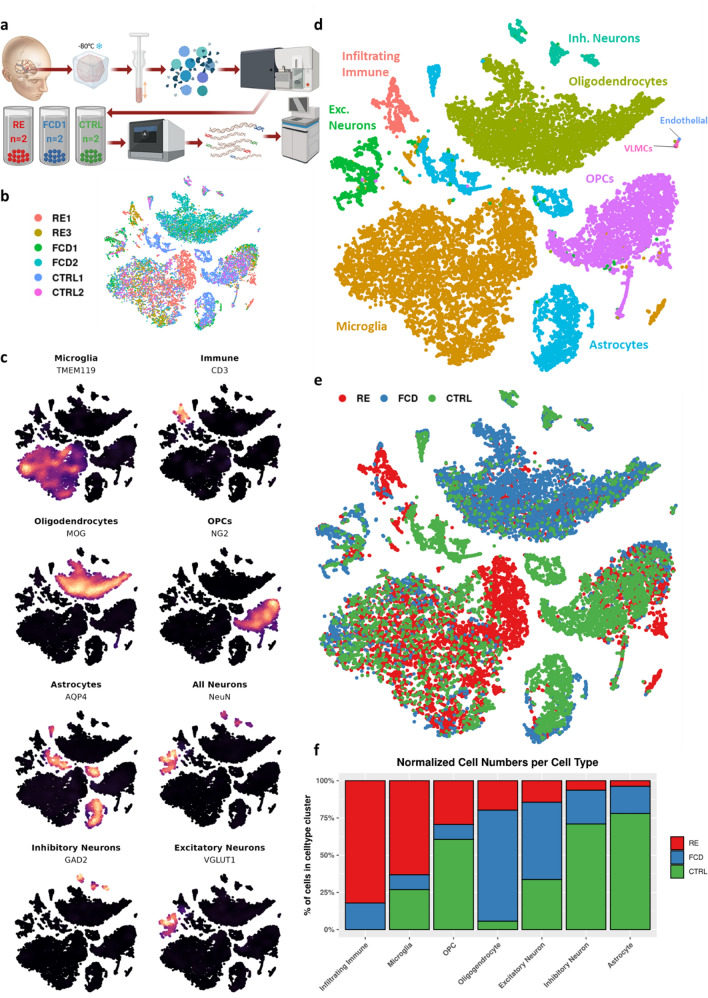
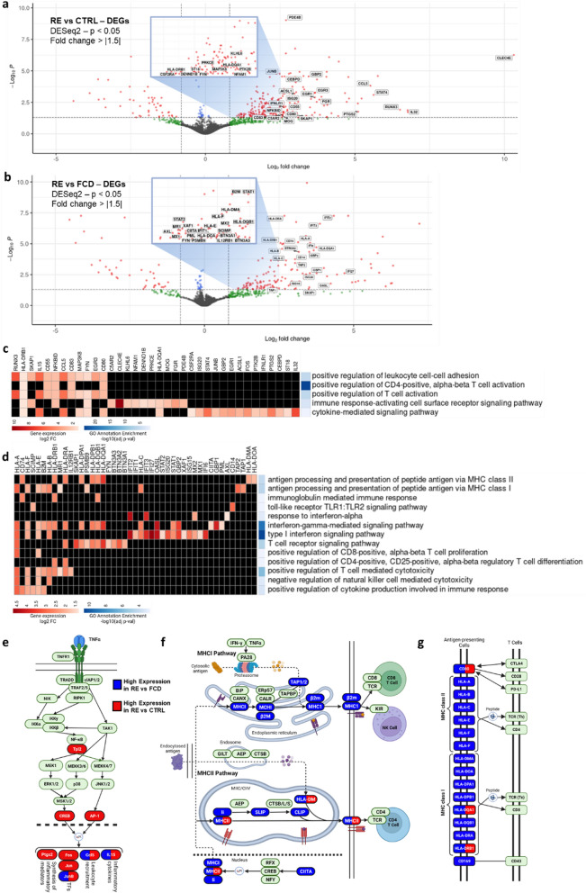
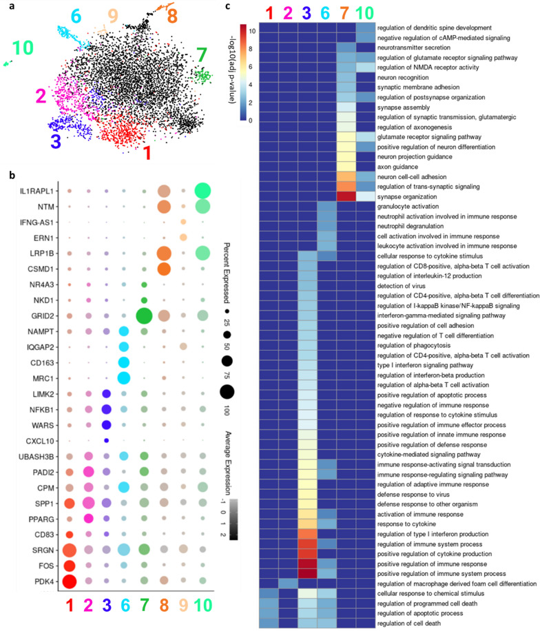
Rasmussen encephalitis (RE) is a rare childhood neurological disease characterized by progressive unilateral loss of function, hemispheric atrophy and drug-resistant epilepsy. Affected brain tissue shows signs of infiltrating cytotoxic T-cells, microglial activation, and neuronal death, implicating an inflammatory disease process. Recent studies have identified molecular correlates of inflammation in RE, but cell-type-specific mechanisms remain unclear. We used single-nucleus RNA-sequencing (snRNA-seq) to assess gene expression across multiple cell types in brain tissue resected from two children with RE. We found transcriptionally distinct microglial populations enriched in RE compared to two age-matched individuals with unaffected brain tissue and two individuals with Type I focal cortical dysplasia (FCD). Specifically, microglia in RE tissues demonstrated increased expression of genes associated with cytokine signaling, interferon-mediated pathways, and T-cell activation. We extended these findings using spatial proteomic analysis of tissue from four surgical resections to examine expression profiles of microglia within their pathological context. Microglia that were spatially aggregated into nodules had increased expression of dynamic immune regulatory markers (PD-L1, CD14, CD11c), T-cell activation markers (CD40, CD80) and were physically located near distinct CD4+ and CD8+ lymphocyte populations. These findings help elucidate the complex immune microenvironment of RE.

Keywords: Cytokine signaling; Encephalitis; Epilepsy; Inflammation; Microglial nodules; Single nucleus RNA-seq; Spatial proteomics.


Conflict of interest statement

The authors declare no competing interests.

Figures

Fig. 1
Fig. 1
Microglial and lymphocytic cell populations are expanded in Rasmussen encephalitis. a Processing of cortical tissue from RE (N = 2), FCD (N = 2), and unaffected (N = 2) individuals for snRNA-seq was performed using 10x Genomics 3’gene expression kit. b tSNE plot highlighting patient contribution to each cluster. c tSNE plots overlaid with feature density for canonical cell type markers (endothelial and VLMC not shown) d tSNE plot colored by cell type, determined using Seurat v.4.0 reference and query workflow (see methods). e tSNE showing contribution of diagnosis by cluster. f Normalized counts of nuclei per cluster by diagnosis
Fig. 2
Fig. 2
Gene expression signatures of microglia in Rasmussen encephalitis. a Volcano plot showing differentially expressed genes in RE vs CTRL (DESeq2 adjusted p-value < 0.05, FC > 1.5). Labeled genes are shown on heatmap in Fig. 2c. b Volcano plot showing differentially expressed genes in RE vs FCD (DESeq2 adjusted p-value < 0.05, FC > 1.5). Labeled genes are shown on heatmap in Fig. 2d. c Heatmap showing individual gene contribution (via positive fold-change) to GO biological process annotation enrichment in RE vs CTRL. d Heatmap showing individual gene contribution (via positive fold-change) to GO biological process annotation enrichment in RE vs FCD. e–g Selections from KEGG Pathways: TNF signaling pathway e, Antigen processing and presentation f and Cell adhesion molecules g highlighting differentially high expression of genes in RE vs both CTRL and FCD controls
Fig. 3
Fig. 3
Microglial heterogeneity visualized via Louvain clustering. a tSNE plot showing eight clusters containing differentially high expression of genes with immune or inflammatory regulatory function. b Dot plot showing percent and average expression of a selection of differentially highly expressed genes in clusters from Fig. 3a with immune or inflammatory regulatory function c Heatmap showing enrichment of GO biological process annotations in clusters with enriched GO terms from Fig. 3a
Fig. 4
Fig. 4
Microglia have distinct expression signatures based on spatial context in Rasmussen encephalitis. a Example ROIs representing a microglial nodule (left, yellow) versus unaggregated microglia (right, light blue) with immunofluorescent staining of microglia (IBA1, green), lymphocytes (CD45, red), and nuclei (SYTO13, blue). b Volcano plot of significant abundantly expressed proteins detected in microglial nodules versus unaggregated microglia. c 20 × micropictograph confirming high expression of CD8 in CD45+ cells near microglial nodules. d-g Relative cell type marker expression d T-cell subtype marker expression e macrophage subtype marker expression f cell activation markers g in CD45+ ROIs for each patient

References

    1. Al Nimer F, Jelcic I, Kempf C, Pieper T, Budka H, Sospedra M, Martin R. Phenotypic and functional complexity of brain-infiltrating T cells in Rasmussen encephalitis. Neurol Neuroimmunol Neuroinflamm. 2018;5:e419. doi: 10.1212/NXI.0000000000000419. - DOI - PMC - PubMed
    1. Alquicira-Hernandez J, Powell JE. Nebulosa recovers single-cell gene expression signals by kernel density estimation. Bioinformatics. 2021;37:2485–2487. doi: 10.1093/bioinformatics/btab003. - DOI - PubMed
    1. Alsegiani AS, Shah ZA. The role of cofilin in age-related neuroinflammation. Neural Regen Res. 2020;15:1451–1459. doi: 10.4103/1673-5374.274330. - DOI - PMC - PubMed
    1. Andoh M, Ikegaya Y, Koyama R. Synaptic pruning by microglia in epilepsy. J Clin Med. 2019;8:2170. doi: 10.3390/jcm8122170. - DOI - PMC - PubMed
    1. Andoh M, Koyama R. Microglia regulate synaptic development and plasticity. Dev Neurobiol. 2021;81:568–590. doi: 10.1002/dneu.22814. - DOI - PMC - PubMed

Publication types

Supplementary concepts